. 2017 Aug 29;114(35):9337-9342.
doi: 10.1073/pnas.1619216114. Epub 2017 Aug 16.

Modular tissue engineering for the vascularization of subcutaneously transplanted pancreatic islets

Affiliations

Modular tissue engineering for the vascularization of subcutaneously transplanted pancreatic islets

Alexander E Vlahos et al. Proc Natl Acad Sci U S A. .

Abstract

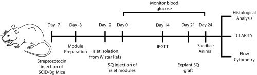
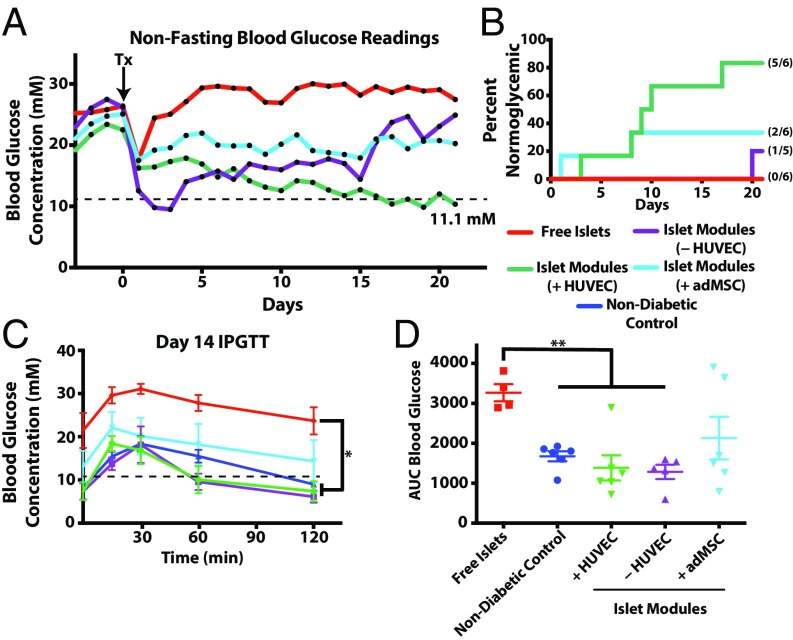
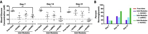
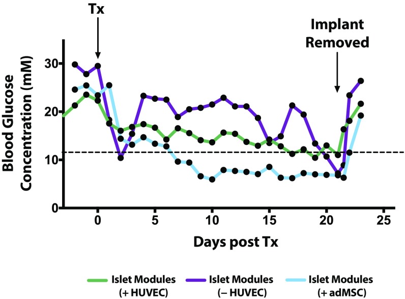
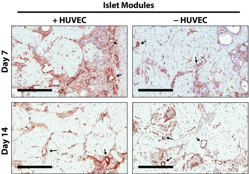
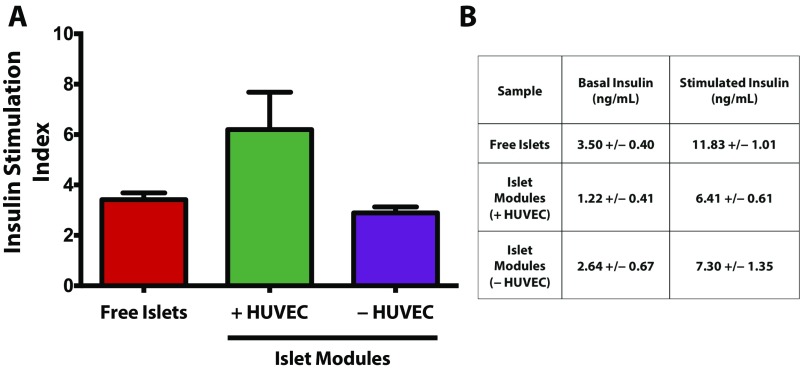
The transplantation of pancreatic islets, following the Edmonton Protocol, is a promising treatment for type I diabetics. However, the need for multiple donors to achieve insulin independence reflects the large loss of islets that occurs when islets are infused into the portal vein. Finding a less hostile transplantation site that is both minimally invasive and able to support a large transplant volume is necessary to advance this approach. Although the s.c. site satisfies both these criteria, the site is poorly vascularized, precluding its utility. To address this problem, we demonstrate that modular tissue engineering results in an s.c. vascularized bed that enables the transplantation of pancreatic islets. In streptozotocin-induced diabetic SCID/beige mice, the injection of 750 rat islet equivalents embedded in endothelialized collagen modules was sufficient to restore and maintain normoglycemia for 21 days; the same number of free islets was unable to affect glucose levels. Furthermore, using CLARITY, we showed that embedded islets became revascularized and integrated with the host's vasculature, a feature not seen in other s.c.

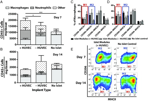
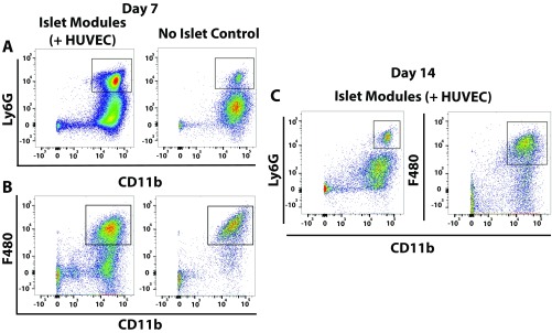
Studies: Collagen-embedded islets drove a small (albeit not significant) shift toward a proangiogenic CD206+MHCII-(M2-like) macrophage response, which was a feature of module-associated vascularization. While these results open the potential for using s.c. islet delivery as a treatment option for type I diabetes, the more immediate benefit may be for the exploration of revascularized islet biology.

Keywords: CLARITY; islet revascularization; pancreatic islets; subcutaneous transplant; tissue engineering.


Conflict of interest statement

The authors declare no conflict of interest.

Figures

Fig. S1.
Fig. S1.
Schematic of the experimental setup. SCID/Bg mice were made diabetic using streptozotocin 7 d before s.c. implantation. Two days before the implantation, Wistar rat islets were isolated and were either embedded in modules (with other cells) or left as free islets. The islet modules (750 IEQ) were then cultured for 1 d before s.c. injection. Blood glucose monitoring was performed daily, and at day 14 an IPGTT was administered. At day 21, implants in animals that had returned to normoglycemia were removed without killing the animal, and animals were followed for an additional 3 d to confirm that the implant was responsible for returning the diabetic mouse to normoglycemia. Explants were analyzed at days 7, 14, and 21/24 using histology, CLARITY, and flow cytometry.
Fig. 1.
Fig. 1.
The s.c. modular islet transplants with HUVEC restored normoglycemia. (A) Shown are average nonfasting blood glucose levels for diabetic mice implanted with free islets (n = 6) or islets in modules with (+HUVEC) (n = 6) or without (−HUVEC; collagen only; n = 5) HUVEC or with adMSC and HUVEC (+adMSC) (n = 6). While all free-islet recipients remained hyperglycemic for the entire experiment, islet modules with HUVEC were able to lower glucose levels to normal in all but one animal. Individual glucose values are shown in Fig. S2. Islet modules without HUVEC or with adMSC and HUVEC had some therapeutic effect on the average glucose levels; however, most of these animals remained hyperglycemic. The dotted line represents the threshold for normoglycemia (11.1 mM). (B) Kaplan–Meier plot showing when animals became normoglycemic. Not shown are animals that became normoglycemic but reverted to hyperglycemia within the 21-d observation period. Mice implanted with islet modules with HUVEC reversed diabetes significantly faster than the free-islet group (P = 0.0043, log-rank, Mantel-cox test) or the group with islet modules without HUVEC (P = 0.0196, log-rank, Mantel–Cox test). Numbers in parentheses indicate the fraction of animals that are shown in this plot. (C) IPGTT glucose profile at day 14. Animals were fasted for 4 h before IPGTT was performed. A repeated-measures ANOVA with a Games–Howell post hoc test revealed a significant difference between the profiles of the free-islet group and the nondiabetic control and the groups with islets embedded in modules with or without HUVEC. (D) AUC analysis confirmed that mice in the free-islet group were intolerant of the glucose challenge and that there was a significant difference between the free-islet group and both groups receiving islets embedded in modules with (n = 6) or without (n = 5) HUVEC. There was no significant difference among the islet module groups. *P < 0.05, **P < 0.01.
Fig. S2.
Fig. S2.
Nonfasting blood glucose readings for each animal were taken at days 7, 14, and 21. (A) There were significant differences in the average nonfasting blood glucose levels in animals transplanted with free islets and those implanted with islet modules with HUVEC at each time point. One or two module recipients were normoglycemic at day 7, but only the islet (+HUVEC) group had the majority of animals becoming normoglycemic by day 14 and sustaining normoglycemia (11.1 mM, represented by the dashed line) to day 21). *P < 0.05, **P < 0.01. (B) The percentage of animals that were normoglycemic at days 7, 14, and 21. While two animals in the islets in collagen gel group (islet modules without HUVEC) were normoglycemic at days 7 and 14, these animals became hyperglycemic before day 21; a third animal became normoglycemic at day 20 and remained so until day 21. Animals transplanted with free islets remained hyperglycemic for the entire 21 d.
Fig. S3.
Fig. S3.
The s.c. islet module implants are responsible for the return to normoglycemia. Animals that had achieved normoglycemia (<11.1 mM) rapidly returned to hyperglycemia after the s.c. implant was removed. Nonfasting blood glucose levels were tracked for an additional 3 d after removal. This graph reports only the subset of animals that had returned to normoglycemia and remained so for the entire observation period of 21 d. Islet modules (+HUVEC) n = 5 (of 6), (−HUVEC) n = 1 (of 5), and (+adMSC) n = 2 (of 6). Remaining animals were not subject to this analysis, since they failed earlier. The graph shows average nonfasting blood glucose levels. Tx, transplantation.
Fig. S4.
Fig. S4.
Insulin-positive islets are present in modules after 21 d in vivo. (A) Masson’s trichrome (Upper Row) and insulin (Lower Row) staining for each of the treatment groups. (Scale bars: 500 μm.) (B) At day 21, the average insulin + pixel density was significantly higher in islet modules with (n = 5) or without (n = 2) HUVEC than in islet modules with adMSC (n = 5). Islet modules without HUVEC could not be found in some of the animals because they had degraded. Free islets could not be found because there was no identifiable implant in the s.c. space. The graph represents average ± SEM; *P < 0.05.
Fig. 2.
Fig. 2.
Module-associated vascularization. (A) Module-associated vasculature was characterized by staining with CD31 and SMA in serial sections at day 21. CD31+ vessels (black arrows, Upper Row) were present in all groups, including the without-HUVEC group, and many of these vessels were also positive for SMA, suggesting that the vessels were mature. (Scale bars: 200 μm.) (B) CD31 vessel counts. There were significantly more vessels when islet modules were coated with HUVEC (n = 6) than without HUVEC (n = 5) at days 7 and 14 (two-sided t test). At day 21 the density of CD31+ vessels was similar between the two module groups (P > 0.05). Islet modules without HUVEC degraded over time, and not many implants could be found at day 21. n = 5 for islet modules with HUVEC and n = 2 for islet modules without HUVEC at day 21. *P < 0.05, **P < 0.01; error bars indicate SEM.
Fig. S5.
Fig. S5.
Representative histology of s.c.-transplanted islet modules. Explants were assessed using CD31, SMA, and insulin staining on serial sections to illustrate the relationship among the stained cells. Mature module-associated vasculature is denoted by arrows, and islets are outlined by dashed lines. (Scale bars: 200 μm.)
Fig. S6.
Fig. S6.
UEA-1 histological analysis of islet module implants. UEA-1 stains human EC, which contribute to graft-derived vessels. (A) UEA-1 staining was absent at day 21, suggesting that all the module-associated vasculature was host-derived. (Scale bars: 300 μm.) (B) At day 7, some UEA-1+ vessels were present (arrows); however by day 14 there was little positive UEA-1 staining within the implants. (Scale bars: 200 μm.) Insets show enlarged views of areas marked by dashed boxes. (Magnification: 25×.)
Fig. 3.
Fig. 3.
Islets in endothelialized modules were revascularized. (A) Islets embedded within endothelialized modules had CD31+ staining within the interior of the islets at day 21. (Scale bars: 200 μm.) (BD) CLARITY-processed islet modules with HUVEC at day 7 (B), day 14 (C), and day 21 (D) show islet revascularization over time. (E) An embedded rat islet (white dashed box in D) was computationally mapped using IMARIS to visualize revascularization of the islet and its vasculature morphology. Movies are shown in Supporting Information. (Scale bars: 150 μm.)
Fig. S7.
Fig. S7.
The percentage of CD31+ pixels within the islet area was calculated for islet modules with (n = 5) or without (n = 2) HUVEC and islet modules with adMSC (n = 5) at day 21. There were no significant differences in the percentage of CD31+ pixels among the three groups using a one-way ANOVA. Few of the islet modules without HUVEC were found at day 21 due to degradation of the graft. Free islets could not be analyzed because there was no identifiable implant in the s.c. space. Error bars indicate SEM.
Fig. S8.
Fig. S8.
Intraislet vasculature at day 21 in islet modules with HUVEC. (A) An example of a z-stack of an individual islet embedded in endothelialized modules at day 21, stained with GSL1 lectin (white). (B) MATLAB was used to convert the image to a binary image that then was used to quantify the vessel volume relative to the total islet volume. (C) Intraislet vessels contributed to 20 ± 3% of the total islet volume (n = 8). Error bars indicate SEM.
Fig. 4.
Fig. 4.
Inflammatory response associated with transplanted islets. (A and B) Many CD45+ cells were seen at day 7 with islet-containing modules; this response decreased by day 14. Shown is the distribution of CD45+ cells found within explants for islet modules with and without HUVEC and the no-islet (+HUVEC) control at day 7 (A) and day 14 (B). There were significantly more neutrophils present in islet modules with HUVEC than in islet modules without HUVEC (*P < 0.05) and in the no-islet control modules (***P < 0.0005) at day 7. (CE) Macrophage polarization was assessed by coexpression of CD206 and MHCII, where M1-like macrophages are CD206MHCII+ and M2-like macrophages are CD206+MHCII. Quantification of macrophage phenotype at day 7 (C) and day 14 (D) with representative flow plots of islet modules with HUVEC and no-islet controls at day 7 and day 14 (E). Error bars represent mean ± SEM, n = 5 for all groups, except in the day 7 no-islet control (n = 3) and in the day 14 islet module with HUVEC and no-islet control (n = 4).
Fig. S9.
Fig. S9.
(A and B) Representative flow plots for neutrophils (A) and macrophages (B) in islet modules with HUVEC and no-islet controls at day 7. (C) Representative flow plots for neutrophils (Left) and macrophages (Right) at day 14 in islet modules with HUVEC.
Fig. S10.
Fig. S10.
Representative histology from SMA-stained slides of islet modules with or without HUVEC at days 7 and 14. At day 7, vessels in both groups were SMA+; however, islet modules with HUVEC were infiltrated with more SMA+ cells not associated with vessels. At day 14, SMA+ cells were predominately associated with the vasculature in islet modules with and without HUVEC (arrows). (Scale bars: 300 μm.)
Fig. S11.
Fig. S11.
(A) Glucose-stimulated insulin secretion assay of free islets and islets embedded in modules with and without HUVEC. There were no significant differences (P = 0.076) in the insulin stimulation index among the three groups after 4 d of culture in vitro. (B) Absolute values of insulin secreted at basal and stimulated glucose solutions. n = 3 in each group; error bars in A indicate SEM.

Similar articles

Cited by

References

    1. Shapiro AMJ, et al. Islet transplantation in seven patients with type 1 diabetes mellitus using a glucocorticoid-free immunosuppressive regimen. N Engl J Med. 2000;343:230–238. - PubMed
    1. Berney T, et al. Long-term insulin-independence after allogeneic islet transplantation for type 1 diabetes: Over the 10-year mark. Am J Transplant. 2009;9:419–423. - PubMed
    1. Markmann JF, et al. Insulin independence following isolated islet transplantation and single islet infusions. Ann Surg. 2003;237:741–749, discussion 749–750. - PMC - PubMed
    1. McCall M, Shapiro AM. Update on islet transplantation. Cold Spring Harb Perspect Med. 2012;2:a007823. - PMC - PubMed
    1. Biarnés M, et al. Beta-cell death and mass in syngeneically transplanted islets exposed to short- and long-term hyperglycemia. Diabetes. 2002;51:66–72. - PubMed

Publication types

Grants and funding