. 2018 Jan;75(1):33-48.
doi: 10.1007/s00018-017-2607-9. Epub 2017 Aug 8.

Rab44, a novel large Rab GTPase, negatively regulates osteoclast differentiation by modulating intracellular calcium levels followed by NFATc1 activation

Affiliations

Rab44, a novel large Rab GTPase, negatively regulates osteoclast differentiation by modulating intracellular calcium levels followed by NFATc1 activation

Yu Yamaguchi et al. Cell Mol Life Sci. 2018 Jan.

Abstract

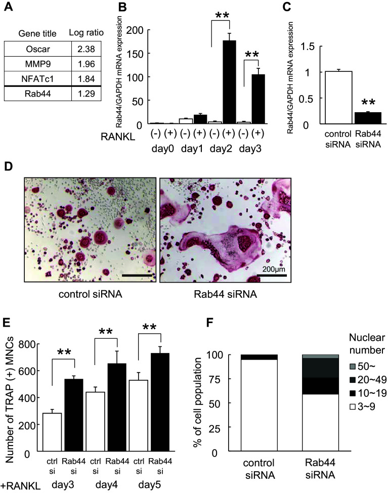
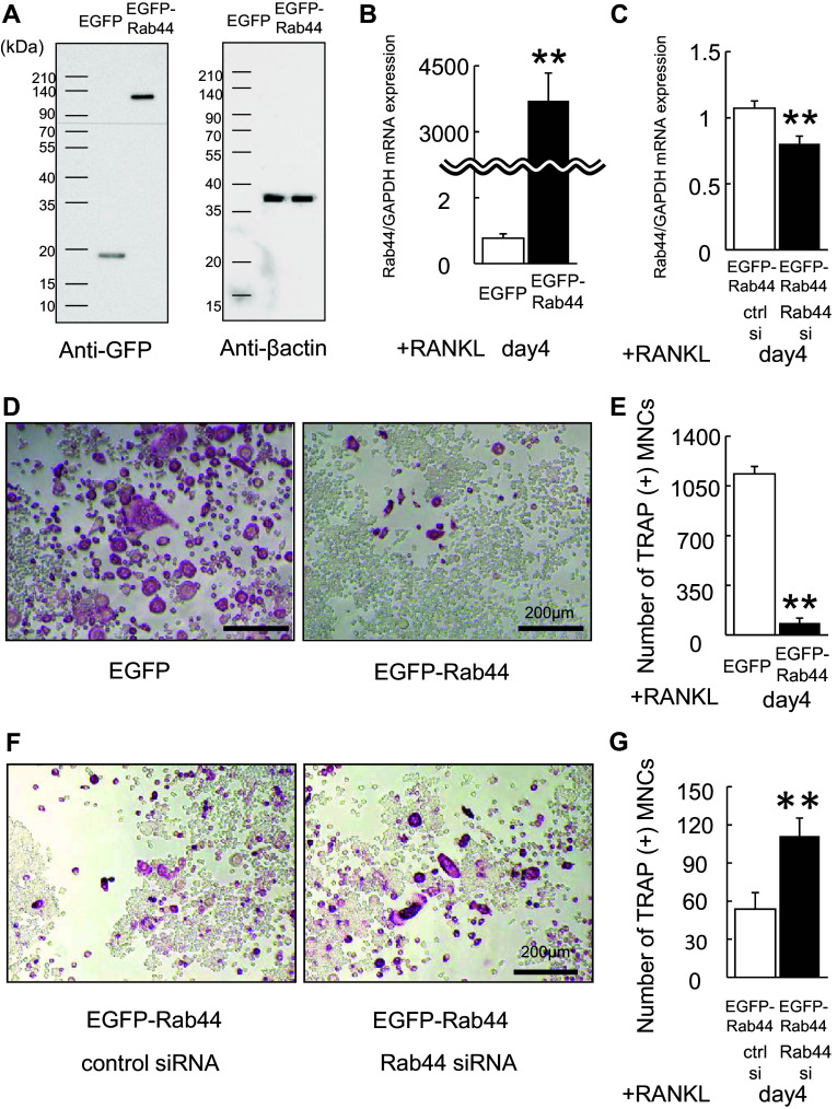
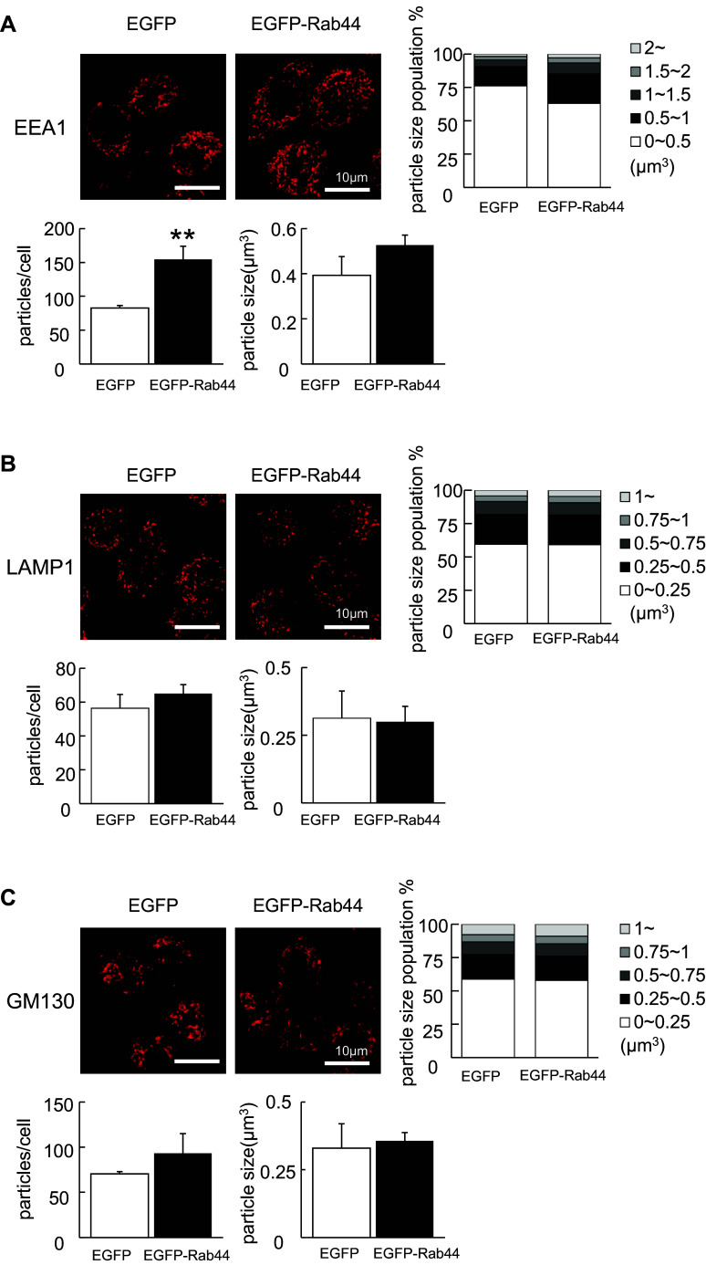
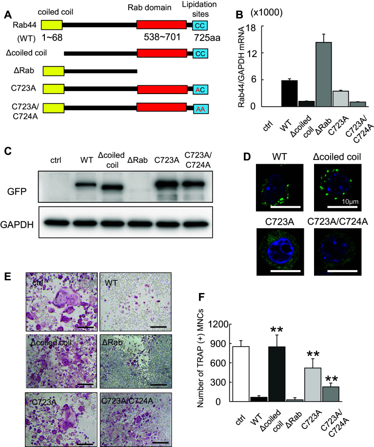
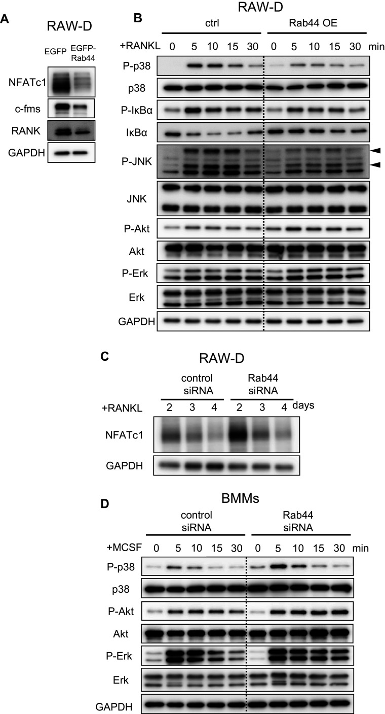
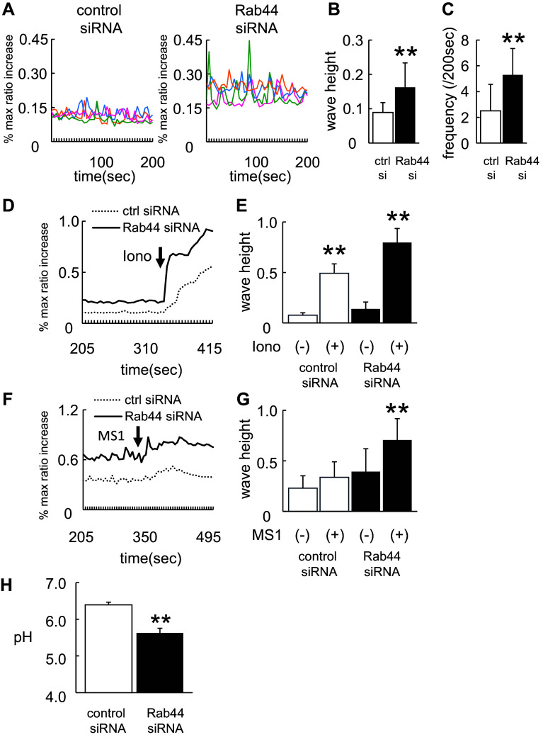
Rab44 is an atypical Rab GTPase that contains some additional domains such as the EF-hand and coiled-coil domains as well as Rab-GTPase domain. Although Rab44 genes have been found in mammalian genomes, no studies concerning Rab44 have been reported yet. Here, we identified Rab44 as an upregulated protein during osteoclast differentiation. Knockdown of Rab44 by small interfering RNA promotes RANKL-induced osteoclast differentiation of the murine monocytic cell line, RAW-D or of bone marrow-derived macrophages (BMMs). In contrast, overexpression of Rab44 prevents osteoclast differentiation. Rab44 was localized in the Golgi complex and lysosomes, and Rab44 overexpression caused an enlargement of early endosomes. A series of deletion mutant studies of Rab44 showed that the coiled-coil domain and lipidation sites of Rab44 is important for regulation of osteoclast differentiation. Mechanistically, Rab44 affects nuclear factor of activated T-cells c1 (NFATc1) signaling in RANKL-stimulated macrophages. Moreover, Rab44 depletion caused an elevation in intracellular Ca2+ transients upon RANKL stimulation, and particularly regulated lysosomal Ca2+ influx. Taken together, these results suggest that Rab44 negatively regulates osteoclast differentiation by modulating intracellular Ca2+ levels followed by NFATc1 activation.

Keywords: Intracellular Ca2+ levels; NFATc1; Osteoclast; Rab GTPase; Rab44.


Figures

Fig. 1
Fig. 1
Upregulation of Rab44 during osteoclastogenesis and knockdown of Rab44 in RAW-D-derived osteoclasts. a List of upregulated transcripts in osteoclasts cultured under rapid differentiation conditions compared to those under slow differentiation conditions. Bone marrow-macrophages were cultured on plastic plate (rapid differentiation conditions) and dentin (slow differentiation conditions) for 72 h in the presence of M-CSF (30 ng/mL) and RANKL (50 ng/mL). Total RNA from these cells was analyzed by Affymetrix Microarray system. b Quantitative RT-PCR analysis of Rab44mRNA expression levels in RANKL-stimulated RAW-D cells. The data are represented as mean ± SD of values from three independent experiments. **P < 0.01; compared with the control cells. c Knockdown efficacy was evaluated by measuring the Rab44 mRNA levels. After incubation with RANKL (100 ng/mL) for 24 h, cells were transfected with control or Rab44-specific siRNA (10 pmol) for an additional 24 h in the presence of RANKL. **P < 0.01; compared with the control cells. d RAW-D cells were transfected with either control or Rab44-specific siRNA. Following stimulation with RANKL (100 ng/mL) for 72 h, osteoclasts were analyzed by TRAP-staining. Bars 200 μm. e The number of TRAP-positive multinucleated osteoclasts per viewing field was counted. **P < 0.01; compared with the control cells. f Total nucleus number of TRAP positive multinucleated osteoclasts, but not TRAP-negative mononucleated cells following a 72 h culture, was counted and classified per viewing field
Fig. 2
Fig. 2
Overexpression of Rab44 in RAW-D cells. a RAW-D cells were transduced with either retrovirus vector containing EGFP-tagged Rab44 or only EGFP (control). The cultured cells were harvested at day 3 and lysates were subjected to western blot analysis with anti-GFP antibody or anti-β actin antibody as a control. b Quantitative RT-PCR analysis of the Rab44 mRNA expression levels in RAW-D cells expressing EGFP or EGFP-Rab44 after 4 days stimulation with RANKL. The data are represented as mean ± SD of values from five independent experiments. **P < 0.01; compared with the control cells. c Knockdown efficacy was evaluated by measuring by Rab44mRNA levels in EGFP-Rab44-expressing RAW-D cells transfected together with control or Rab44 siRNA after 4 days RANKL stimulation. **P < 0.01; compared with the control cells. d The EGFP and EGFP-Rab44-overexpressing RAW-D cells were cultured with RANKL (100 ng/mL) for the 4 days. The cells were fixed and stained for TRAP. Bars 200 μm. e The number of TRAP-positive MNCs in control and Rab44-overexpressing cells was counted at indicated day. **P < 0.01; compared with the control cells. F Rab44-overexpressing RAW-D cells were transfected with control or Rab44-specific siRNA for 24 h, and stimulated with RANKL (100 ng/mL) for the 4 days. The cells were fixed and stained for TRAP. Bars 200 μm. G The number of TRAP-positive MNCs in Rab44-overexpressing cells transfected with control or Rab44 siRNA was counted at the indicated day. **P < 0.01; compared with the control cells
Fig. 3
Fig. 3
Subcellular localization of EGFP-Rab44-overexpressing RAW-D cells. The cells on cover glasses were fixed, permeabilized with 0.2% Triton X-100 in PBS, and then allowed to react with antibodies against a KDEL (marker for ER), b GM130 (marker for the Golgi complex), c LAMP1 (marker for late endosomes/lysosomes), and d EEA1 (marker for early endosomes). After washing, the samples were incubated with a fluorescence-labeled secondary antibody and then were visualized by confocal laser microscopy. Bar 10 μm
Fig. 4
Fig. 4
Comparison of organelle size between EGFP-Rab44-overexpressing and control RAW-D cells. The cells on cover glasses were fixed, permeabilized with 0.2% Triton X-100 in PBS, and then allowed to react with antibodies against a EEA1 b LAMP1, and c GM130 on the cover glasses. After washing, the samples were incubated with a fluorescence-labeled secondary antibody and then were visualized by confocal laser microscopy. The data are representative of five independent experiments. Bar 10 μm. The number of these organelles as particles per a cell was quantified from images obtained by confocal microscopy using IMARIS 6.0 software (Bitplane, Zurich, Switzerland). Data are the means and standard deviation from three independent experiments. The particle size was measured by (μm3) using IMARIS and classified. **P < 0.01; compared with the control cells
Fig. 5
Fig. 5
Effects of expression of Rab44 mutants on osteoclast differentiation. a Schematic representation of mouse Rab44 and its mutants used in this study. Rab44 consists of a coiled-coil domain, and a Rab domain. Lipidation sites are at amino acid residues of 723 and 724. b Quantitative RT-PCR analysis of Rab44 mRNA expression levels of osteoclasts expressing EGFP-tagged Rab44 mutants. c Cell lysates (same protein amounts) were subjected to SDS–PAGE followed by western blotting with antibodies against GFP or GAPDH (control). d Typical EGFP fluorescence images of RAW-D cells expressing EGFP-Rab44 mutants and WT. The cells on cover glasses were fixed, and then allowed to DAPI staining. Bars 10 μm. e Typical TRAP-staining images of osteoclasts expressing EGFP-Rab44 mutants, WT and EGFP alone as a control. RAW-D cells expressing the control or Rab44 were cultured with RANKL (100 ng/mL) for 4 days. TRAP staining was performed. Bars 200 μm. f The number of TRAP-positive MNCs per viewing field was counted. **P < 0.01; compared with the WT cells
Fig. 6
Fig. 6
Effects of Rab44 on NFATc1 signaling in RANKL-stimulated macrophages. a Control or Rab44-expressing RAW-D cells were cultured with RANKL (100 ng/mL) for 1 day. Cell lysates (same protein amounts) were subjected to SDS-PAGE followed by western blotting with antibodies against NFATc1, c-fms, RANK, and GAPDH (control). b Control or Rab44-expressing RAW-D cells were pre-incubated for 2 h in serum-free media in the absence of RANKL. After adding RANKL, the cells were incubated for the indicated times, consequently harvested. Cell lysates (same protein amounts) were subjected to SDS-PAGE followed by western blotting with antibodies to p-p-38, p-38, p-IκBα, IκBα, p-JNK, JNK, p-Akt, Akt, p-Erk, Erk, and GAPDH (control). c The protein levels of NFATc1 in the control and Rab44-knockdown cells after RANKL stimulation. Control or Rab44-knockdown cells were cultured with RANKL (100 ng/mL) for 2–4 days. Cell lysates (same protein amounts) were subjected to SDS-PAGE followed by western blotting with antibodies against NFATc1, and GAPDH (control). d The control or Rab44-knockdown bone marrow-macrophages (BMMs) were pre-incubated for 2 h in serum free media in the absence of M-CSF. After adding M-CSF (50 ng/mL), the cells were incubated for the indicated time periods, and subsequently harvested. Cell lysates (equivalent protein amounts) were subjected to SDS-PAGE followed by western blotting with antibodies to p-p-38, p-38, p-Akt, Akt, p-Erk, Erk, and GAPDH (control)
Fig. 7
Fig. 7
Effects of Rab44 on lysosomal Ca2+ influx and acidic pH in the endosomes/lysosomes. a Ca2+ oscillations in control and Rab44-knockdown RAW-D cells after RANKL treatment. Cells were stimulated with RANKL (100 ng/mL) for 24 h, and then transfected with control or Rab44 siRNA for further 24 h. The cells were washed and then loaded with 3 μM Fluo 4-AM, or 3 μM Fura Red AM for 1 h in serum-free α-MEM. The cells were then washed and analyzed by confocal laser-scanning microscopy. Data show the results obtained from four cells. b Mean wave height of Ca2+ oscillations in control and Rab44-knockdown RAW-D cells after RANKL-treatment. Each column indicates mean ± SD. Results are representative of ten cells. **P < 0.01; compared with the control cells. c Mean frequency of Ca2+ oscillations in the control and Rab44-knockdown cells after RANKL-treatment. Each wave height 0.05 or more (ratio) were counted. Each column indicates mean ± SD. Results are representative of 10 cells. **P < 0.01. d, f Effects of ionomycin or ML-SA1 on the RANKL-induced Ca2+ levels. Cells were stimulated with RANKL for 24 h, and then was transfected with control or Rab44 siRNA for 24 h. Subsequently, cells were loaded with 3 μM Fluo 4-AM, or 3 μM Fura Red AM for 1 h in serum-free α-MEM. Cells were stimulated with 10 μM ionomycin (d) or 20 μM ML-SA1 (f), and consequently analyzed by confocal laser-scanning microscopy. Results are representative of 30 cells, and are expressed as mean ± SD for five independent experiments. Arrow indicates the point of addition of 10 μM ionomycin (d) or 20 μM ML-SA1 (f). e, g Mean wave height of Ca2+ oscillations in the control and Rab44-knockdown cells after RANKL-treatment in the absence (−) or presence (+) of 10 μM ionomycin (e) or 20 μM ML-SA1 (g). Each column indicates mean ± SD. Number of cells studied was 30 cells. **P < 0.01. Results are representative of five independent experiments. h Measurement of lysosomal pH in control and Rab44-knockdown cells. Cells were stimulated with RANKL for 24 h, and then were transfected with control or Rab44 siRNA, and concomitantly incubated with LysoSensor Yellow/Blue dextran in control or Rab44-knockdown cells for 24 h. After washing with PBS, the fluorescence in each cell type was measured by the emission intensity ratio at 430 and 535 nm using an excitation at 340 nm. **P < 0.01; compared with the control cells

Similar articles

Cited by

References

    1. Stenmark H. Rab GTPases as coordinators of vesicle traffic. Nat Rev Mol Cell Biol. 2009;10:513–525. doi: 10.1038/nrm2728. - DOI - PubMed
    1. Klopper TH, Kienle N, Fasshauer D, Munro S. Untangling the evolution of Rab G proteins: implications of a comprehensive genomic analysis. BMC Biol. 2012;10:71. doi: 10.1186/1741-7007-10-71. - DOI - PMC - PubMed
    1. Diekmann Y, Seixas E, Gouw M, Tavares-Cadete F, Seabra MC, Pereira-Leal JB. Thousands of rab GTPases for the cell biologist. PLoS Comput Biol. 2011;7(10):e1002217. doi: 10.1371/journal.pcbi.1002217. - DOI - PMC - PubMed
    1. Surkont J, Diekmann Y, Pereira-Leal JB. Rabifier2: an improved bioinformatic classifier of Rab GTPases. Bioinformatics (Oxford, England) 2016 - PubMed
    1. Srikanth S, Woo JS, Gwack Y. A large Rab GTPase family in a small GTPase world. Small GTPases. 2016 - PMC - PubMed

Publication types

MeSH terms

LinkOut - more resources