. 2017 Aug 2;95(3):591-607.e10.
doi: 10.1016/j.neuron.2017.07.004.

Molecular Mechanisms of Synaptic Vesicle Priming by Munc13 and Munc18

Affiliations

Molecular Mechanisms of Synaptic Vesicle Priming by Munc13 and Munc18

Ying Lai et al. Neuron. .

Abstract

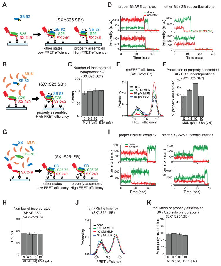
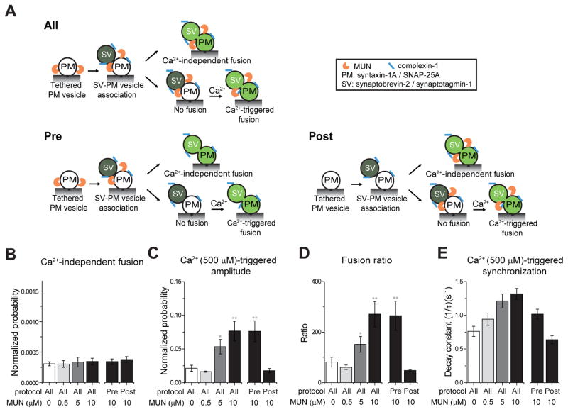
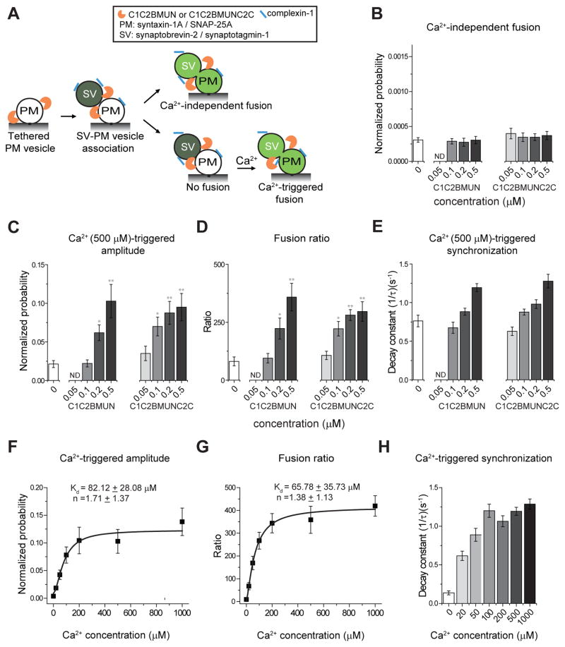
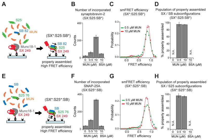
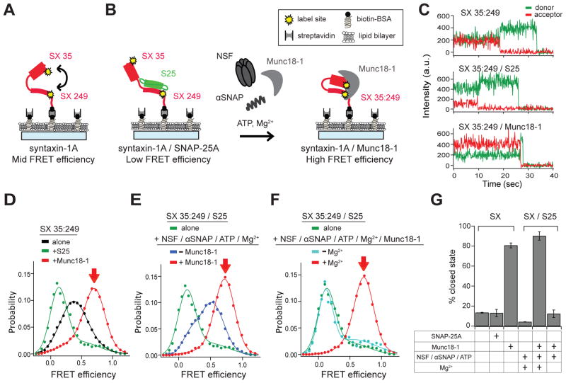
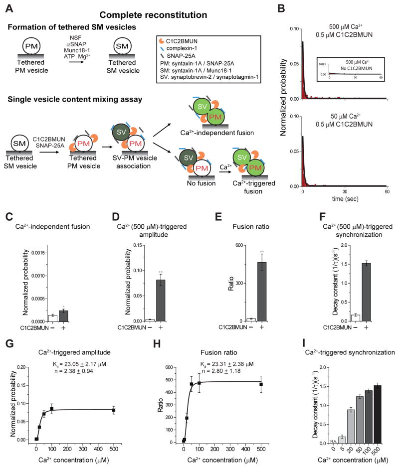
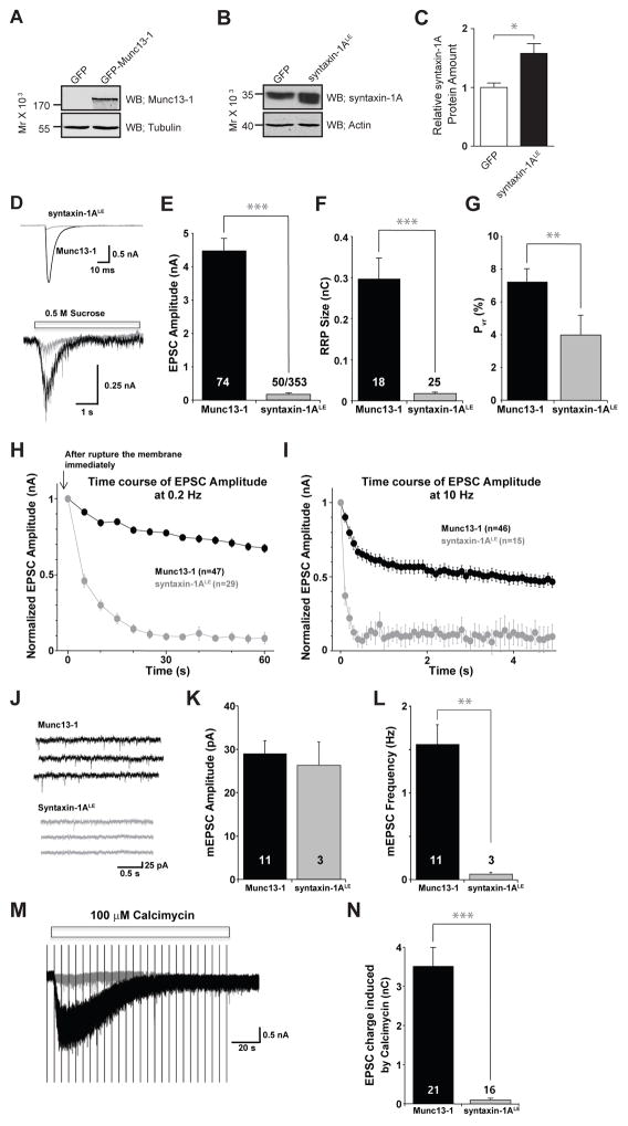
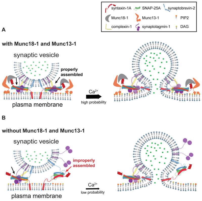
Munc13 catalyzes the transit of syntaxin from a closed complex with Munc18 into the ternary SNARE complex. Here we report a new function of Munc13, independent of Munc18: it promotes the proper syntaxin/synaptobrevin subconfiguration during assembly of the ternary SNARE complex. In cooperation with Munc18, Munc13 additionally ensures the proper syntaxin/SNAP-25 subconfiguration. In a reconstituted fusion assay with SNAREs, complexin, and synaptotagmin, inclusion of both Munc13 and Munc18 quadruples the Ca2+-triggered amplitude and achieves Ca2+ sensitivity at near-physiological concentrations. In Munc13-1/2 double-knockout neurons, expression of a constitutively open mutant of syntaxin could only minimally restore neurotransmitter release relative to Munc13-1 rescue. Together, the physiological functions of Munc13 may be related to regulation of proper SNARE complex assembly.

Keywords: Munc13; Munc18; complexin; neuronal SNAREs; neurotransmitter release; plasticity; priming; synaptotagmin.


Figures

Figure 1
Figure 1. The MUN domain alone promotes the proper syntaxin-1A / synaptobrevin-2 subconfiguration, but not the syntaxin-1A / SNAP-25A subconfiguration
(A, B, G) The syntaxin-1A (SX) / synaptobrevin-2 (SB) and the syntaxin-1A / SNAP-25A (S25) subconfigurations of the ternary SNARE complex were probed by single molecule experiments. The yellow dots and the asterisks (*) indicate fluorescently labeled residues. The cytoplasmic domain of SX was tethered on a PEG-passivated microscope slide via streptavidin-biotin linkage and labeled with Alexa 647 at residue 249. The cytoplasmic domain of SB was labeled with Alexa 555 at residue 82 and S25 was labeled with Alexa 555 at residue 76. The thin arrows represent parallel or antiparallel SX / SB (A, B) or SX / S25 (G) subconfigurations. (A, B) Beginning with surface-tethered SX, the binary complex with S25 was first formed, and then SB, was added in the absence (A) or in the presence (B) of the MUN domain at the specified concentration or BSA control. (G) Beginning with surface-tethered SX, the ternary SNARE complex was formed by adding S25 and SB at the same time in the presence of the MUN domain at the specified concentration or BSA control. (C, H) Mean counts from three fields of view ± SD (Table S1) of SB (C) or S25 (H) incorporation into the ternary SNARE complex in presence of the MUN domain at the specified concentrations or BSA control. (D, I) Representative fluorescence intensity time traces of properly (left panels) and improperly (right panels) assembled SNARE complexes that were formed starting from surface-tethered SX / S25 complex (D) or from surface-tethered SX molecules (I) in the absence of the MUN domain. (E, J) smFRET efficiency histograms using labeled SB (E), or S25 (J). (F, K) Percent of properly assembled SX / SB (F), or SX / S25 (K) subconfigurations. Shown are means ± SD for the two subsets of an equal partition of the data (Table S2).
Figure 2
Figure 2. The MUN domain improves the efficiency of Ca2+-triggered fusion
(A) Single vesicle content mixing assay. PM: plasma membrane mimic vesicles with reconstituted syntaxin-1A and SNAP-25A; SV: synaptic vesicle mimic with reconstituted synaptobrevin-2 and synaptotagmin-1. After SV-PM vesicle association, vesicle pairs either undergo Ca2+-independent fusion or remain associated until fusion is triggered by Ca2+ addition. The MUN domain is only present during the specified stages. “0 μM MUN” refers to the absence of the MUN domain in all stages. In protocol “Pre” the MUN domain is only present before and during SV-PM vesicle association. In protocol “Post” the MUN domain is present only after SV-PM vesicle association. (B–E) The bar graphs show the effects of MUN domain on the average probability of Ca2+-independent fusion events per second (B), the amplitude of the first 1-sec time bin upon 500 μM Ca2+-injection (C), the ratio of the Ca2+-triggered amplitude to the average probably of Ca2+-independent fusion per second (D), and the decay rate (1/τ) of the Ca2+-triggered fusion histogram (E). The fusion probabilities and amplitudes were normalized to the number of analyzed SV-PM vesicle pairs (Table S3). Individual histograms are in Figure S2. Panels B–D show means ± SD for multiple independent repeat experiments (Table S33). (E) Decay constants and error estimates computed from the covariance matrix upon fitting the corresponding histograms with a single exponential decay function using the Levenberg-Marquardt algorithm. * p < 0.05, ** p < 0.01 by Student’s t-test, compared to the experiment without the MUN domain.
Figure 3
Figure 3. The C1C2BMUN and C1C2BMUNC2C fragments improve the efficiency of Ca2+-triggered fusion
(A) The experimental scheme is identical to that shown in Figure 2A, protocol “all”, except that the C1C2BMUN or the C1C2BMUNC2C fragment is used instead of the MUN domain. (B–E) The bar graphs show the effects of the C1C2BMUN or the C1C2BMUNC2C fragment on the average probability of Ca2+-independent fusion events per second (B), the amplitude of the first 1-sec time bin upon Ca2+-injection (C), the ratio of the Ca2+-triggered amplitude to the average probably of Ca2+-independent fusion per second (D), and the decay rate (1/τ) of the histogram upon Ca2+-injection (E). (F–H) Ca2+ concentration dependence of triggered fusion in the presence of 500 nM C1C2BMUN fragment. Shown are the amplitude of the first 1-sec time bin upon 500 μM Ca2+-injection (F), the ratio of the Ca2+-triggered amplitude and the average probably of Ca2+-independent fusion per second (G). F and G were fit to Hill functions yielding the Ca2+ concentration at half occupation Kd and the Hill coefficient n. (H) decay rate (1/τ) of fusion histograms as a function of Ca2+. The fusion probabilities and amplitudes were normalized to the number of analyzed SV-PM vesicle pairs (Table S4). Individual histograms are in Figure S2. Panels B–D and F–G show means ± SD (Table S4). Panels E and H show decay constants and error estimates computed from the covariance matrix upon fitting the corresponding histograms with a single exponential decay function using the Levenberg-Marquardt algorithm. * p < 0.05, ** p < 0.01 by Student’s t-test, compared to the experiment without the C1C2BMUN fragment.
Figure 4
Figure 4. Munc13-1 and Munc18-1 promote proper assembly of the ternary SNARE complex
(A) The configurations of the ternary SNARE complex were probed by single molecule experiments. The cytoplasmic domain of syntaxin-1A (SX) was tethered on a PEG-passivated microscope slide via streptavidin-biotin linkage. Munc18-1 was added to assemble the SX / Munc18-1 complex. SNAP-25A (S25), the cytoplasmic domain of synaptobrevin-2 (SB) were then added in the presence of the MUN domain at the specified concentration or BSA control. The yellow dots and the asterisks (*) indicate the fluorescently labeled residues. The thin arrows represent parallel SX / SB (A) or SX / S25 (E) subconfigurations. The labeling sites are SX residue 249, SB residue 82, and S25 residue 76. (B) Beginning with surface tethered SX / Munc18-1 complex, SB incorporation into the ternary SNARE complex was measured by counting the number of labeled (Alexa 555) SB molecules in presence of unlabeled S25 and the MUN domain at the specified concentrations or BSA control. (C) smFRET efficiency histograms for labels attached to SB (Alexa 555) and SX (Alexa 647) in the presence MUN at the specified concentrations. (D) Percent of properly assembled ternary SNARE subconfigurations. (E–H) Similar to panels (A–D), starting from surface tethered SX / Munc18-1 complex and forming ternary SNARE complex in the presence of unlabeled SB, labeled (Alexa 555) S25, and the MUN domain at the specified concentration or BSA control. Panels B, F show means of the counts from three fields of view ± SD; panels D, H show means ± SD for the two subsets of an equal partition of the data (Tables S1 and S2).
Figure 5
Figure 5. Formation of syntaxin-1A / Munc18-1 complex starting from syntaxin-1A / SNAP-25A complex
smFRET experiments starting from surface-tethered cytoplasmic domain of syntaxin-1A (SX) alone (A) or SX / SNAP-25A (S25) complex (B). SX / S25 or SX / Munc18-1 complexes were formed by incubating S25 or Munc18-1 to surface-tethered SX, respectively. The yellow stars indicate residues that are used for labeling with fluorescent dyes. To form SX / Munc18-1 complex starting from SX / S25 complex, 1 μM Munc18-1 was added in presence of the disassembly factors (1 μM NSF, 10 μM αSNAP, 1 mM ATP, 1 mM Mg2+). (C) Representative fluorescence intensity time traces for smFRET efficiency measurements on surface-tethered SX (top panel), in complex with S25 (middle panel) or Munc18-1 (bottom panel). (D) smFRET efficiency histograms of surface-tethered SX (black line), in SX / S25 complex (green line), or SX / Munc18-1 complex (red line). (E) smFRET efficiency histograms starting from SX / S25 complex (green line) in the presence (red line) and absence (blue line) of 1 μM Munc18-1. (F) smFRET efficiency histograms starting from SX / S25 complex (green line) in presence of 1 μM Munc18-1 in the presence (red line) or absence (cyan line) of 1 mM Mg2+. (G) The bar chart shows means ± SD of the percent closed state for the two subsets of an equal partition of the data (Table S5). In each smFRET histogram, the red arrow indicates the closed state of SX.
Figure 6
Figure 6. Fusion experiments with complete reconstitution
(A) Single vesicle content mixing assay with complete reconstitution. PM and SV defined in Figure 2; SM: vesicles with reconstituted syntaxin-1A / Munc18-1 complex. The C1C2BMUN fragment was added after formation of the SM vesicles and during all subsequent stages. (B) Representative histograms for triggered fusion at 50 μM, 500 μM Ca2+. Inset, triggered fusion without C1C2BMUN (note that without C1C2BMUN, ternary SNARE complex cannot form, thus little fusion is observed). (C–F) The bar graphs show the effects of C1C2BMUN fragment on the average probability of Ca2+-independent fusion events per second (C), the amplitude of the first 1-sec time bin upon 500 μM Ca2+-injection (D), the ratio of the Ca2+-triggered amplitude to the average probably of Ca2+-independent fusion per second (E), and the decay rate (1/τ) of the histogram upon Ca2+-injection (F). (G–I) Ca2+ concentration dependence of triggered fusion in the presence of 500 nM C1C2BMUN fragment. Shown are the amplitude of the first 1-sec time bin upon Ca2+-injection (G), the ratio of the Ca2+-triggered amplitude to the average probably of Ca2+-independent fusion per second (H), and the decay rate (1/τ) of the histogram upon Ca2+-injection (I) as a function of Ca2+-concentration. Hill functions were fit yielding the Ca2+ concentration at half occupation Kd and the Hill coefficient n. Individual histograms are in Figure S2. Panels C–E and G–H show means ± SD for multiple independent repeat experiments (Table S7). Panels F and I show decay constants and error estimates computed from the covariance matrix upon fitting the corresponding histograms with a single exponential decay function using the Levenberg-Marquardt algorithm. * p < 0.05, ** p < 0.01 by Student’s t-test.
Figure 7
Figure 7. Syntaxin-1ALE mutant overexpression in Munc13-1/2 double-knockout neurons
Experiments in hippocampal autaptic neurons cultured from Munc13-1/2 double-knockout mice. Neurons were infected with recombinant Semliki Forest virus expressing either wild-type Munc13-1 or syntaxin-1ALE mutant. (A) Western blot using an anti-Munc13-1 antibody showed the level of expression of Munc13-1 in Munc13-1/2 double-knockout hippocampal neurons infected with Semliki Forest virus expressing GFP only, or Munc13-1 (upper panel). Tubulin was used as a loading control (bottom panel). (B) Western blot using an anti-syntaxin-1A antibody showed the level of expression of syntaxin-1A/syntaxin-1ALE in Munc13-1/2 double-knockout hippocampal neurons expressing GFP only syntaxin-1ALE (upper panel). Actin was used as a loading control (C) Relative quantification of syntaxin-1A protein amounts in Munc13-1/2 double-knockout hippocampal neurons. Signals were normalized to the level of endogenous syntaxin-1A in GFP-only overexpressing neurons (white bar; 1.0 + 0.1, n=4). Overexpression of syntaxin-1ALE increase the signal 1.7 ± 0.2 fold (black bar; n=3, p=0.037 (Student’s t-test)). (D) EPSCs were recorded in whole-cell patch-clamp mode. Representative traces of EPSCs (top) and postsynaptic currents evoked by 0.5 M sucrose (bottom). (E) Average values of EPSC amplitudes of the first three stimuli of a 0.2 Hz series of stimuli in neurons expressing Munc13-1 (black; from 74 neurons from 5 mice) and syntaxin-1ALE mutant (grey; from 50 neurons from 5 mice) that showed a response out of 353 that expressed GFP). (F) Apparent readily releasable vesicle pool (RRP) size calculated by the integral of the sucrose response over 4 seconds in Munc13-1 (black; from 18 neurons from 3 mice) and syntaxin-1ALE mutant neurons (grey; from 25 neurons from 3 mice). (G) Release probability calculated as the ratio of the EPSC amplitude to that of the RRP in Munc13-1 (black; from 18 neurons from 3 mice) and syntaxin-1ALE mutant neurons (grey; from 25 neurons from 3 mice). (H) Normalized EPSC amplitudes in Munc13-1 wild type (black) and syntaxin-1ALE mutant neurons (grey) during 0.2 Hz stimulation. (I) Normalized EPSC amplitudes during 10 Hz stimulation. (J) Representative traces of miniature EPSCs (mEPSC) in neurons with overexpressed Munc13-1 (top) or syntaxin-1ALE mutant (bottom) in the presence of 200 nM TTX. (K) Average mEPSC amplitudes of Munc13-1 (black; from 11 neurons from 1 mouse) and syntaxin-1ALE mutant neurons (grey; from 3 neurons from 1 mouse). (L) Average mEPSC frequencies of Munc13-1 (black) and syntaxin-1ALE mutant neurons (grey). (M) Example traces of neurons in the presence of 100 μM calcimycin. The vertical lines are stimulation artifacts resulting from intermittent monitoring of evoked EPSCs. (N) EPSC charge induced by calcimycin in Munc13-1 (from 21 neurons from 3 mice) and syntaxin-1ALE mutant neurons (from 16 neurons from 4 mice). Note * p<0.05, ** p<0.01, and *** p < 0.001.
Figure 8
Figure 8. Model of concerted Munc18-1 / Munc13-1 action to properly assemble ternary SNARE complex
Model of the combined effect of Munc13-1 and Munc18-1 on Ca2+-triggered fusion. (A) Munc13-1 and Munc18-1 cooperate to produce proper trans ternary SNARE complex. (B) Without Munc13-1 and Munc18-1, trans ternary SNARE complex assembly is error prone resulting in much reduced Ca2+-triggered fusion probability.

Similar articles

Cited by

References

    1. Aravamudan B, Fergestad T, Davis WS, Rodesch CK, Broadie K. Drosophila UNC-13 is essential for synaptic transmission. Nat Neurosci. 1999;2:965–971. - PubMed
    1. Ashery U, Betz A, Xu T, Brose N, Rettig J. An efficient method for infection of adrenal chromaffin cells using the Semliki Forest virus gene expression system. Eur J Cell Biol. 1999;78:525–532. - PubMed
    1. Augustin I, Rosenmund C, Südhof TC, Brose N. Munc13-1 is essential for fusion competence of glutamatergic synaptic vesicles. Nature. 1999;400:457–461. - PubMed
    1. Baker RW, Jeffrey PD, Zick M, Phillips BP, Wickner WT, Hughson FM. A direct role for the Sec1/Munc18-family protein Vps33 as a template for SNARE assembly. Science (80– ) 2015;349:1111–1114. - PMC - PubMed
    1. Basu J, Shen N, Dulubova I, Lu J, Guan R, Guryev O, Grishin NV, Rosenmund C, Rizo J. A minimal domain responsible for Munc13 activity. Nat Struct Mol Biol. 2005;12:1017–1018. - PubMed

MeSH terms