

The Zahn drawings: new illustrations of Xenopus embryo and tadpole stages for studies of craniofacial development
- PMID: 28765211
- PMCID: PMC5560046
- DOI: 10.1242/dev.151308
The Zahn drawings: new illustrations of Xenopus embryo and tadpole stages for studies of craniofacial development
Abstract
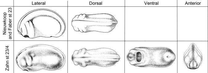
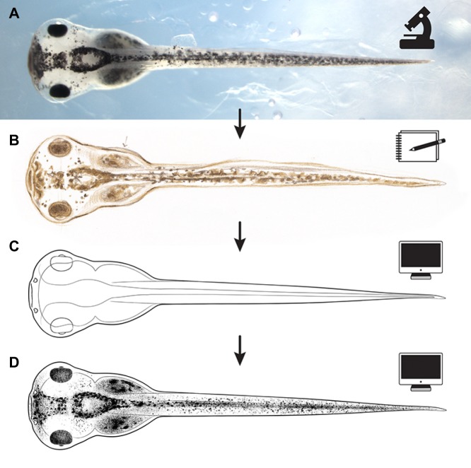
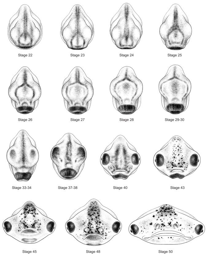
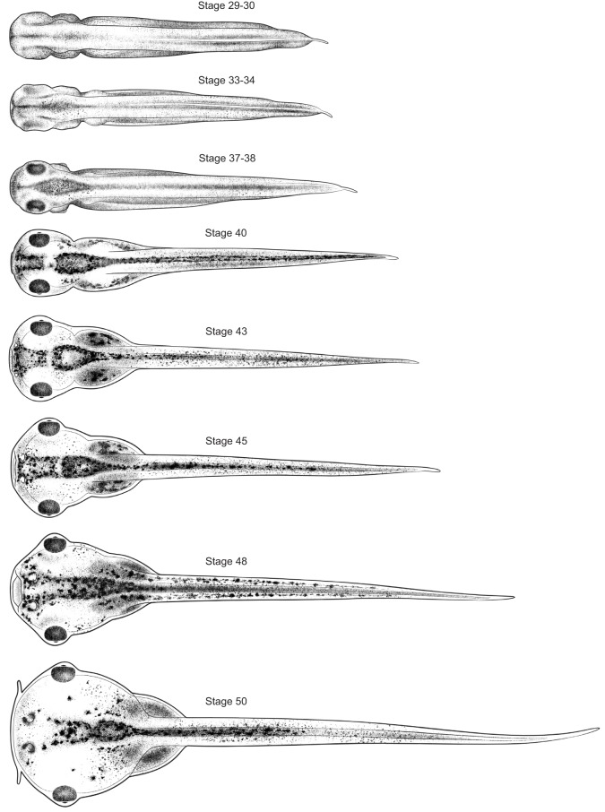
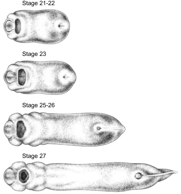
The embryos and tadpoles of the frog Xenopus are increasingly important subjects for studies of the development of the head and face - studies that are providing novel and crucial insight into the causes and prevention of a suite of devastating birth defects, as well as basic evolutionary and developmental biology. However, many studies are conducted on a range of embryonic stages that are not fully represented in the beloved Xenopus resource, Nieuwkoop and Faber's classic Normal Table of Xenopus laevis (Daudin) The lack of standardized images at these stages acts as a barrier to the efficient and accurate representation and communication of experimental methodology and expression data. To fill this gap, we have created 27 new high-quality illustrations. Like their oft-used predecessors from Nieuwkoop and Faber, these drawings can be freely downloaded and used, and will, we hope, serve as an essential resource for this important model system.
Keywords: Craniofacial patterning; Embryonic development; Stage series; Xenopus laevis.
© 2017. Published by The Company of Biologists Ltd.
Conflict of interest statement
Competing interestsThe authors declare no competing or financial interests.
Figures
Similar articles
-
Normal Table of Xenopus development: a new graphical resource.Development. 2022 Jul 15;149(14):dev200356. doi: 10.1242/dev.200356. Epub 2022 Jul 14. Development. 2022. PMID: 35833709 Free PMC article.
-
Effects of depleted uranium on survival, growth, and metamorphosis in the African clawed frog (Xenopus laevis).J Toxicol Environ Health A. 2005 Jun 11-25;68(11-12):951-65. doi: 10.1080/15287390590912595. J Toxicol Environ Health A. 2005. PMID: 16020186
-
Metamorphosis delay in Xenopus laevis (Daudin) tadpoles exposed to a 50 Hz weak magnetic field.Int J Radiat Biol. 2010 Jan;86(1):37-46. doi: 10.3109/09553000903137687. Int J Radiat Biol. 2010. PMID: 20070214
-
Xenopus tropicalis: an ideal experimental animal in amphibia.Exp Anim. 2010;59(4):395-405. doi: 10.1538/expanim.59.395. Exp Anim. 2010. PMID: 20660986 Review.
-
Xenopus laevis as a model for studying thyroid hormone signalling: from development to metamorphosis.Mol Cell Endocrinol. 2008 Oct 10;293(1-2):71-9. doi: 10.1016/j.mce.2008.06.012. Epub 2008 Jun 28. Mol Cell Endocrinol. 2008. PMID: 18657589 Review.
Cited by
-
Xenopus retinal ganglion cell axon extension is unaffected by 5-HT 1B/D receptor activation during visual system development.MicroPubl Biol. 2023 Dec 4;2023:10.17912/micropub.biology.001076. doi: 10.17912/micropub.biology.001076. eCollection 2023. MicroPubl Biol. 2023. PMID: 38116474 Free PMC article.
-
The Many Faces of Xenopus: Xenopus laevis as a Model System to Study Wolf-Hirschhorn Syndrome.Front Physiol. 2019 Jun 26;10:817. doi: 10.3389/fphys.2019.00817. eCollection 2019. Front Physiol. 2019. PMID: 31297068 Free PMC article.
-
Technological Approach to Mind Everywhere: An Experimentally-Grounded Framework for Understanding Diverse Bodies and Minds.Front Syst Neurosci. 2022 Mar 24;16:768201. doi: 10.3389/fnsys.2022.768201. eCollection 2022. Front Syst Neurosci. 2022. PMID: 35401131 Free PMC article.
-
Navigating Xenbase: An Integrated Xenopus Genomics and Gene Expression Database.Methods Mol Biol. 2018;1757:251-305. doi: 10.1007/978-1-4939-7737-6_10. Methods Mol Biol. 2018. PMID: 29761462 Free PMC article.
-
Advancing genetic and genomic technologies deepen the pool for discovery in Xenopus tropicalis.Dev Dyn. 2019 Aug;248(8):620-625. doi: 10.1002/dvdy.80. Epub 2019 Jul 9. Dev Dyn. 2019. PMID: 31254427 Free PMC article. Review.
References
Publication types
MeSH terms
Grants and funding
LinkOut - more resources
Full Text Sources
Other Literature Sources
Miscellaneous
