. 2017 Aug 7;214(8):2387-2404.
doi: 10.1084/jem.20162152. Epub 2017 Jul 10.

Monocyte-derived alveolar macrophages drive lung fibrosis and persist in the lung over the life span

Affiliations

Monocyte-derived alveolar macrophages drive lung fibrosis and persist in the lung over the life span

Alexander V Misharin et al. J Exp Med. .

Abstract

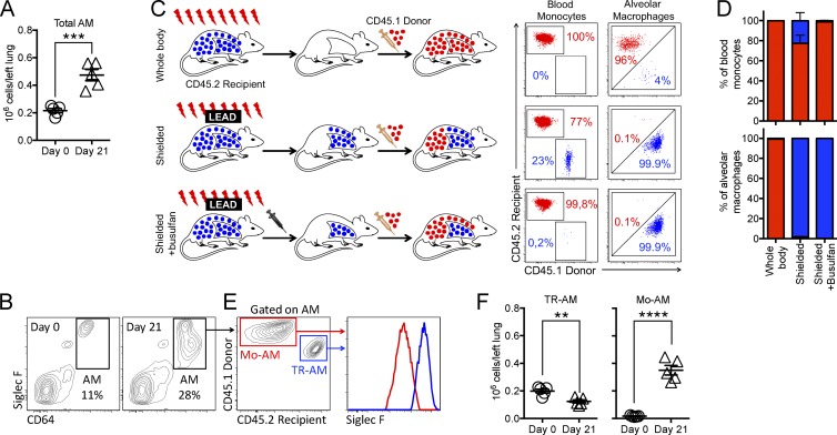
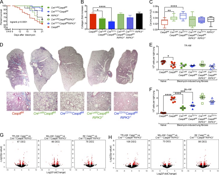
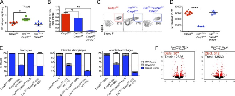
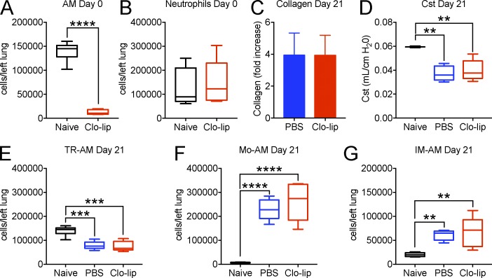
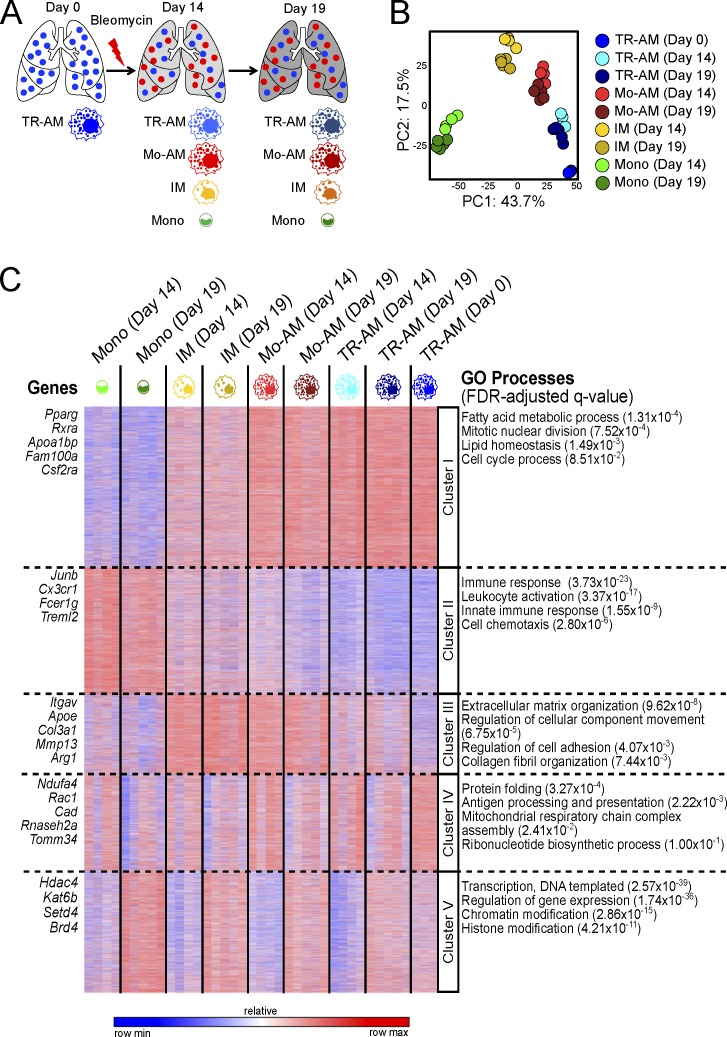
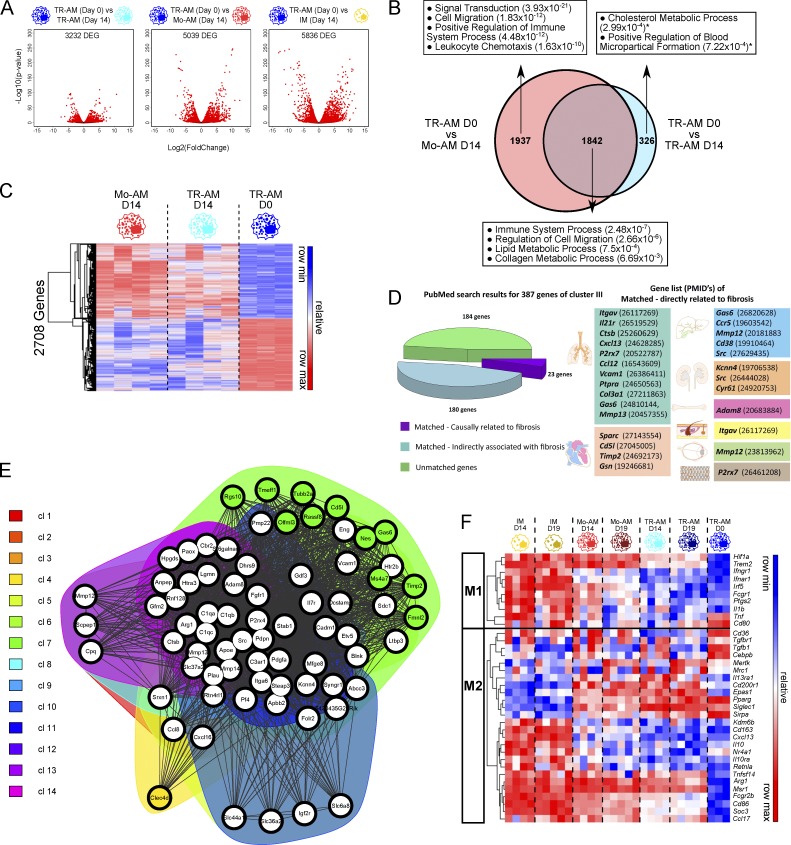
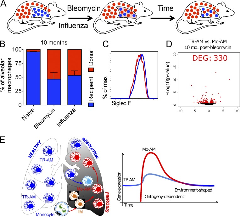
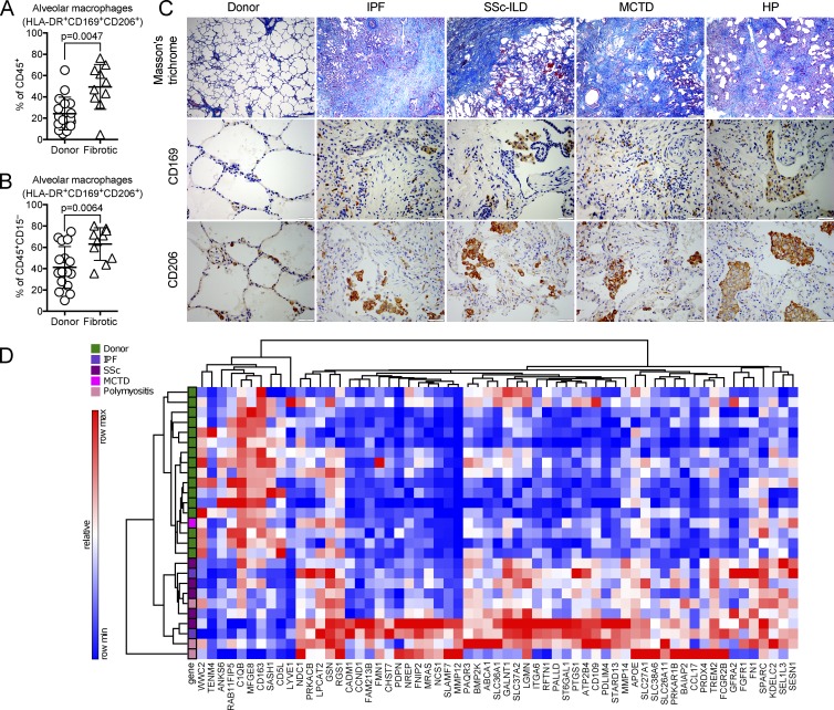
Little is known about the relative importance of monocyte and tissue-resident macrophages in the development of lung fibrosis. We show that specific genetic deletion of monocyte-derived alveolar macrophages after their recruitment to the lung ameliorated lung fibrosis, whereas tissue-resident alveolar macrophages did not contribute to fibrosis. Using transcriptomic profiling of flow-sorted cells, we found that monocyte to alveolar macrophage differentiation unfolds continuously over the course of fibrosis and its resolution. During the fibrotic phase, monocyte-derived alveolar macrophages differ significantly from tissue-resident alveolar macrophages in their expression of profibrotic genes. A population of monocyte-derived alveolar macrophages persisted in the lung for one year after the resolution of fibrosis, where they became increasingly similar to tissue-resident alveolar macrophages. Human homologues of profibrotic genes expressed by mouse monocyte-derived alveolar macrophages during fibrosis were up-regulated in human alveolar macrophages from fibrotic compared with normal lungs. Our findings suggest that selectively targeting alveolar macrophage differentiation within the lung may ameliorate fibrosis without the adverse consequences associated with global monocyte or tissue-resident alveolar macrophage depletion.


Figures

Figure 1.
Figure 1.
TR-AMs are depleted during lung fibrosis and replaced with Mo-AMs. (A and B) The alveolar macrophage pool is expanded 21 d after the intratracheal administration of bleomycin when fibrosis is maximal because of the appearance of a novel population of cells characterized by lower expression of Siglec F (n = 5 per group, paired t test; data are shown as mean ± SEM; ***, P < 0.001). (C) Schematic explaining the technique for the generation of bone marrow chimeras with thoracic shielding and busulfan and representative dot plots reflecting chimerism in peripheral blood monocytes and alveolar macrophages in the lung 8 wk after irradiation and bone marrow transfer. Whole-body irradiation leads to complete replacement of recipient monocytes and alveolar macrophages with donor-derived cells (top). Irradiation with thoracic shielding protects alveolar macrophages but results in incomplete chimerism among circulating monocytes (middle). The addition of busulfan conditioning postirradiation allows complete elimination of recipient’s monocytes while preserving alveolar macrophages (bottom). (D) Percentage of monocytes and alveolar macrophages of donor and recipient origin 8 wk after the generation of chimeras (n = 3–5 per group; data are shown as mean ± SEM and are representative of >100 mice). (E) The new population of alveolar macrophages that emerges during bleomycin-induced fibrosis (CD45+CD11b+/−CD11c+CD64+SiglecFlow) is monocyte derived. Representative contour plots show expansion of the alveolar macrophage pool; numbers indicate percentage of the parent population (gated on singlets/live/CD45+ cells). (F) Expansion of the alveolar macrophage pool during bleomycin-induced lung fibrosis is caused by recruitment of Mo-AMs, whereas the number of TR-AMs is decreased (n = 5 per group, paired t test; data are shown as mean ± SEM and are representative of more than five independent experiments; **, P < 0.01; ****, P < 0.0001).
Figure 2.
Figure 2.
Necroptosis of Mo-AMs attenuates bleomycin-induced lung fibrosis. (A–D) Mice with deficiency of Casp8 in alveolar macrophages have (A) improved survival (16–101 mice per group; combined data from 10 independent experiments; Mantel-Cox log-rank test), (B) lower levels of collagen, (C) improved static lung compliance, and (D) less extent histological fibrosis compared with controls, whereas simultaneous deletion of RIPK3 rescues bleomycin-induced lung fibrosis in CD11cCreCasp8flox/flox and LysMCreCasp8flox/flox mice (n = 9–45 mice per group, data expressed as mean ± SEM, one-way ANOVA with Dunnett’s test for multiple comparisons; *, P < 0.05; **, P < 0.01; ****, P < 0.0001). (E and F) Protection from lung fibrosis is associated with the loss of Mo-AMs, but not TR-AMs, in Casp8-deficient mice, whereas RIPK3 deficiency rescues Mo-AMs (n = 4–5 mice per group; data are expressed as mean ± SEM; one-way ANOVA with Dunnett’s test for multiple comparisons; *, P < 0.05; ***, P < 0.001; ****, P < 0.0001; the experiment was performed at least two times). (G and H) The loss of RIPK3 does not alter the response of alveolar or interstitial macrophages to fibrosis in CD11cCreCasp8flox/floxRIPK3−/− and LysMCreCasp8flox/floxRIPK3−/− mice. Volcano plot showing number of differentially expressed genes (red, FDR q value < 0.05; n = 2–5 mice per group). DEG, differentially expressed genes.
Figure 3.
Figure 3.
TR-AMs in LysMCreCasp8flox/flox and CD11cCreCasp8flox/flox mice have preserved expression of Casp8, suggesting they escape Cre-mediated recombination. (A) CD11cCreCasp8flox/flox and LysMCreCasp8flox/flox mice have a normal number of TR-AMs in a steady state (n = 5 mice per group; data are shown as mean ± SEM; one-way ANOVA with Dunnett’s test for multiple comparisons; *, P < 0.05; the experiment was performed three times). (B) Casp8 expression in flow-sorted TR-AMs measured by quantitative PCR (n = 3–5 mice per group; data are shown as mean ± SEM; one-way ANOVA with Dunnett’s test for multiple comparisons; **, P < 0.01; the experiment was performed two times). (C and D) Bone marrow from CD11cCreCasp8flox/flox mice fails to reconstitute the alveolar niche 8 wk after irradiation, as indicated by ongoing recruitment of the immature Mo-AMs with low expression of Siglec F. Repopulation of the alveolar niche was rescued in CD11cCreCasp8flox/floxRIPK3−/− mice (n = 4–5 mice per group; data are expressed as mean ± SEM; one-way ANOVA with Dunnett’s test for multiple comparisons; ****, P < 0.0001). (E) Alveolar macrophages derived from bone marrow from CD11cCreCasp8flox/flox mice fail to reconstitute the alveolar niche 8 wk after irradiation, as shown by decreased expression of Siglec F on alveolar macrophages. Mice were lethally irradiated, followed by reconstitution with bone marrow from control mice (Casp8flox/flox) or bone marrow from mice with deletion of Casp8 in cells expressing CD11c (CreCD11cCasp8flox/flox), either alone or in combination with bone marrow from wild-type mice (50% mixture of the two genotypes). Monocytes, interstitial macrophages, and alveolar macrophages were identified by flow cytometry of lung homogenates 8 wk later (CD45.1 or CD45.2 mice were used for lineage tracing). Although ∼50% of monocytes in both Casp8flox/flox and CreCD11cCasp8flox/flox mice were wild-type cells, the majority of interstitial macrophages and alveolar macrophages in mice reconstituted with CreCD11cCasp8flox/flox bone marrow, but not Casp8flox/flox mice, were of wild-type origin, suggesting the loss of Casp8 during differentiation results in a selective disadvantage in the differentiation of monocytes in to alveolar macrophages; data are shown as mean ± SEM. (F) Cells that escaped Cre-mediated recombination behave similarly to wild-type cells during fibrosis (day 14) with <1% differentially expressed genes (DEG). Volcano plot showing number of differentially expressed genes (red, FDR q value <0.05, n = 2–5 mice per group).
Figure 4.
Figure 4.
Depletion of TR-AMs does not protect from bleomycin-induced lung fibrosis. (A and B) Intratracheal administration of 50 μl clodronate-loaded liposomes (Clo-lip) results in nearly complete depletion of TR-AMs 3 d later without recruitment of neutrophils. (C and D) Mice with depleted TR-AMs showed the same severity of bleomycin-induced lung fibrosis, as indicated by collagen levels (C) and static lung compliance (D) 21 d later. (E–G) Numbers of TR-AMs (E), Mo-AMs (F), and interstitial macrophages (IMs; G) did not differ between the clodronate-loaded liposome–treated and control (PBS) groups (n = 5–6 mice per group; data are expressed as mean ± SEM; one-way ANOVA with Dunnett’s test for multiple comparisons; **, P < 0.01; ***, P < 0.001; ****, P < 0.0001; the experiment was performed one time).
Figure 5.
Figure 5.
TR-AMs and Mo-AMs differ in their response to bleomycin-induced lung fibrosis. (A–C) Transcriptional profiling of macrophage populations over the course of bleomycin-induced lung fibrosis. (A) Experimental outline. (B) Principal-component analysis (PCA) of the transcriptomes of FACS-sorted monocytes, interstitial macrophages (IM), Mo-AMs, and TR-AMs during the course of bleomycin-induced lung fibrosis shows that a significant fraction of the variance in the dataset can be explained by principal component 1 (PC1; 43.7%, differentiation) and principal component 2 (PC2; 17.5%, fibrosis time course). PCA was performed on differentially expressed genes identified from a generalized linear model to perform an ANOVA-like test for differential expression between any conditions in the dataset (FDR step-up procedure q-value < 0.05). (C) k-means clustering of all identified genes revealed clusters of genes associated with monocyte into alveolar macrophage differentiation (clusters I and II), genes differentially expressed over the course of bleomycin-induced lung fibrosis (clusters IV and V), and genes specifically up-regulated in IM and Mo-AMs, but not in monocytes or TR-AMs (cluster III; optimal k of 5 determined from within-group and between-group sum of squares analysis; Fig. S3 C). The characteristic genes and GO processes associated with each cluster are shown on the left and the right sides, correspondingly (see Table S3).
Figure 6.
Figure 6.
Monocyte-derived and tissue-resident alveolar macrophages differ in their response to bleomycin-induced lung injury. (A) Volcano plots demonstrating the number of differentially expressed genes (DEG) for the selected comparisons 14 d after instillation of bleomycin (FDR step-up procedure q-value < 0.05). (B) Comparison of DEG and associated GO processes reveals shared and unique response between Mo-AMs and TR-AMs. (C) Hierarchical clustering of the 2708 DEG shared by Mo-AMs and TR-AMs during the response to bleomycin-induced lung fibrosis. (D) Association between genes in cluster III and fibrosis. Differentially expressed genes from cluster III were taken; term “AND Fibrosis” was added to the gene names and the resulting term was used as input term in the PubMed search engine to search abstracts and full text using an in-house Python script. The abstracts were then manually reviewed for evidence of a genetic association with fibrosis (gene knockouts were protected from fibrosis or transgenic overexpression of the gene increased fibrosis). Of the 387 differentially expressed genes in cluster III (FDR q-value < 0.05), 203 were linked with fibrosis in PubMed and 23 were causally related to the development of fibrosis in different organs (as indicated by images in figure) in genetic mouse models. The numbers in parenthesis are PubMed IDs. (E) Functional gene network analysis was performed on the top 100 of differentially expressed genes between Mo-AMs and TR-AMs from cluster III using GeneTerm Linker and visualized using FGNet tool. White circles indicate hub genes that belong to the multiple metagroups. For metagroup annotations, see Table S4. (F) Heat map of M1/M2 genes that were differentially expressed in our dataset (FDR step-up q-value < 0.05; see also Fig. S4 C).
Figure 7.
Figure 7.
Monocyte-derived alveolar macrophages persist after the resolution of lung injury and fibrosis. (A) Schematic of the experimental approach. Shielded bone marrow chimeras were harvested 10 mo after challenge with either bleomycin or with influenza A virus. (B) Percentage of cells identified as TR-AMs (CD45.2) and Mo-AMs (CD45.1) in untreated shielded chimeric mice, mice that received bleomycin, or mice infected with 100 PFU influenza A virus (A/WSN/33) as young adults (n = 3–5 mice per group; data are expressed as mean ± SEM). (C and D) 10 mo after the initial challenge, Mo-AMs and TR-AMs are no longer distinguishable by flow cytometry, and transcriptional profiling revealed only 330 differentially expressed genes (DEG; FDR step-up procedure q-value < 0.05; see Table S5). (E) Schematic illustration of the differential role of TR-AMs and Mo-AMs during the different stages of the lung injury and fibrosis.
Figure 8.
Figure 8.
Alveolar macrophages are expanded in patients with lung fibrosis and exhibit a profibrotic gene expression signature similar to mouse Mo-AMs during lung fibrosis. (A–C) The population of alveolar macrophages is increased in patients with fibrotic lung disease. At the time of lung transplantation, small distal lung biopsy specimens were harvested from the donor lung (n = 16) and the recipient lung (n = 10) and processed for flow cytometry and immunohistochemistry. (A) Alveolar macrophages were identified as CD206+CD169+HLA-DR+ cells out of all hematopoietic CD45+ cells from donor lung tissue and from patients with lung fibrosis (Mann–Whitney test; data are expressed as mean ± SD). (B) The same analysis was repeated after excluding neutrophils (Mann–Whitney test; data are expressed as mean ± SD). (C, top) Masson’s trichrome staining; bar, 500 µm. (C, middle) Immunohistochemistry for CD169; bar, 50 µm. (C, bottom) Immunohistochemistry for CD206; bar, 50 µm. Representative images from healthy donor, patients with idiopathic pulmonary fibrosis (IPF), systemic sclerosis-associated interstitial lung disease (SSc-ILD), mixed connective tissue disease (MCTD), and hypersensitivity pneumonitis (HP) are shown. (D) Of the differentially expressed genes in cluster III (Table S2), homologues of 61 were differentially expressed (FDR step-up procedure q-value < 0.05) between alveolar macrophages isolated from human fibrotic lung explants compared with donor lungs; 51 were up-regulated in patients with lung fibrosis, and 10 were down-regulated.

Similar articles

Cited by

References

    1. Agassandian M., Tedrow J.R., Sembrat J., Kass D.J., Zhang Y., Goncharova E.A., Kaminski N., Mallampalli R.K., and Vuga L.J.. 2015. VCAM-1 is a TGF-β1 inducible gene upregulated in idiopathic pulmonary fibrosis. Cell. Signal. 27:2467–2473. 10.1016/j.cellsig.2015.09.003 - DOI - PMC - PubMed
    1. Aibar S., Fontanillo C., Droste C., and De Las Rivas J.. 2015. Functional Gene Networks: R/Bioc package to generate and analyse gene networks derived from functional enrichment and clustering. Bioinformatics. 31:1686–1688. 10.1093/bioinformatics/btu864 - DOI - PMC - PubMed
    1. Aschner Y., Khalifah A.P., Briones N., Yamashita C., Dolgonos L., Young S.K., Campbell M.N., Riches D.W., Redente E.F., Janssen W.J., et al. . 2014. Protein tyrosine phosphatase α mediates profibrotic signaling in lung fibroblasts through TGF-β responsiveness. Am. J. Pathol. 184:1489–1502. 10.1016/j.ajpath.2014.01.016 - DOI - PMC - PubMed
    1. Bharat A., Bhorade S.M., Morales-Nebreda L., McQuattie-Pimentel A.C., Soberanes S., Ridge K., DeCamp M.M., Mestan K.K., Perlman H., Budinger G.R., and Misharin A.V.. 2016. Flow cytometry reveals similarities between lung macrophages in humans and mice. Am. J. Respir. Cell Mol. Biol. 54:147–149. 10.1165/rcmb.2015-0147LE - DOI - PMC - PubMed
    1. Brodeur T.Y., Robidoux T.E., Weinstein J.S., Craft J., Swain S.L., and Marshak-Rothstein A.. 2015. IL-21 promotes pulmonary fibrosis through the induction of profibrotic CD8+ T cells. J. Immunol. 195:5251–5260. 10.4049/jimmunol.1500777 - DOI - PMC - PubMed