

Tetrandrine, an Activator of Autophagy, Induces Autophagic Cell Death via PKC-α Inhibition and mTOR-Dependent Mechanisms
- PMID: 28642707
- PMCID: PMC5462963
- DOI: 10.3389/fphar.2017.00351
Tetrandrine, an Activator of Autophagy, Induces Autophagic Cell Death via PKC-α Inhibition and mTOR-Dependent Mechanisms
Abstract
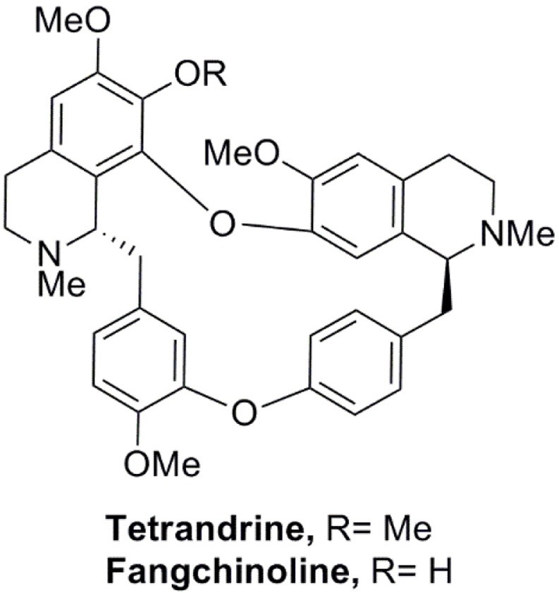
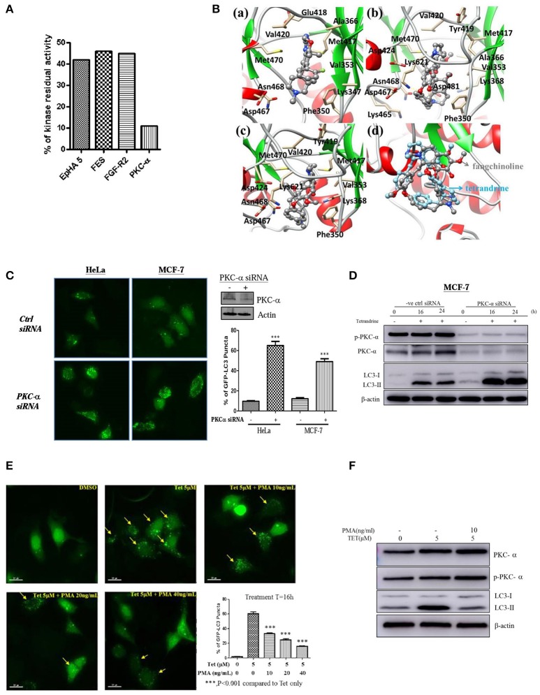
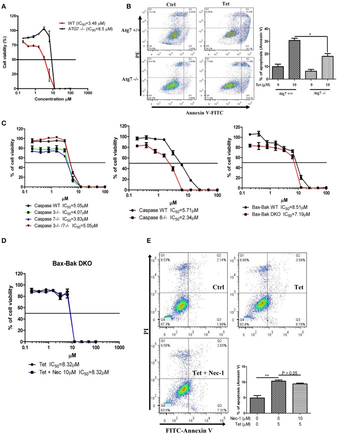
Emerging evidence suggests the therapeutic role of autophagic modulators in cancer therapy. This study aims to identify novel traditional Chinese medicinal herbs as potential anti-tumor agents through autophagic induction, which finally lead to autophagy mediated-cell death in apoptosis-resistant cancer cells. Using bioactivity-guided purification, we identified tetrandrine (Tet) from herbal plant, Radix stephaniae tetrandrae, as an inducer of autophagy. Across a number of cancer cell lines, we found that breast cancer cells treated with tetrandrine show an increase autophagic flux and formation of autophagosomes. In addition, tetrandrine induces cell death in a panel of apoptosis-resistant cell lines that are deficient for caspase 3, caspase 7, caspase 3 and 7, or Bax-Bak respectively. We also showed that tetrandrine-induced cell death is independent of necrotic cell death. Mechanistically, tetrandrine induces autophagy that depends on mTOR inactivation. Furthermore, tetrandrine induces autophagy in a calcium/calmodulin-dependent protein kinase kinase-β (CaMKK-β), 5' AMP-activated protein kinase (AMPK) independent manner. Finally, by kinase profiling against 300 WT kinases and computational molecular docking analysis, we showed that tetrandrine is a novel PKC-α inhibitor, which lead to autophagic induction through PKC-α inactivation. This study provides detailed insights into the novel cytotoxic mechanism of an anti-tumor compound originated from the herbal plant, which may be useful in promoting autophagy mediated- cell death in cancer cell that is resistant to apoptosis.
Keywords: PKC-α; apoptosis-resistant; autophagy; mTOR; tetrandrine.
Figures


Similar articles
-
Autophagy induction enhances tetrandrine-induced apoptosis via the AMPK/mTOR pathway in human bladder cancer cells.Oncol Rep. 2017 Nov;38(5):3137-3143. doi: 10.3892/or.2017.5988. Epub 2017 Sep 21. Oncol Rep. 2017. PMID: 29048631
-
Saikosaponin-d, a novel SERCA inhibitor, induces autophagic cell death in apoptosis-defective cells.Cell Death Dis. 2013 Jul 11;4(7):e720. doi: 10.1038/cddis.2013.217. Cell Death Dis. 2013. PMID: 23846222 Free PMC article.
-
Alisol B, a novel inhibitor of the sarcoplasmic/endoplasmic reticulum Ca(2+) ATPase pump, induces autophagy, endoplasmic reticulum stress, and apoptosis.Mol Cancer Ther. 2010 Mar;9(3):718-30. doi: 10.1158/1535-7163.MCT-09-0700. Epub 2010 Mar 2. Mol Cancer Ther. 2010. PMID: 20197400
-
Radix Stephaniae Tetrandrine: An Emerging Role for Management of Breast Cancer.Curr Pharm Des. 2020;26(1):25-36. doi: 10.2174/1381612826666200110143706. Curr Pharm Des. 2020. PMID: 31924150 Review.
-
Tetrandrine, a Chinese plant-derived alkaloid, is a potential candidate for cancer chemotherapy.Oncotarget. 2016 Jun 28;7(26):40800-40815. doi: 10.18632/oncotarget.8315. Oncotarget. 2016. PMID: 27027348 Free PMC article. Review.
Cited by
-
Sestrin2 drives ER-phagy in response to protein misfolding.Dev Cell. 2024 Aug 19;59(16):2035-2052.e10. doi: 10.1016/j.devcel.2024.07.004. Epub 2024 Aug 1. Dev Cell. 2024. PMID: 39094564 Free PMC article.
-
mTORC1 controls lysosomal Ca2+ release through the two-pore channel TPC2.Sci Signal. 2018 Apr 10;11(525):eaao5775. doi: 10.1126/scisignal.aao5775. Sci Signal. 2018. PMID: 29636391 Free PMC article.
-
Exploring the five different genes associated with PKCα in bladder cancer based on gene expression microarray.J Cell Mol Med. 2021 Feb;25(3):1759-1770. doi: 10.1111/jcmm.16284. Epub 2021 Jan 15. J Cell Mol Med. 2021. PMID: 33452764 Free PMC article.
-
Design, Synthesis and Anticancer Evaluation of Fangchinoline Derivatives.Molecules. 2017 Nov 8;22(11):1923. doi: 10.3390/molecules22111923. Molecules. 2017. PMID: 29117113 Free PMC article.
-
Benzyl Isothiocyanate-Induced Cytotoxicity via the Inhibition of Autophagy and Lysosomal Function in AGS Cells.Biomol Ther (Seoul). 2022 Jul 1;30(4):348-359. doi: 10.4062/biomolther.2022.019. Biomol Ther (Seoul). 2022. PMID: 35768332 Free PMC article.
References
LinkOut - more resources
Full Text Sources
Other Literature Sources
Research Materials
Miscellaneous