

Comparative Gene Expression Analysis of Lymphocytes Treated with Exosomes Derived from Ovarian Cancer and Ovarian Cysts
- PMID: 28620375
- PMCID: PMC5451634
- DOI: 10.3389/fimmu.2017.00607
Comparative Gene Expression Analysis of Lymphocytes Treated with Exosomes Derived from Ovarian Cancer and Ovarian Cysts
Abstract
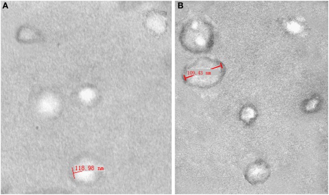
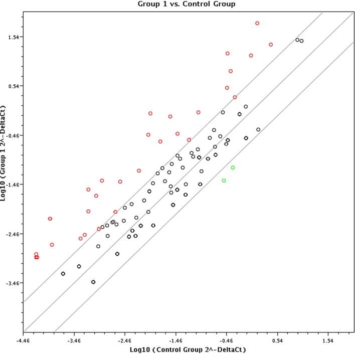
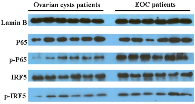
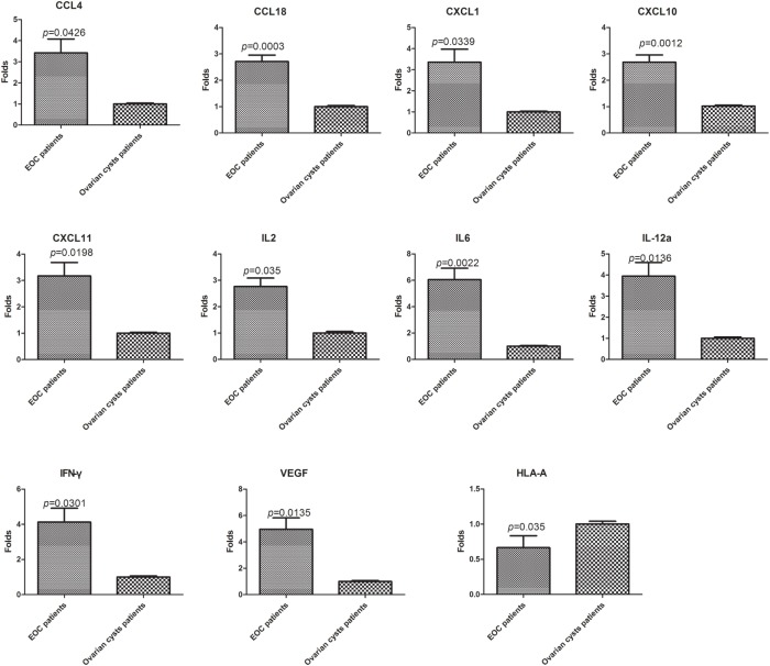
Cancer cells employ many strategies to evade immune defense and to facilitate tumor growth and angiogenesis. As a novel mode of intercellular communication, cancer-derived exosomes contribute to the recruitment and mediation of lymphocytes within the tumor environment. However, the mechanisms and key molecules mediating the effect of exosomes on lymphocytes are unclear. We treated healthy peripheral blood lymphocytes with exosomes from ovarian cancer and ovarian cysts and screened for differentially expressed genes using the RT2 Profiler Cancer Inflammation and Immunity Crosstalk PCR Array. A total of 26 upregulated genes (mainly pro-inflammatory genes and immunostimulatory and immunosuppressive factor) and two downregulated genes (antigen presentation HLA-A/B) were identified. Western blotting using lymphocytes from malignant ascites and peritoneal washings of benign ovarian cysts suggested that the interferon and NF-κB signaling pathway were involved in the immune regulation of malignant exosomes. Out of 28 differentially expressed genes detected using the array, 11 were validated by real-time PCR using lymphocytes within ovarian cancer (n = 27) and ovarian cyst (n = 9) environments. In conclusion, our findings indicate that malignant cells secrete exosomes in the tumor microenvironment to recruit lymphocytes in order to suppress antitumor immunity (IL10, Foxp3, and HLA-A/B) and enhance tumor invasion, angiogenesis, and dissemination of proinflammatory cytokines (such as IL6 and VEGFA) via the interferon and NF-κB signaling pathways. These results clarify lymphocyte-cancer cell cross talk via exosomes and may facilitate the development of effective immunotherapeutic strategies for ovarian cancer.
Keywords: exosomes; lymphocytes; ovarian cancer; ovarian cysts; tumor microenvironment.
Figures
Similar articles
-
Lung tumor exosomes induce a pro-inflammatory phenotype in mesenchymal stem cells via NFκB-TLR signaling pathway.J Hematol Oncol. 2016 Apr 18;9:42. doi: 10.1186/s13045-016-0269-y. J Hematol Oncol. 2016. PMID: 27090786 Free PMC article.
-
Malignant ascite-derived extracellular vesicles inhibit T cell activity by upregulating Siglec-10 expression.Cancer Manag Res. 2019 Jul 29;11:7123-7134. doi: 10.2147/CMAR.S210568. eCollection 2019. Cancer Manag Res. 2019. PMID: 31534365 Free PMC article.
-
Exosomes and their roles in immune regulation and cancer.Semin Cell Dev Biol. 2015 Apr;40:72-81. doi: 10.1016/j.semcdb.2015.02.009. Epub 2015 Feb 25. Semin Cell Dev Biol. 2015. PMID: 25724562 Review.
-
The limited capacity of malignant glioma-derived exosomes to suppress peripheral immune effectors.J Neuroimmunol. 2016 Jan 15;290:103-8. doi: 10.1016/j.jneuroim.2015.11.025. Epub 2015 Nov 28. J Neuroimmunol. 2016. PMID: 26711578
-
Role of exosomes in the immune microenvironment of ovarian cancer.Oncol Lett. 2021 May;21(5):377. doi: 10.3892/ol.2021.12638. Epub 2021 Mar 15. Oncol Lett. 2021. PMID: 33777201 Free PMC article. Review.
Cited by
-
Microphysiological systems: What it takes for community adoption.Exp Biol Med (Maywood). 2021 Jun;246(12):1435-1446. doi: 10.1177/15353702211008872. Epub 2021 Apr 25. Exp Biol Med (Maywood). 2021. PMID: 33899539 Free PMC article. Review.
-
The role of small extracellular vesicle-miRNAs in endometriosis.Hum Reprod. 2023 Dec 4;38(12):2296-2311. doi: 10.1093/humrep/dead216. Hum Reprod. 2023. PMID: 37877421 Free PMC article. Review.
-
Extracellular vesicles, news about their role in immune cells: physiology, pathology and diseases.Clin Exp Immunol. 2019 Jun;196(3):318-327. doi: 10.1111/cei.13274. Epub 2019 Mar 11. Clin Exp Immunol. 2019. PMID: 30756386 Free PMC article. Review.
-
Extracellular vesicles as mediators of the progression and chemoresistance of pancreatic cancer and their potential clinical applications.Mol Cancer. 2018 Jan 5;17(1):2. doi: 10.1186/s12943-017-0755-z. Mol Cancer. 2018. PMID: 29304816 Free PMC article. Review.
-
Apoptotic exosome-like vesicles regulate endothelial gene expression, inflammatory signaling, and function through the NF-κB signaling pathway.Sci Rep. 2020 Jul 28;10(1):12562. doi: 10.1038/s41598-020-69548-0. Sci Rep. 2020. PMID: 32724121 Free PMC article.
References
LinkOut - more resources
Full Text Sources
Other Literature Sources
Research Materials