

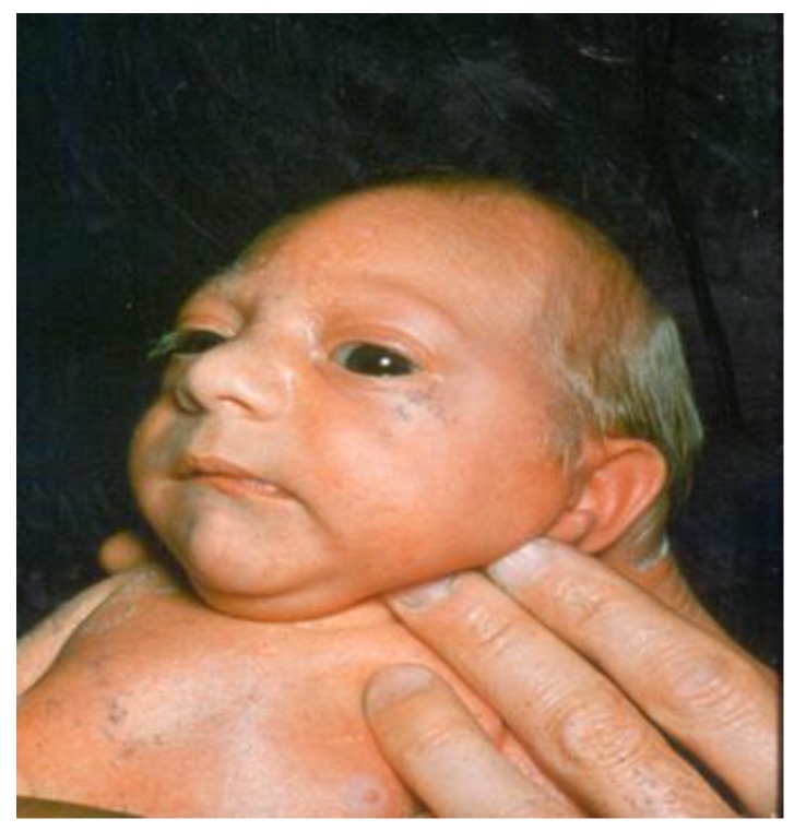
Microcephaly
- PMID: 28598357
- PMCID: PMC5483622
- DOI: 10.3390/children4060047
Microcephaly
Abstract
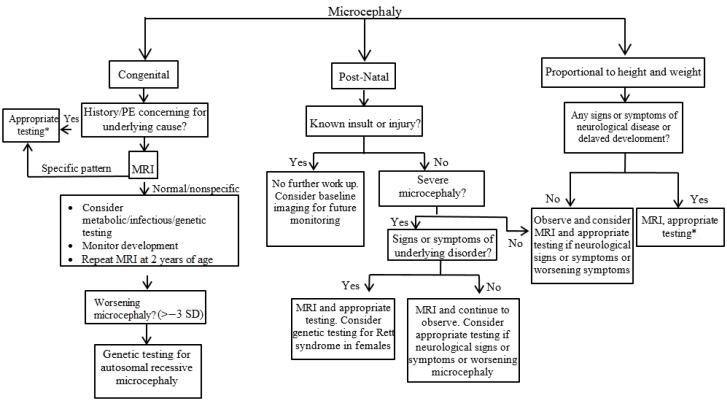
Microcephaly is defined as a head circumference more than two standard deviations below the mean for gender and age. Congenital microcephaly is present at birth, whereas postnatal microcephaly occurs later in life. Genetic abnormalities, syndromes, metabolic disorders, teratogens, infections, prenatal, perinatal, and postnatal injuries can cause both congenital and postnatal microcephaly. Evaluation of patients with microcephaly begins with a thorough history and physical examination. In cases of worsening microcephaly or neurological signs or symptoms, neuroimaging, metabolic, or genetic testing should be strongly considered. Any further studies and workup should be directed by the presence of signs or symptoms pointing to an underlying diagnosis and are usually used as confirmatory testing for certain conditions. Neuroimaging with magnetic resonance imaging (MRI) is often the first diagnostic test in evaluating children with microcephaly. Genetic testing is becoming more common and is often the next step following neuroimaging when there is no specific evidence in the history or physical examination suggesting a diagnosis. Microcephaly is a lifelong condition with no known cure. The prognosis is usually worse for children who experienced an intrauterine infection or have a chromosomal or metabolic abnormality. Zika virus has rapidly spread since 2015, and maternal infection with this virus is associated with microcephaly and other serious brain abnormalities. Microcephaly has become much more prevalent in the news and scientific community with the recent emergence of Zika virus as a cause of congenital microcephaly.
Keywords: Zika virus; genetic abnormalities; head circumference; microcephaly; neuroimaging; syndromes.
Conflict of interest statement
The authors declare no conflict of interest.
Figures
Similar articles
-
Clinical assessment and brain findings in a cohort of mothers, fetuses and infants infected with ZIKA virus.Am J Obstet Gynecol. 2018 Apr;218(4):440.e1-440.e36. doi: 10.1016/j.ajog.2018.01.012. Epub 2018 Jan 17. Am J Obstet Gynecol. 2018. PMID: 29353032
-
Serial Head and Brain Imaging of 17 Fetuses With Confirmed Zika Virus Infection in Colombia, South America.Obstet Gynecol. 2017 Jul;130(1):207-212. doi: 10.1097/AOG.0000000000002105. Obstet Gynecol. 2017. PMID: 28594771 Free PMC article.
-
Progressive lesions of central nervous system in microcephalic fetuses with suspected congenital Zika virus syndrome.Ultrasound Obstet Gynecol. 2017 Dec;50(6):717-722. doi: 10.1002/uog.17303. Epub 2017 Nov 8. Ultrasound Obstet Gynecol. 2017. PMID: 27644020
-
Testing for Zika virus infection in pregnancy: key concepts to deal with an emerging epidemic.Am J Obstet Gynecol. 2017 Mar;216(3):209-225. doi: 10.1016/j.ajog.2017.01.020. Epub 2017 Jan 23. Am J Obstet Gynecol. 2017. PMID: 28126366 Review.
-
Prenatal imaging findings in fetal Zika virus infection.Curr Opin Obstet Gynecol. 2017 Apr;29(2):95-105. doi: 10.1097/GCO.0000000000000345. Curr Opin Obstet Gynecol. 2017. PMID: 28134669 Review.
Cited by
-
Investigating the effects of a single ASPM variant (c.8508_8509) on brain architecture among siblings in a consanguineous Pakistani family.Mol Biol Rep. 2024 Jan 15;51(1):104. doi: 10.1007/s11033-023-09161-2. Mol Biol Rep. 2024. PMID: 38224417
-
Modeling human neurodevelopmental diseases with brain organoids.Cell Regen. 2022 Jan 4;11(1):1. doi: 10.1186/s13619-021-00103-6. Cell Regen. 2022. PMID: 34982276 Free PMC article. Review.
-
A Fetus with Congenital Microcephaly, Microphthalmia and Cataract Was Detected with Biallelic Variants in the OCLN Gene: A Case Report.Diagnostics (Basel). 2021 Aug 30;11(9):1576. doi: 10.3390/diagnostics11091576. Diagnostics (Basel). 2021. PMID: 34573918 Free PMC article.
-
Brain MRI segmentation of Zika-Exposed normocephalic infants shows smaller amygdala volumes.PLoS One. 2023 Jul 28;18(7):e0289227. doi: 10.1371/journal.pone.0289227. eCollection 2023. PLoS One. 2023. PMID: 37506075 Free PMC article.
-
Head CT: Toward Making Full Use of the Information the X-Rays Give.AJNR Am J Neuroradiol. 2021 Aug;42(8):1362-1369. doi: 10.3174/ajnr.A7153. Epub 2021 Jun 17. AJNR Am J Neuroradiol. 2021. PMID: 34140278 Free PMC article. Review.
References
-
- Alamo-Junquera D., Sunyer J., Iniguez C., Ballester F., Garcia-Esteban R., Forns J., Turner M.C., Lertxundi A., Lertxundi N., Fernandez-Somoano A., et al. Prenatal head growth and child neuropsychological development at age 14 months. Am. J. Obstet. Gynecol. 2015;212:e1–e11. doi: 10.1016/j.ajog.2014.12.001. - DOI - PubMed
-
- Von der Hagen M., Pivarcsi M., Liebe J., von Bernuth H., Didonato N., Hennermann J.B., Buhrer C., Wieczorek D., Kaindl A.M. Diagnostic approach to microcephaly in childhood: A two-center study and review of the literature. Dev. Med. Child Neurol. 2014;56:732–741. doi: 10.1111/dmcn.12425. - DOI - PubMed
Publication types
LinkOut - more resources
Full Text Sources
Other Literature Sources
