. 2017 May;98(5):1058-1072.
doi: 10.1099/jgv.0.000736.

Human cytomegalovirus microRNAs are carried by virions and dense bodies and are delivered to target cells

Affiliations

Human cytomegalovirus microRNAs are carried by virions and dense bodies and are delivered to target cells

Abdul-Aleem Mohammad et al. J Gen Virol. 2017 May.

Abstract

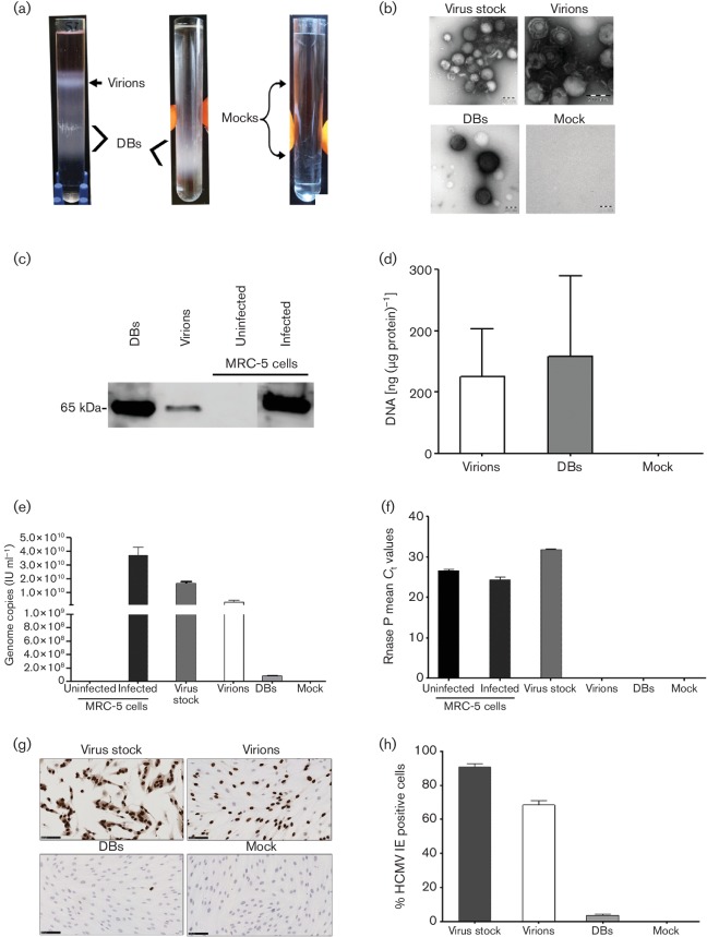
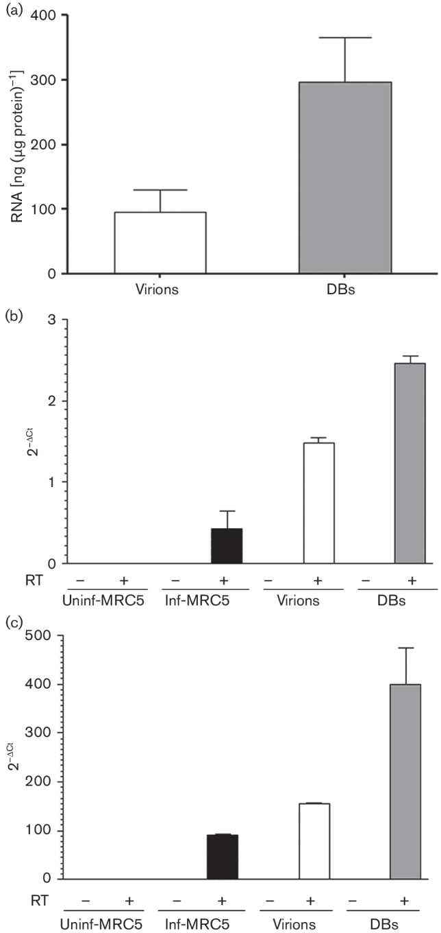
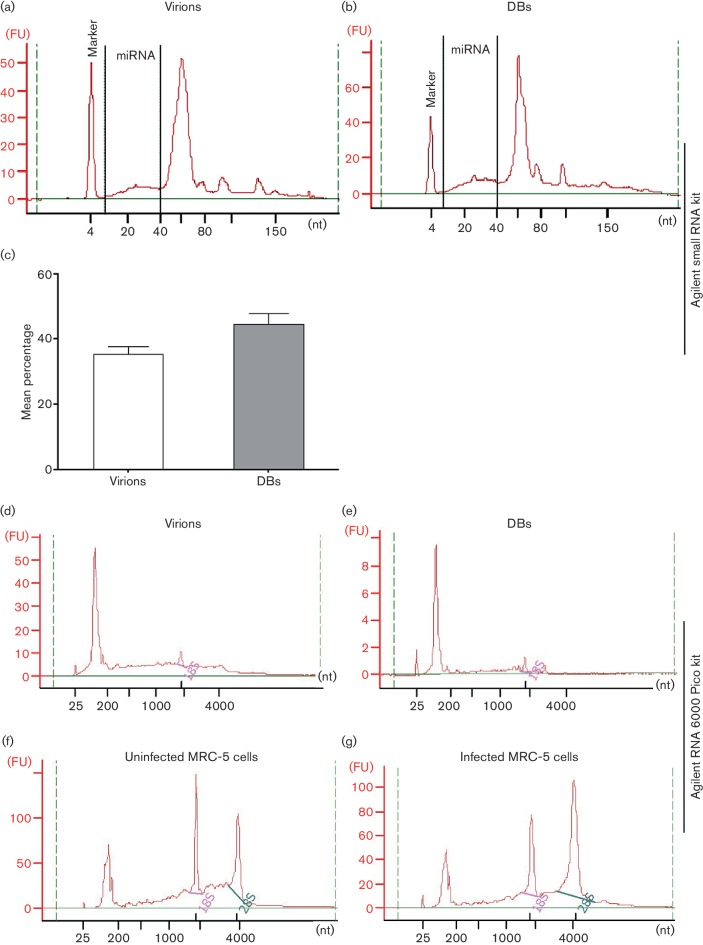
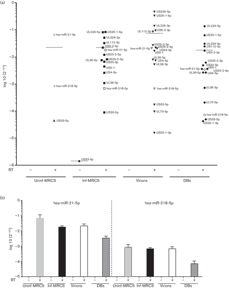
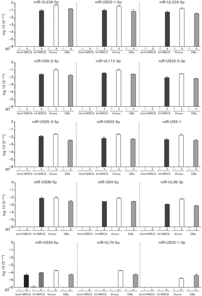
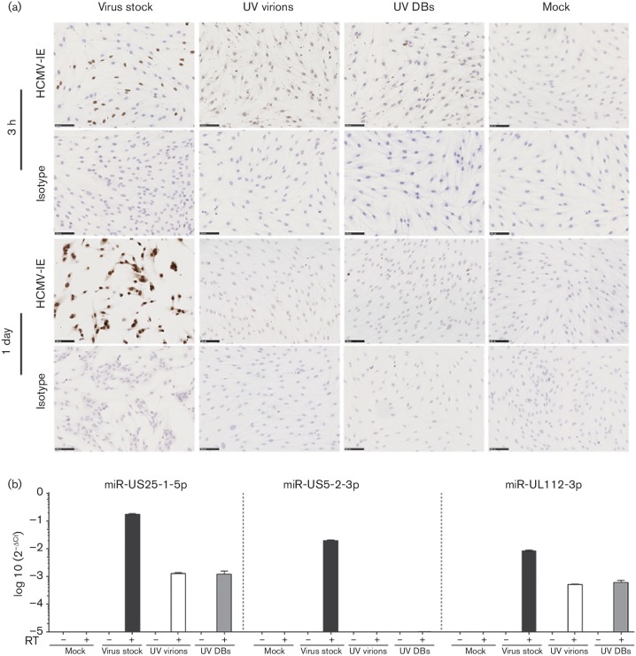
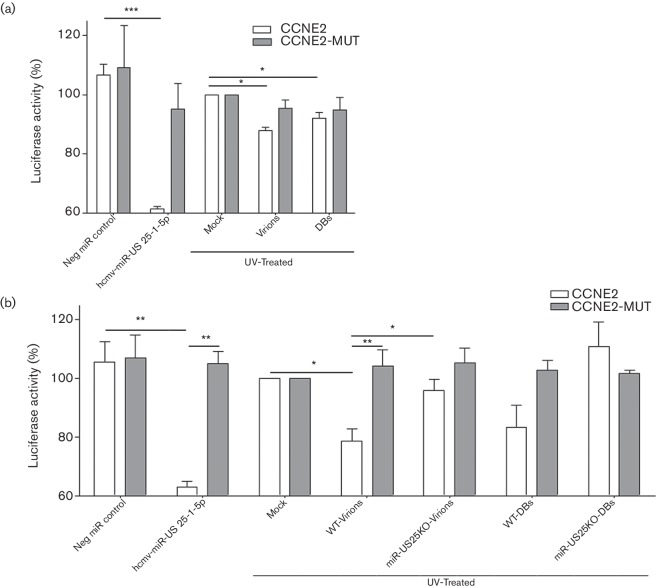
Human cytomegalovirus (HCMV) infection results in the production of virions, dense bodies (DBs) and non-infectious enveloped particles, all of which incorporate proteins and RNAs that can be transferred to host cells. Here, we investigated whether virions and DBs also carry microRNAs (miRNAs) and assessed their delivery and functionality in cells. Human lung fibroblasts (MRC-5) were infected with the HCMV strain AD169, and conditioned cell culture medium was collected and centrifuged. The pellets were treated with RNase-ONE, and the virions and DBs were purified with a potassium tartrate-glycerol gradient and dialysed. The virions and DBs were incubated with micrococcal nuclease, DNA and RNA were extracted and then analysed with TaqMan PCR assays, while the proteins were examined with Western blots. To assess the delivery of miRNAs to cells and their functionality, virions and DBs were irradiated with UV light. The purity of the virions and DBs was confirmed by typical morphology, the presence of the structural protein pp65 and the HCMV genome, the ability to infect MRC-5 cells and the absence of the host genome. RNA analysis revealed the presence of 14 HCMV-encoded miRNAs (UL22A-5p, US25-1-5p, UL22A-3p, US5-2-3p, UL112-3p, US25-2-3p, US25-2-5p, US33-3p, US5-1, UL36-5p, US4-5p, UL36-3p, UL70-5p and US25-1-3p), HCMV immediate-early mRNA and long non-coding RNA2.7, moreover, two host-encoded miRNAs (hsa-miR-218-5p and hsa-miR-21-5p) and beta-2-microglobulin RNA. UV-irradiated virions and DBs delivered viral miRNAs (US25-1-5p and UL112-3p) to the host cells, and miR-US25-1-5p was functional in a luciferase reporter assay. We conclude that virions and DBs carry miRNAs that are biologically functional and can be delivered to cells, which may affect cellular processes.


Figures

Fig. 1.
Fig. 1.
Isolation and characterization of virions and dense bodies (DBs). Media from MRC-5 cells infected with HCMV strain AD169 was collected and concentrated to pellets (viral stock), which were treated with RNase-ONE, and the virions and DBs were then purified by using potassium tartrate–glycerol gradient ultracentrifugation. (a) Representative tubes after gradient ultracentrifugation. (Left) Light-scattering upper band containing virions, lower band containing DBs. (Middle) A second ultracentrifugation was performed to further purify the DBs. (Right) No bands were seen in the mock samples. (b) Negative-staining electron microscopy showed virions (in upper band of gradient tubes), DBs (in lower band of gradient tubes), and some cellular debris in the viral stock; no particles were found in the mock samples. (c) Representative Western blot of lysed proteins from purified virions and DBs identified the HCMV structural protein pp65. Infected cells (5 days p.i., m.o.i. 0.1) and uninfected cells were used as positive and negative controls, respectively. (d) Concentration of DNA isolated from virions and DBs. (e) The number of HCMV genome copies [determined with World Health Organization (WHO) standards and gB Taqman] in the viral stocks, virions, DBs and mock samples; uninfected and infected MRC-5 cells were used as assay controls. (f) RNase P of host genome was found in the viral stock but not in the virions or DBs, confirming the purification of the virions and DBs; infected and uninfected MRC-5 cells were used as assay controls. (g) MRC-5 cells were infected with purified virions, DBs, virus stock and mock samples for 1 day, and analysed by immunocytochemistry. Brown, HCMV IE proteins; blue, nuclei; cells infected with viral stock served as a positive control. (h) Quantification of HCMV IE protein-positive cells. Values are mean±sem of three experiments performed in triplicate.
Fig. 2.
Fig. 2.
Analysis of RNA isolated from purified virions and dense bodies (DBs). Virions and DBs were purified from RNase-ONE-treated virus stocks, dialysed and treated with micrococcal nuclease; RNA was extracted, and during the RNA extraction the column was treated with DNase-I. (a) RNA concentrations of virions and DBs. Presence of (b) viral IE RNA transcripts and (c) lncRNA2.7 transcripts determined by TaqMan PCR assays. Values are mean±sem of three experiments performed in triplicate. RT, reverse transcriptase.
Fig. 3.
Fig. 3.
Bioanalyser-based analysis of RNA isolated from virions and dense bodies (DBs). To examine the small RNA species in virions (a) and DBs (b), a small RNA analysis kit (Agilent) was used. Representative electropherograms of RNA from virions and DBs show both miRNAs and small RNAs. (c) Mean percentages of miRNAs in virions and DBs; values are mean±sem of three experiments performed in triplicate. To inspect the total RNA species in virions (d) and DBs (e), an Agilent RNA 6000 Pico kit was used. Representative electropherograms of RNA from virions (d) and DBs (e) show very little 18S and almost no 28S rRNA, in contrast to the uninfected (f) and infected (g) MRC-5 cells. FU, fluorescence units; nt, number of nucleotides. Representative data from three experiments performed in triplicate are shown.
Fig. 4.
Fig. 4.
HCMV- and host-cell-encoded miRNAs in virions and dense bodies (DBs). To detect the miRNAs present in the virions and DBs, miRNA specific cDNA was synthesised in the presence or absence of reverse transcriptase enzyme (RT), and TaqMan PCR was performed. (a) TaqMan PCR analysis of 15 HCMV-encoded miRNAs and two host miRNAs in virions, DBs, and uninfected and infected MRC-5 cells. (b) Two host encoded miRNAs detected in virions, DBs, infected and uninfected control MRC-5 cells. The data were analysed with the 2−ΔCt method, where ΔC t=miRNA of interest C t value – Cel-miR39-3p spiked-in control C t value. Values are mean±sem of three experiments performed in triplicate.
Fig. 5.
Fig. 5.
HCMV encoded miRNAs in virions, dense bodies (DBs), and infected and uninfected MRC-5 cells. To detect the miRNAs present in the virions and DBs, miRNA-specific cDNA was synthesised in the presence or absence of reverse transcriptase enzyme (RT), and TaqMan PCR was performed. TaqMan PCR analysis of 15 HCMV-encoded miRNAs, organised from the most abundant (top left) to the least abundant (bottom right), in virions, DBs, and uninfected and infected MRC-5 cells. The data were analysed with the 2−ΔCt method, where ΔC t=miRNA of interest C t value – Cel-miR39-3p spiked-in control C t value. Values are mean±sem of three experiments performed in triplicate.
Fig. 6.
Fig. 6.
HCMV-encoded miRNAs from virions and DBs can be delivered to host cells. UV-irradiated virions, or DBs, or mock samples were added to MRC-5 cell monolayers; non-UV-treated virus stock was used as a positive control. (a) Immunocytochemistry staining for HCMV IE to validate the effectiveness of UV treatment of virions and DBs. UV-irradiated virions and DBs failed to infect the MRC-5 cells; non-UV-irradiated viral stocks resulted in infection of MRC-5 cells. Grey, HCMV IE staining; blue, haematoxylin staining of nuclei. (b) Two HCMV-encoded miRNAs, miR-US25-1-5p and miR-UL112-3p, were detected in cells treated with UV-irradiated virions and DBs. The data were analysed with the 2−ΔCt method, where ΔC t=miRNA of interest C t value – RNU48 C t value. Values are mean±sem of three experiments performed in triplicate.
Fig. 7.
Fig. 7.
Virions and dense bodies (DBs) delivered miRNA to host cells may affect cellular functions, as shown by luciferase reporter assay. (a, b) HCMV-miR-US25-1-5p luciferase reporter assay – HEK-293 cells transfected with luciferase reporter constructs containing CCNE2 5′ untranslated region (CCNE2) or its mutated control (CCNE2-MUT) together with Renilla vector as internal control. (a) First luciferase experiment: on the transfected cells UV-irradiated virions, or DBs, or mock samples were added. (b) Second luciferase experiment: to further strengthen the data obtained from the first luciferase reporter assay experiments and improve the specificity of the assay. Virions and DBs were purified from the miR-US25 knockout (miR-US25KO) and its control wild-type (WT) virus stocks, and particles were UV irradiated and added to the HEK-293 cells transfected with CCNE2 or CCNE2-MUT luciferase reporters (the amount of particles was doubled in the second luciferase experiment compared to the first). In both the first and the second luciferase experiments as positive and negative miRNA controls hcmv-miR-US25-1-5p mimic or scramble miRNA mimics were co-transfected with the luciferase constructs, respectively. Cells were lysed 20 h later and luciferase activity was quantified. Firefly luciferase activity was normalized to Renilla luciferase; values are percentage mean±sem of three experiments performed in triplicate. P<0.05 was considered statistically significant. *P<0.05; **P<0.01; ***P<0.001.

Similar articles

Cited by

References

    1. Krech U, Tobin J. A collaborative study of cytomegalovirus antibodies in mothers and young children in 19 countries. Bull World Health Organ. 1981;59:605–610. - PMC - PubMed
    1. Damato EG, Winnen CW. Cytomegalovirus infection: perinatal implications. J Obstet Gynecol Neonatal Nurs. 2002;31:86–92. doi: 10.1111/j.1552-6909.2002.tb00026.x. - DOI - PubMed
    1. Söderberg-Nauclér C. Treatment of cytomegalovirus infections beyond acute disease to improve human health. Expert Rev Anti Infect Ther. 2014;12:211–222. doi: 10.1586/14787210.2014.870472. - DOI - PubMed
    1. Gatherer D, Seirafian S, Cunningham C, Holton M, Dargan DJ, et al. High-resolution human cytomegalovirus transcriptome. Proc Natl Acad Sci USA. 2011;108:19755–19760. doi: 10.1073/pnas.1115861108. - DOI - PMC - PubMed
    1. Pfeffer S, Zavolan M, Grässer FA, Chien M, Russo JJ, et al. Identification of virus-encoded microRNAs. Science. 2004;304:734–736. doi: 10.1126/science.1096781. - DOI - PubMed