. 2017 May 22:8:572.
doi: 10.3389/fimmu.2017.00572. eCollection 2017.

PD-1 Blockade Promotes Emerging Checkpoint Inhibitors in Enhancing T Cell Responses to Allogeneic Dendritic Cells

Affiliations

PD-1 Blockade Promotes Emerging Checkpoint Inhibitors in Enhancing T Cell Responses to Allogeneic Dendritic Cells

Carmen Stecher et al. Front Immunol. .

Abstract

Immune checkpoint inhibitors, which target coinhibitory T cell molecules to promote anticancer immune responses, are on the rise to become a new pillar of cancer therapy. However, current immune checkpoint-based therapies are successful only in a subset of patients and acquired resistances pose additional challenges. Finding new targets and combining checkpoint inhibitors might help to overcome these limitations. In this study, human T cells stimulated with allogeneic dendritic cells (DCs) were used to compare immune checkpoint inhibitors targeting TIM-3, BTLA, LAG-3, CTLA-4, and TIGIT alone or in combination with a PD-1 antibody. We found that PD-1 blockade bears a unique potency to enhance T cell proliferation and cytokine production. Other checkpoint inhibitors failed to significantly augment T cell responses when used alone. However, antibodies to TIM-3, BTLA, LAG-3, and CTLA-4 enhanced T cell proliferation in presence of a PD-1 antibody. Upregulation of coinhibitory T cell receptors upon PD-1 blockade was identified as a potential mechanism for synergistic effects between checkpoint inhibitors. Donor-specific variation in response to immune checkpoint inhibitors was attributed to the T cells rather than DCs. Additionally, we analyzed the regulation of checkpoint molecules and their ligands on T cells and allogeneic DCs in coculture, which suggested a PD-1 blockade-dependent crosstalk between T cells and APC. Our results indicate that several immune checkpoint inhibitors have the capacity to enhance T cell responses when combined with PD-1 blockade. Additional in vitro studies on human T cells will be useful to identify antibody combinations with the potential to augment T cell responses in cancer patients.

Keywords: BTLA; CD160; CTLA-4; LAG-3; PD-1; TIM-3; coinhibitory receptors; immune checkpoint.


Figures
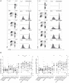
Figure 1
Figure 1
Influence of checkpoint inhibitors on T cell proliferation during the stimulation of human T cells with allogeneic mature dendritic cells (DCs). 1 × 105 CFSE-labeled human T cells were stimulated with 6 × 103 mature allogeneic human monocyte-derived DCs in the presence of blocking antibodies to the indicated molecules. After 6 days of coculture, the cells were stained for CD4 and CD8 and analyzed by flow cytometry. 7AAD+ cells were excluded from the analysis. (A) T cell proliferation during allo-MLR of a representative T cell donor, showing dot plots and histograms of isotype, PD-1 antibody, and PD-1 + BTLA antibody conditions for CD4 and CD8 T cells. Dot plots show CFSE versus CD4 (left panel) or CD8 (right panel) in live cells. Histograms show the percentage of CFSElow cells gated on live CD4 or CD8 T cells, respectively. (B) Normalized proliferation scores (as described in Section “Materials and Methods”) of CD4 T cells (left panel) and CD8 T cells (right panel) of 26 healthy T cell donors are shown. Each data point represents the mean of triplicates of one T cell donor (mean ± 95% CI). Stars indicate significant differences compared to IgG1 isotype control (single antibody conditions) or PD-1 antibody (PD-1 antibody containing conditions). The PD-1 dataset was included in both analyses. All P values were calculated using Dunn’s multiple comparison post hoc test following a Friedman ANOVA (*P < 0.05, **P < 0.01, and ***P < 0.001).
Figure 2
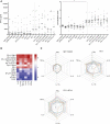
Figure 2
Effect of blocking antibodies to coinhibitory molecules on the cytokine content in the culture supernatants. Culture supernatants of T cell proliferation assays were collected and analyzed using a Luminex™-based multiplexing assay. Data from 14 T cell donors cocultured with 6 × 103 mature allogeneic dendritic cells are shown. (A) The IFN-γ content in the culture supernatant increases with the addition of a PD-1-blocking antibody. The median of the absolute amount of IFN-γ (left panel) and the deduced normalization values (mean ± 95% CI, number of SDs above the median of the isotype control replicates) are shown (right panel). (B) Summary of the cytokines simultaneously measured in the culture supernatants, depicted as a heat map. The mean normalization values of 14 donors are shown for IL-2, IL-17a, TNF-α, IL-13, IL-10, and IFN-γ. (C) Radar plots showing the IL-2, IL-17a, TNF-α, IL-13, IL-10, and IFN-γ content for the IgG1 isotype control, PD-1, and PD-1 + BTLA antibody conditions. Each line represents normalized data of one individual donor (n = 14).
Figure 3
Figure 3
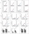
Expression of coinhibitory molecules on CD4 and CD8 T cells during the course of an MLR. T cells were left unstimulated or cocultured with mature allogeneic dendritic cells for 2–6 days in presence or absence of a PD-1-blocking antibody. At the indicated time points, CD4 and CD8 T cells were analyzed for CFSE dilution (A), expression of the activation marker CD25 (B), and the coinhibitory molecules PD-1 (C), TIGIT (D), TIM-3 (E), LAG-3 (F), and PD-L1 (G). Line graphs represent mean ± SEM of technical triplicates from one representative donor. Panels (E–G) additionally show cumulative data from day 6 of three to six donors and the percentage of CD4 and CD8 T cells expressing the indicated marker in the CFSEhigh and CFSElow subset.
Figure 4
Figure 4
Cytotoxic capacity of T cells stimulated by allogeneic dendritic cells (DCs) in presence of checkpoint inhibitors. T cells were stimulated with allogeneic DCs for 6 days without antibodies or in presence of blocking antibodies to the indicated coinhibitory receptors. Subsequently, their capacity to kill target cells harboring a membrane-bound anti-CD3 antibody fragment was assessed by incubating them with a 1:1 mixture of Bwwt and BwaCD3 target cells. The relative loss of CD14+ BwaCD3 target cells in the coculture was analyzed by flow cytometry and used to calculate the percentage of specific killing. (A) Representative FACS plots from one donor showing allo-stimulated T cells in coculture with Bw cells. The murine Bw cells were detected using an antibody to murine CD45, and a CD14 antibody was used to detect the target cells via the CD14 stem of the anti-CD3 antibody fragment. (B) Representative data from one donor showing the abundance of CD14+ BwaCD3 target cells and the calculated percentage of specific killing. (C) Cumulative data from six donors of three independent experiments are shown. Data represent mean ± SEM.
Figure 5
Figure 5
Effect of dendritic cell (DC) number and maturation status on T cell proliferation, and changes of DC marker expression during coculture. (A) Comparison of the effect of different amounts of immature and mature DCs on T cell proliferation. Data are expressed as %CFSElow cells of live CD4+ or CD8+ T cells. Each dot represents data from one antibody condition of one of 10 T cell donors. Mean with 95% CI is shown. Significances were calculated using RM one-way ANOVA with Tukey’s multiple comparisons post hoc test. (B) Immature DC expression of the costimulatory markers CD80, CD83, and CD86 and the coinhibitory markers CD155, PD-L1, and PD-L2 before coculture with allogeneic T cells, and after 24 or 48 h of coculture. Data from one donor, representative of at least three different experiments, are shown. (C) Measurement of costimulatory markers on immature and mature DCs after 24–48 h of coculture with allogeneic T cells, with (red lines) or without (blue lines) the addition of a PD-1-blocking antibody. Dotted lines represent the expression of the respective marker on DCs before coculture with T cells. Data from one donor (mean ± SEM of triplicates) are shown.
Figure 6
Figure 6
The contribution of T cells versus dendritic cells (DCs) on the effect of checkpoint inhibitors on T cell proliferation. T cells derived from four donors were stimulated for 6 days with mature DCs from three allogeneic donors in the presence of blocking antibodies to the indicated molecules. The data were both analyzed by plotting proliferation data from different T cell donors that were stimulated by an individual DC donor, and vice versa. (A) The data points in each line represent proliferation of T cells derived from four different donors upon stimulation with DCs derived from one individual donor. (B) The data points in each line represent proliferation of T cells derived from one individual donor in response to stimulation with DCs from three different donors. (A,B) The percentages of CFSElow CD4 T cells (left panels) and CFSElow CD8 T cells (right panels) are shown. Data represent mean ± SEM. Differences between donors were calculated by a Dunn’s post hoc test following a Kruskal–Wallis test.

Similar articles

Cited by

References

    1. Coley WB. The treatment of malignant tumors by repeated inoculations of erysipelas. With a report of ten original cases. 1893. Clin Orthop (1991) (262):3–11. - PubMed
    1. Parish CR. Cancer immunotherapy: the past, the present and the future. Immunol Cell Biol (2003) 81:106–13.10.1046/j.0818-9641.2003.01151.x - DOI - PubMed
    1. Hodi FS, O’Day SJ, McDermott DF, Weber RW, Sosman JA, Haanen JB, et al. Improved survival with ipilimumab in patients with metastatic melanoma. N Engl J Med (2010) 363:711–23.10.1056/NEJMoa1003466 - DOI - PMC - PubMed
    1. Robert C, Thomas L, Bondarenko I, O’Day S, Weber J, Garbe C, et al. Ipilimumab plus dacarbazine for previously untreated metastatic melanoma. N Engl J Med (2011) 364:2517–26.10.1056/NEJMoa1104621 - DOI - PubMed
    1. Rothschild SI, Thommen DS, Moersig W, Müller P, Zippelius A. Cancer immunology – development of novel anti-cancer therapies. Swiss Med Wkly (2015) 145:w14066.10.4414/smw.2015.14066 - DOI - PubMed