. 2017 Jul 15;130(14):2405-2415.
doi: 10.1242/jcs.200477. Epub 2017 Jun 5.

Arf6 and Rab22 mediate T cell conjugate formation by regulating clathrin-independent endosomal membrane trafficking

Affiliations

Arf6 and Rab22 mediate T cell conjugate formation by regulating clathrin-independent endosomal membrane trafficking

Debra L Johnson et al. J Cell Sci. .

Abstract

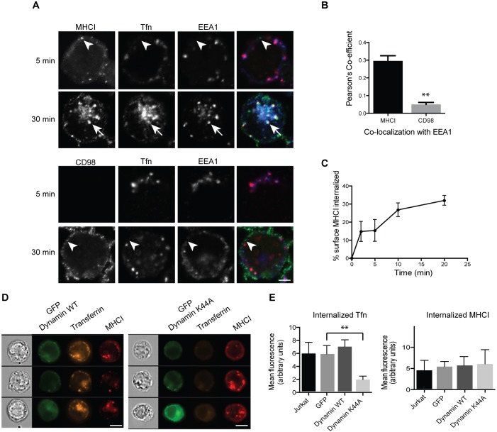
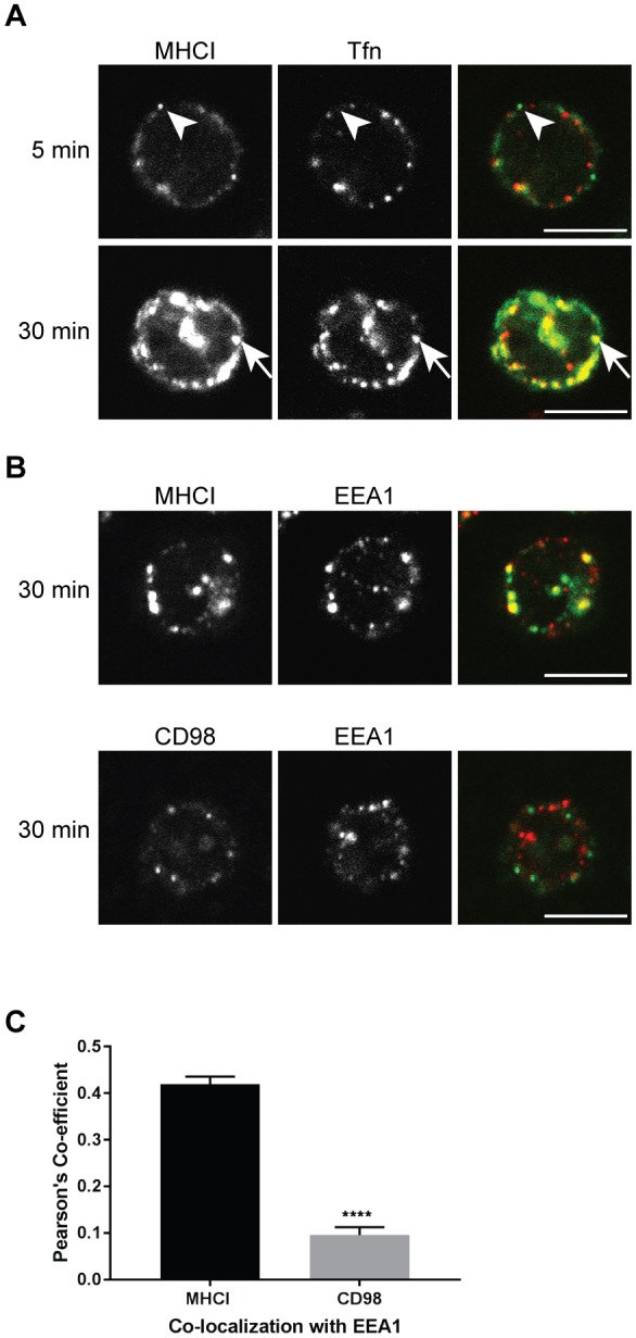
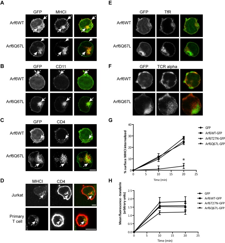
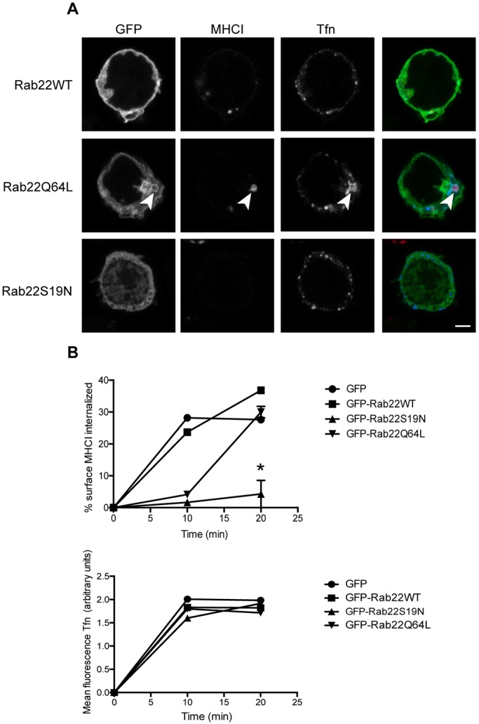
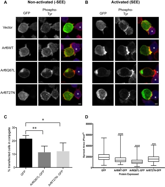
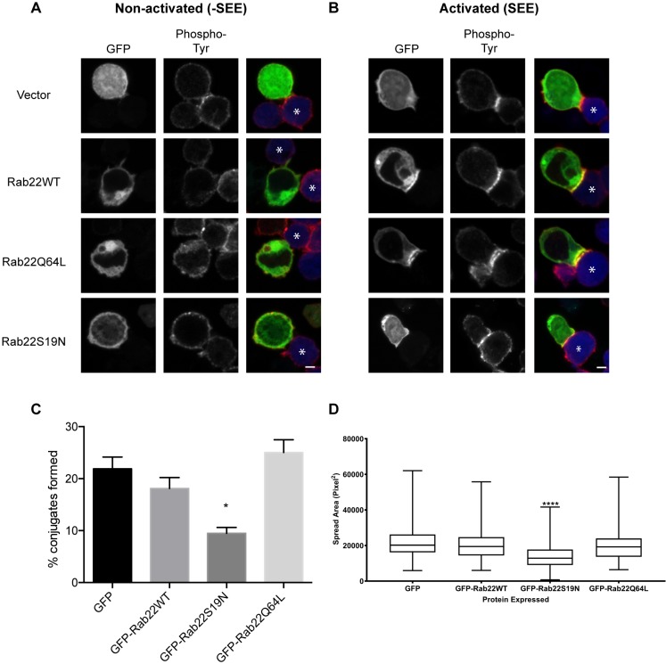
Endosomal trafficking can influence the composition of the plasma membrane and the ability of cells to polarize their membranes. Here, we examined whether trafficking through clathrin-independent endocytosis (CIE) affects the ability of T cells to form a cell-cell conjugate with antigen-presenting cells (APCs). We show that CIE occurs in both the Jurkat T cell line and primary human T cells. In Jurkat cells, the activities of two guanine nucleotide binding proteins, Arf6 and Rab22 (also known as Rab22a), influence CIE and conjugate formation. Expression of the constitutively active form of Arf6, Arf6Q67L, inhibits CIE and conjugate formation, and results in the accumulation of vacuoles containing lymphocyte function-associated antigen 1 (LFA-1) and CD4, molecules important for T cell interaction with the APC. Moreover, expression of the GTP-binding defective mutant of Rab22, Rab22S19N, inhibits CIE and conjugate formation, suggesting that Rab22 function is required for these activities. Furthermore, Jurkat cells expressing Rab22S19N were impaired in spreading onto coverslips coated with T cell receptor-activating antibodies. These observations support a role for CIE, Arf6 and Rab22 in conjugate formation between T cells and APCs.

Keywords: Arf6; Clathrin-independent endocytosis; Immunological synapse; Rab22; Rab22a; T cell.


Conflict of interest statement

Competing interestsThe authors declare no competing or financial interests.

Figures

Fig. 1.
Fig. 1.
Jurkat T cells internalize CIE cargo separately from transferrin, in a dynamin-independent manner, at a rate of 2% per minute. (A) Cells were incubated at 37°C with antibodies to MHCI or CD98 and Alexa Fluor 633-conjugated transferrin for 5 or 30 min. Bound surface antibodies were removed by treating cells with a low pH buffer for 8 s prior to fixation. Cells were immunolabeled with antibodies to EEA1, and fluorescent secondary antibodies were used to detect internalized MHCI and CD98 antibody. MHCI is in puncta that are distinct from transferrin-containing endosomes (arrowheads) at 5 min, indicating an entry mechanism distinct from CME. Note that at 30 min, MHCI and transferrin meet in a common endosome (arrows) and in the ERC. CD98 is in a distinct endosome (arrowhead) that does not colocalize with transferrin. Scale bar: 3 µm. (B) The Pearson's correlation coefficient shows a significant difference between the correlation of MHCI with EEA1, as compared to CD98 with EEA1, at the 30 min time point. Shown is the mean±s.e.m. of three experiments, with at least 42 cells measured in each experiment. **P<0.01 (unpaired t-test). (C) Cells were incubated at 4°C with antibodies to MHCI. Cells were washed, returned to warm medium, fixed at indicated time points, and remaining surface antibody detected by fluorescent secondary antibodies and imaging flow cytometry. MHCI enters cells at a rate of ∼2% per minute. Shown is the mean±s.e.m. of four experiments. (D,E) Cells were transfected with GFP-tagged wild-type (WT) or K44A mutant dynamin, and then incubated at 37°C with antibodies to MHCI and Alexa Fluor 594-conjugated transferrin for 30 min. Bound surface antibodies were removed by treating cells with a low pH buffer for 8 s prior to fixation. (D) Sample images from imaging flow cytometry showing that there is a decrease in internalized transferrin in cells expressing dynamin K44A. Scale bars: 7 µm. (E) Quantification of internalized transferrin (Tfn) and MHCI by flow cytometry. Shown is the mean±s.e.m. of four experiments (2000 cells scored/treatment). GFP–dynamin-K44A significantly reduces transferrin internalization but not MHCI internalization. **P<0.01 (unpaired t-test).
Fig. 2.
Fig. 2.
CIE occurs in naive human T cells. (A) Naive, CD4+ human T cells were incubated with antibodies to MHCI and Alexa Fluor 594-conjugated transferrin for 5 or 30 min, and then treated with low pH buffer prior to fixation and labeling of internalized MHCI with Alexa Fluor 488-conjugated secondary antibody. Arrowhead highlights MHCI without transferrin; arrow highlights MHCI with transferrin. (B) T cells were incubated with antibodies to MHCI or CD98 for 30 min and then treated with low pH buffer prior to fixation and subsequent labeling for EEA1, and use of Alexa Fluor 488-conjugated secondary antibodies for detection of MHCI and CD98 and Alexa Fluor 594-conjugated secondary antibodies for EEA1. Scale bars: 5 µm. (C) The Pearson's correlation coefficient shows a significant difference between the correlation of MHCI with EEA1 as compared to CD98 with EEA1 at the 30 min time point. Shown is the mean±s.e.m. of three experiments, with at least 30 cells measured in each experiment. ****P<0.0001 (unpaired t-test).
Fig. 3.
Fig. 3.
Arf6 colocalizes with CIE cargo proteins that are important for T cell activation and Arf6Q67L inhibits CIE in Jurkat T cells. Cells were transiently transfected with Arf6WT–GFP or Arf6Q67L–GFP. After fixation, the steady state distribution of cargo proteins was assessed with antibodies, followed by labeling with Alexa Fluor 594-conjugated secondary antibodies. MHCI (A), CD11 (LFA-1) (B), and CD4 (C) colocalize with Arf6WT and Arf6Q67L. CME cargo proteins transferrin receptor (E) and T cell receptor α (F) do not colocalize with Arf6-positive membranes. Scale bars: 3 µm. (D) Jurkat T cells and primary T cells were incubated with antibodies to MHCI for 30 min, treated with a low pH wash, then fixed and stained with antibodies to CD4, followed by use of Alexa Fluor 488-secondary antibody to detect MHCI and Alexa Fluor 594-secondary antibodies to detect CD4. Scale bars: 5 µm. Arrows highlight cargo-containing endosomes. (G,H) Cells were chilled to 4°C and incubated with antibodies to MHCI (G) or fluorescently conjugated transferrin (H). Cells were rinsed and warmed for the indicated times to allow internalization of antibody-bound MHCI and transferrin. Remaining surface MHCI and internalized transferrin were measured by using imaging flow cytometry. Expression of Arf6Q67L inhibits internalization of the CIE cargo protein MHCI but not the CME cargo protein transferrin receptor, as measured by determining transferrin internalization. Shown are the mean±s.e.m. for four experiments. For the 20 min time point, cells expressing Arf6Q67L internalized less MHCI than GFP controls. *P<0.05 (unpaired t-test).
Fig. 4.
Fig. 4.
Rab22 is required for internalization of MHCI. Cells were transiently transfected with GFP-tagged Rab22WT, Rab22Q64L or Rab22S19N and incubated at 37°C with antibodies against MHCI and Alexa Fluor 647-conjugated transferrin to allow endocytosis. Surface antibody was removed by a low pH rinse and then cells were fixed after 30 min and primary antibody was detected with Alexa Fluor 594 secondary antibodies. (A) Confocal imaging shows that expression of Rab22Q64L results in the formation of large endosomes that are positive for MHCI (arrowheads). MHCI is not internalized in cells expressing Rab22S19N. Scale bar: 3 µm. (B) Imaging flow cytometry was used to quantify uptake of CIE and CME cargo. Rab22S19N inhibits internalization of MHCI but not of transferrin. Shown are the mean±s.e.m. for three experiments. For the 20 min time point, cells expressing Rab22S19N internalized less MHCI than GFP controls. *P<0.05 (unpaired t-test).
Fig. 5.
Fig. 5.
Arf6 polarizes to the immunological synapse, and expression of Arf6T27N or Arf6Q67L reduces formation of stable T cell/APC conjugates. Jurkat T cells were transfected with GFP-tagged Arf6WT, Arf6Q67L or Arf6T27N. Jurkat cells were then incubated with APCs or with APCs that had been exposed to SEE. Cells were fixed, and phosphorylated tyrosine was detected with antibodies. (A) The non-activated control (-SEE) shows no polarization of Arf6 at the site of contact with the APC and there is only a small amount of phosphorylated tyrosine in the Arf6WT- and Arf6T27N-expressing cells. (B) In the activated sample (+SEE), Arf6WT and Arf6Q67L polarize towards the site of contact and colocalize with phosphorylated tyrosine. Arf6T27N does not polarize toward the site of contact and alters the phosphorylated tyrosine pattern. Scale bar: 3 µm. Asterisks mark APCs. (C) Cells expressing GFP alone, Arf6T27N or Arf6Q67L were incubated with APCs that had been exposed to SEE. After 1.5 h, cells were fixed and conjugate formation was quantified by using imaging flow cytometry. Shown are the mean±s.d. for four experiments. *P<0.05, **P<0.01 (unpaired t-test). (D) Jurkat cells expressing GFP-tagged Arf6WT, Arf6Q67L or Arf6T27N, or GFP alone, were plated on coverslips coated with antibodies against the T cell receptor (anti-CD3 antibody) for 3 min and then fixed and stained with a plasma membrane cell mask. The area of cell spread was quantified by using the image analysis software package Metamorph. Data is presented as box-and-whisker plots representing the median and the 25th and 75th percentiles. Whiskers show the minimum to maximum. n>150 cells over three independent experiments. ****P<0.0001 (one-way Anova with Kruskal–Wallis test).
Fig. 6.
Fig. 6.
Rab22 polarizes to the immunological synapse and is required for the formation of stable T cell–APC conjugates. Jurkat cells were transfected with GFP-tagged Rab22WT, Rab22Q64L or Rab22S19N. Jurkat cells were then incubated with APCs or with APCs that had been exposed to SEE. Cells were fixed and phosphorylated tyrosine was detected with antibodies. (A) The non-activated control (-SEE) shows no polarization of Rab22 at the site of contact with the APC and phosphorylated tyrosine is low. (B) In the activated sample (+SEE), Rab22WT and Rab22Q64L polarize toward the site of contact and colocalize with phosphorylated tyrosine on the plasma membrane. Rab22S19N does not polarize toward the site of contact and reduces phosphorylated tyrosine at the site of contact. Asterisks mark APCs. Scale bars: 3 µm. (C) Cells expressing GFP or GFP-tagged Rab22 were incubated with APCs that had been exposed to SEE. After 1 h, cells were fixed and conjugate formation was quantified by using imaging flow cytometry. Shown are mean±s.d. from three experiments. *P<0.05 (unpaired t-test). (D) Jurkat cells expressing GFP-tagged Rab22WT, Rab22Q64L or Rab22S19N, or GFP alone, were plated on coverslips coated with antibodies against the T cell receptor (anti-CD3 antibody) for 3 min and then fixed and stained with a plasma membrane cell mask. The area the cell spread was quantified by using the image analysis software package Metamorph. Data is presented as box-and-whisker plots representing the median and the 25th and 75th percentiles. Whiskers show the minimum and maximum. n>150 cells over at least three independent experiments. ****P<0.0001 (one-way Anova with Kruskal–Wallis test).

Similar articles

Cited by

References

    1. Balagopalan L., Barr V. A. and Samelson L. E. (2009). Endocytic events in TCR signaling: focus on adapters in microclusters. Immunol. Rev. 232, 84-98. 10.1111/j.1600-065X.2009.00840.x - DOI - PMC - PubMed
    1. Barral D. C., Cavallari M., McCormick P. J., Garg S., Magee A. I., Bonifacino J. S., De Libero G. and Brenner M. B. (2008). CD1a and MHC class I follow a similar endocytic recycling pathway. Traffic 9, 1446-1457. 10.1111/j.1600-0854.2008.00781.x - DOI - PMC - PubMed
    1. Blander J. M. (2016). The comings and goings of MHC class I molecules herald a new dawn in cross-presentation. Immunol. Rev. 272, 65-79. 10.1111/imr.12428 - DOI - PMC - PubMed
    1. Bouchet J., del Rio-Iñiguez I., Lasserre R., Agüera-Gonzalez S., Cuche C., Danckaert A., McCaffrey M. W., Di Bartolo V. and Alcover A. (2016). Rac1-Rab11-FIP3 regulatory hub coordinates vesicle traffic with actin remodeling and T-cell activation. EMBO J. 35, 1160-1174. 10.15252/embj.201593274 - DOI - PMC - PubMed
    1. Brown F. D., Rozelle A. L., Yin H. L., Balla T. and Donaldson J. G. (2001). Phosphatidylinositol 4,5-bisphosphate and Arf6-regulated membrane traffic. J. Cell Biol. 154, 1007-1017. 10.1083/jcb.200103107 - DOI - PMC - PubMed

MeSH terms