

Biochemical Underpinnings of Immune Cell Metabolic Phenotypes
- PMID: 28514672
- PMCID: PMC5660630
- DOI: 10.1016/j.immuni.2017.04.013
Biochemical Underpinnings of Immune Cell Metabolic Phenotypes
Abstract
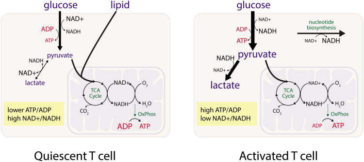
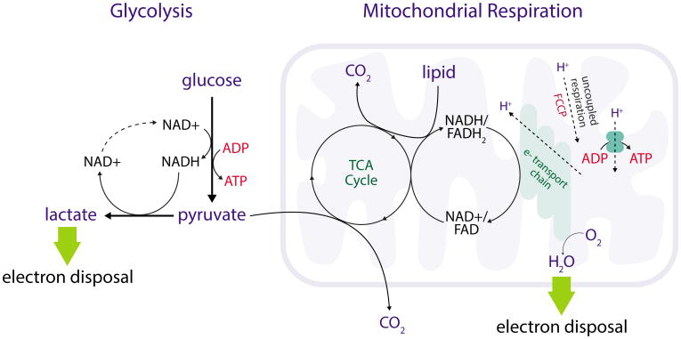
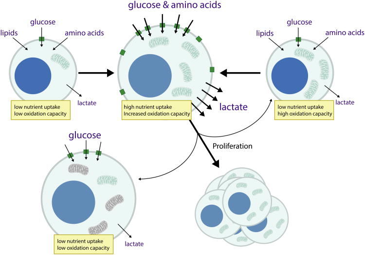
The metabolism of immune cells affects their function and influences host immunity. This review explores how immune cell metabolic phenotypes reflect biochemical dependencies and highlights evidence that both the metabolic state of immune cells and nutrient availability can alter immune responses. The central importance of oxygen, energetics, and redox homeostasis in immune cell metabolism, and how these factors are reflected in different metabolic phenotypes, is also discussed. Linking immune cell metabolic phenotype to effector functions is important to understand how altering metabolism can impact the way in which immune cells meet their metabolic demands and affect the immune response in various disease contexts.
Copyright © 2017 Elsevier Inc. All rights reserved.
Figures
Similar articles
-
Mitochondria as central hub of the immune system.Redox Biol. 2019 Sep;26:101255. doi: 10.1016/j.redox.2019.101255. Epub 2019 Jun 15. Redox Biol. 2019. PMID: 31247505 Free PMC article. Review.
-
Extracellular flux analysis to monitor glycolytic rates and mitochondrial oxygen consumption.Methods Enzymol. 2014;542:125-49. doi: 10.1016/B978-0-12-416618-9.00007-8. Methods Enzymol. 2014. PMID: 24862264
-
How Mitochondrial Metabolism Contributes to Macrophage Phenotype and Functions.J Mol Biol. 2018 Oct 19;430(21):3906-3921. doi: 10.1016/j.jmb.2018.07.003. Epub 2018 Jul 10. J Mol Biol. 2018. PMID: 30006265 Review.
-
Metabolic adaptations of tissue-resident immune cells.Nat Immunol. 2019 Jul;20(7):793-801. doi: 10.1038/s41590-019-0407-0. Epub 2019 Jun 18. Nat Immunol. 2019. PMID: 31213715 Review.
-
Metabolic Regulation of the Immune Humoral Response.Immunity. 2017 May 16;46(5):743-755. doi: 10.1016/j.immuni.2017.04.009. Immunity. 2017. PMID: 28514675 Free PMC article. Review.
Cited by
-
Deciphering the Interplay between the Epithelial Barrier, Immune Cells, and Metabolic Mediators in Allergic Disease.Int J Mol Sci. 2024 Jun 24;25(13):6913. doi: 10.3390/ijms25136913. Int J Mol Sci. 2024. PMID: 39000023 Free PMC article. Review.
-
Single-cell metabolic profiling of human cytotoxic T cells.Nat Biotechnol. 2021 Feb;39(2):186-197. doi: 10.1038/s41587-020-0651-8. Epub 2020 Aug 31. Nat Biotechnol. 2021. PMID: 32868913 Free PMC article.
-
Norovirus NS1/2 protein increases glutaminolysis for efficient viral replication.PLoS Pathog. 2024 Jul 8;20(7):e1011909. doi: 10.1371/journal.ppat.1011909. eCollection 2024 Jul. PLoS Pathog. 2024. PMID: 38976719 Free PMC article.
-
Impact of Exercise on Immunometabolism in Multiple Sclerosis.J Clin Med. 2020 Sep 21;9(9):3038. doi: 10.3390/jcm9093038. J Clin Med. 2020. PMID: 32967206 Free PMC article. Review.
-
Role of Polyamines in Immune Cell Functions.Med Sci (Basel). 2018 Mar 8;6(1):22. doi: 10.3390/medsci6010022. Med Sci (Basel). 2018. PMID: 29517999 Free PMC article. Review.
References
-
- Adams WC, Chen YH, Kratchmarov R, Yen B, Nish SA, Lin WHW, Rothman NJ, Luchsinger LL, Klein U, Busslinger M, Rathmell JC, Snoeck HW, Reiner SL. Anabolism-Associated Mitochondrial Stasis Driving Lymphocyte Differentiation over Self-Renewal. Cell Rep. 2016;17:3142–3152. doi: 10.1016/j.celrep.2016.11.065. - DOI - PMC - PubMed
-
- Angelin A, Gil-de-Gómez L, Dahiya S, Jiao J, Guo L, Levine MH, Wang Z, Quinn WJ, III, Kopinski PK, Wang L, Akimova T, Liu Y, Bhatti TR, Han R, Laskin BL, Baur JA, Blair IA, Wallace DC, Hancock WW, Beier UH. Foxp3 Reprograms T Cell Metabolism to Function in Low-Glucose, High-Lactate Environments. Cell Metab. 2017 doi: 10.1016/j.cmet.2016.12.018. - DOI - PMC - PubMed
-
- Banh RS, Iorio C, Marcotte R, Xu Y, Cojocari D, Rahman AA, Pawling J, Zhang W, Sinha A, Rose CM, Isasa M, Zhang S, Wu R, Virtanen C, Hitomi T, Habu T, Sidhu SS, Koizumi A, Wilkins SE, Kislinger T, Gygi SP, Schofield CJ, Dennis JW, Wouters BG, Neel BG. PTP1B controls non-mitochondrial oxygen consumption by regulating RNF213 to promote tumour survival during hypoxia. Nat Cell Biol. 2016;18:803–813. doi: 10.1038/ncb3376. - DOI - PMC - PubMed
-
- Beier UH, Angelin A, Akimova T, Wang L, Liu Y, Xiao H, Koike MA, Hancock SA, Bhatti TR, Han R, Jiao J, Veasey SC, Sims CA, Baur JA, Wallace DC, Hancock WW. Essential role of mitochondrial energy metabolism in Foxp3+ T-regulatory cell function and allograft survival. FASEB J. 2015;29:2315–2326. doi: 10.1096/fj.14-268409. - DOI - PMC - PubMed
Publication types
MeSH terms
Substances
Grants and funding
LinkOut - more resources
Full Text Sources
Other Literature Sources
Medical
