miR-205 is a critical regulator of lacrimal gland development
- PMID: 28511845
- PMCID: PMC9276161
- DOI: 10.1016/j.ydbio.2017.05.012
miR-205 is a critical regulator of lacrimal gland development
Abstract
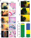
The tear film protects the terrestrial animal's ocular surface and the lacrimal gland provides important aqueous secretions necessary for its maintenance. Despite the importance of the lacrimal gland in ocular health, molecular aspects of its development remain poorly understood. We have identified a noncoding RNA (miR-205) as an important gene for lacrimal gland development. Mice lacking miR-205 fail to properly develop lacrimal glands, establishing this noncoding RNA as a key regulator of lacrimal gland development. Specifically, more than half of knockout lacrimal glands never initiated, suggesting a critical role of miR-205 at the earliest stages of lacrimal gland development. RNA-seq analysis uncovered several up-regulated miR-205 targets that may interfere with signaling to impair lacrimal gland initiation. Supporting this data, combinatorial epistatic deletion of Fgf10, the driver of lacrimal gland initiation, and miR-205 in mice exacerbates the lacrimal gland phenotype. We develop a molecular rheostat model where miR-205 modulates signaling pathways related to Fgf10 in order to regulate glandular development. These data show that a single microRNA is a key regulator for early lacrimal gland development in mice and highlights the important role of microRNAs during organogenesis.
Keywords: Fgf10; Lacrimal gland; MiR-205; MicroRNAs.
Copyright © 2017 Elsevier Inc. All rights reserved.
Figures


Similar articles
-
Lacrimal gland development and Fgf10-Fgfr2b signaling are controlled by 2-O- and 6-O-sulfated heparan sulfate.J Biol Chem. 2011 Apr 22;286(16):14435-44. doi: 10.1074/jbc.M111.225003. Epub 2011 Feb 28. J Biol Chem. 2011. PMID: 21357686 Free PMC article.
-
Molecular regulation of ocular gland development.Semin Cell Dev Biol. 2019 Jul;91:66-74. doi: 10.1016/j.semcdb.2018.07.023. Epub 2018 Sep 25. Semin Cell Dev Biol. 2019. PMID: 30266427 Review.
-
Barx2 and Fgf10 regulate ocular glands branching morphogenesis by controlling extracellular matrix remodeling.Development. 2011 Aug;138(15):3307-17. doi: 10.1242/dev.066241. Development. 2011. PMID: 21750040 Free PMC article.
-
FGF signaling activates a Sox9-Sox10 pathway for the formation and branching morphogenesis of mouse ocular glands.Development. 2014 Jul;141(13):2691-701. doi: 10.1242/dev.108944. Epub 2014 Jun 12. Development. 2014. PMID: 24924191 Free PMC article.
-
Is the main lacrimal gland indispensable? Contributions of the corneal and conjunctival epithelia.Surv Ophthalmol. 2016 Sep-Oct;61(5):616-27. doi: 10.1016/j.survophthal.2016.02.006. Epub 2016 Mar 9. Surv Ophthalmol. 2016. PMID: 26968256 Review.
Cited by
-
MIR205HG Is a Long Noncoding RNA that Regulates Growth Hormone and Prolactin Production in the Anterior Pituitary.Dev Cell. 2019 May 20;49(4):618-631.e5. doi: 10.1016/j.devcel.2019.03.012. Epub 2019 Apr 11. Dev Cell. 2019. PMID: 30982661 Free PMC article.
-
Unveiling the ups and downs of miR-205 in physiology and cancer: transcriptional and post-transcriptional mechanisms.Cell Death Dis. 2020 Nov 15;11(11):980. doi: 10.1038/s41419-020-03192-4. Cell Death Dis. 2020. PMID: 33191398 Free PMC article. Review.
-
Epithelial Markers aSMA, Krt14, and Krt19 Unveil Elements of Murine Lacrimal Gland Morphogenesis and Maturation.Front Physiol. 2017 Sep 26;8:739. doi: 10.3389/fphys.2017.00739. eCollection 2017. Front Physiol. 2017. PMID: 29033846 Free PMC article.
-
Metazoan MicroRNAs.Cell. 2018 Mar 22;173(1):20-51. doi: 10.1016/j.cell.2018.03.006. Cell. 2018. PMID: 29570994 Free PMC article. Review.
-
Generation of 3D lacrimal gland organoids from human pluripotent stem cells.Nature. 2022 May;605(7908):126-131. doi: 10.1038/s41586-022-04613-4. Epub 2022 Apr 20. Nature. 2022. PMID: 35444274
References
-
- Abràmoff MD, Magalhães PJ and Ram SJ (2004). Image processing with imageJ. Biophotonics Int. 11, 36–41.
Publication types
MeSH terms
Substances
Grants and funding
LinkOut - more resources
Full Text Sources
Other Literature Sources
Molecular Biology Databases

