. 2017 Jun 26;91(14):e00450-17.
doi: 10.1128/JVI.00450-17. Print 2017 Jul 15.

A Naturally Occurring Recombinant Enterovirus Expresses a Torovirus Deubiquitinase

Affiliations

A Naturally Occurring Recombinant Enterovirus Expresses a Torovirus Deubiquitinase

Pengcheng Shang et al. J Virol. .

Abstract

Enteroviruses (EVs) are implicated in a wide range of diseases in humans and animals. In this study, a novel enterovirus (enterovirus species G [EVG]) (EVG 08/NC_USA/2015) was isolated from a diagnostic sample from a neonatal pig diarrhea case and identified by using metagenomics and complete genome sequencing. The viral genome shares 75.4% nucleotide identity with a prototypic EVG strain (PEV9 UKG/410/73). Remarkably, a 582-nucleotide insertion, flanked by 3Cpro cleavage sites at the 5' and 3' ends, was found in the 2C/3A junction region of the viral genome. This insertion encodes a predicted protease with 54 to 68% amino acid identity to torovirus (ToV) papain-like protease (PLP) (ToV-PLP). Structural homology modeling predicts that this protease adopts a fold and a catalytic site characteristic of minimal PLP catalytic domains. This structure is similar to those of core catalytic domains of the foot-and-mouth disease virus leader protease and coronavirus PLPs, which act as deubiquitinating and deISGylating (interferon [IFN]-stimulated gene 15 [ISG15]-removing) enzymes on host cell substrates. Importantly, the recombinant ToV-PLP protein derived from this novel enterovirus also showed strong deubiquitination and deISGylation activities and demonstrated the ability to suppress IFN-β expression. Using reverse genetics, we generated a ToV-PLP knockout recombinant virus. Compared to the wild-type virus, the ToV-PLP knockout mutant virus showed impaired growth and induced higher expression levels of innate immune genes in infected cells. These results suggest that ToV-PLP functions as an innate immune antagonist; enterovirus G may therefore gain fitness through the acquisition of ToV-PLP from a recombination event.IMPORTANCE Enteroviruses comprise a highly diversified group of viruses. Genetic recombination has been considered a driving force for viral evolution; however, recombination between viruses from two different orders is a rare event. In this study, we identified a special case of cross-order recombination between enterovirus G (order Picornavirales) and torovirus (order Nidovirales). This naturally occurring recombination event may have broad implications for other picornaviral and/or nidoviral species. Importantly, we demonstrated that the exogenous ToV-PLP gene that was inserted into the EVG genome encodes a deubiquitinase/deISGylase and potentially suppresses host cellular innate immune responses. Our results provide insights into how a gain of function through genetic recombination, in particular cross-order recombination, may improve the ability of a virus to evade host immunity.

Keywords: deubiquitinase; enterovirus G; genetic recombination; papain-like protease; torovirus.


Figures

FIG 1
FIG 1
Schematic diagram of the genome organization of EVG 08/NC_USA/2015. The single ORF is flanked by a long 5′ UTR (812 nucleotides) and a short 3′ UTR (72 nucleotides), followed by a poly(A) tail. Secondary structural elements in the 5′ UTR were adapted as previously described (93). The ToV-PLP gene is presented as a red box that is inserted at the 2C/3A cleavage junction. The 5′- and 3′-flanking sequences of 3C protease cleavage sites are shown in blue. Vertical lines indicate the polyprotein processing site by the 3C protease. IRES, internal ribosome entry site. An, poly(A) tail.
FIG 2
FIG 2
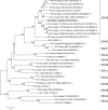
Phylogenetic analysis of enterovirus full-length genome nucleotide sequences. The phylogenetic tree was constructed by the maximum likelihood method using the best-fitting general time-reversible model with a gamma distribution. The numbers on branches are bootstrap values (percent) from 500 replicates. Bootstrap values of <50% are not shown. GenBank accession numbers are given in parentheses after the name of each isolate. Enterovirus, rhinovirus, porcine enterovirus, ovine enterovirus, simian enterovirus, coxsackievirus, and poliovirus are denoted EV, RV, PEV, OEV, SEV, CV, and PV, respectively.
FIG 3
FIG 3
Phylogenetic analysis of coronaviral and picornaviral papain-like proteases. The phylogenetic tree was constructed by the maximum likelihood method using the best-fitting algorithm Whelan-and-Goldman model. The numbers on branches are bootstrap values (percent) from 500 replicates. Bootstrap values of <50% are not shown. Amino acid sequences of PLPs are acquired from coronaviral, toroviral, and picornaviral polyproteins, with GenBank accession numbers shown in parentheses after the name of each protein entry.
FIG 4
FIG 4
Amino acid sequence alignment of the EVG ToV-PLP protein with other known papain-like proteases. Shown is structure- and sequence-guided alignment of EVG ToV-PLP with the corresponding catalytic domains of the FMDV leader protease, coronaviral PLPs, and the human deubiquitinating/deISGylating enzyme USP18. Sequences and numbering correspond to the following GenBank protein accession numbers: CAA25416.1 for the FMDV leader protease, AEA10817.1 for SARS coronavirus PLP, YP_009047202.1 for MERS coronavirus PLP, AAU06353.1 for murine hepatitis virus (MHV) PLP2, AFD98833.1 for human coronavirus NL63 PLP2, ABI26421.1 for infectious bronchitis virus (IBV) PLP, and AAD49967.1 for human USP18. The secondary structures of ToV-PLP and FMDV-Lpro are shown in black above the sequences. The secondary structure of an inserted β-sheet domain that is not present in the core fold of FMDV-Lpro but found in the other sequences is shown above the alignment in green. The catalytic-triad residues are marked with purple stars, and a DUB-specific “signature” motif in the protease catalytic sites is indicated with a light blue box (21). Hs., Homo sapiens.
FIG 5
FIG 5
Structural modeling and comparison of the EVG ToV-PLP protein with other known papain-like proteases. Structures of the homology model of the core ToV-PLP catalytic domain were compared with those of the catalytic domains of representative viral proteases from FMDV (44) and SARS-CoV (83) and the eukaryotic USP18 deubiquitinating enzyme (94). Each representation is colored by sequence progression from N to C termini (blue to red) and is shown in an approximately equivalent orientation. The side chains of the catalytic Cys, His, and Asp residues from each enzyme are circled and shown in a stick representation. PDB accession numbers are shown in parentheses.
FIG 6
FIG 6
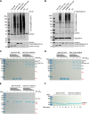
Deubiquitination and deISGylation activities of the recombinant ToV-PLP protease. (A and B) Effect of PLP expression on Ub or ISG15 conjugation. (A) HEK-293T cells were cotransfected with plasmid DNAs expressing HA-Ub and FLAG-tagged ToV-PLP or PRRSV-PLP2 or their catalytic-site mutants (ToV-PLP mut and PRRSV-PLP2 mut, respectively). HA-Ub-conjugated cellular proteins were visualized by Western blotting using anti-HA MAb. The expression of FLAG-tagged PLPs was detected by anti-FLAG MAb M2. (B). HEK-293T cells were cotransfected with plasmid DNAs expressing ISG15 and its conjugation enzyme E1/E2/E3, ToV-PLP, or PRRSV-PLP2 or their catalytic-site mutants. ISG15-conjugated cellular proteins were detected with an ISG15-specific MAb by Western blot analysis. For both panels A and B, the empty pCAGGS vector plasmid was included as a control, while the expression of the GAPDH housekeeping gene was detected as a loading control. (C to E) Ubiquitin cleavage activity of recombinant ToV-PLP on K48/K63/M1-linked polyubiquitin chains. Cell-free deubiquitination activity was analyzed by using K48-linked polyubiquitin chains (Ub3–7) (C), K63-linked polyubiquitin chains (D), or M1-linked polyubiquitin chains (E) as the substrates and incubation with serially diluted GST-PLP (1 μg, 0.5 μg, and 0.25 μg) or equal amounts of the catalytic-site mutant (GST-PLP C/H/D>A) at 37°C for 2 h. Cleaved ubiquitin products were visualized by SDS-PAGE and are labeled in red. Note that the molecular mass of Ub6 (51 kDa) is close to that of GST-PLP (50.4 kDa). WT, wild type. (F) DeISGylation activities of GST-tagged recombinant ToV-PLP (left) and its catalytic-site mutant (right). The ISG15 precursor (proISG15) was incubated with 40 nM GST-PLP or equal amounts of the catalytic-site mutant (GST-PLP C/H/D>A) at 37°C for a series of time points (1 min, 2 min, 5 min, and 10 min). The mature form of ISG15 was visualized by SDS-PAGE and is labeled in red.
FIG 7
FIG 7
Effect of ToV-PLP expression on IFN-β expression. (A) ToV-PLP affects reporter gene expression derived by the IFN-β promoter. HEK-293T cells were transfected with a plasmid expressing ToV-PLP, the PRRSV-PLP2 domain, or the catalytic-site mutant (PLP mut and PLP2 mut, respectively), along with the firefly luciferase reporter plasmid (p125-Luc) and Renilla luciferase expression plasmid pRL-SV40. Cells were stimulated by SeV at 24 h posttransfection. Luciferase activity was measured at 16 h poststimulation. Relative luciferase activity is defined as the ratio of firefly luciferase reporter activity to Renilla luciferase activity. Each data point represents the mean value from three experiments. Error bars show standard deviations of the normalized data. The expression of PLPs was detected by Western blotting using anti-FLAG MAb M2. The expression of the GAPDH housekeeping gene was detected as a loading control. RLU, relative light units. (B) ToV-PLP affects IFN-β mRNA expression. HEK-293T cells were transfected with a plasmid expressing ToV-PLP or PRRSV-PLP2 (wild type or mutant). At 24 h posttransfection, cells were stimulated with SeV. At 16 h poststimulation, quantitative RT-PCR was performed to evaluate the innate immune response. The relative mRNA expression level of IFN-β was acquired by normalization to the value for the TBP housekeeping gene. Mean values with standard deviations are shown. P values of <0.01 (**), <0.001 (***), and <0.0001 (****) are indicated.
FIG 8
FIG 8
Construction of the EVG 08/NC_USA/2015 infectious clone and rescue of recombinant viruses. (A) Strategy for assembling the full-length cDNA infectious clone of EVG 08/NC_USA/2015. Two RT-PCR-amplified genomic fragments and a synthesized HDV ribozyme gene were assembled into the pACYC177 vector by using the NEBuilder HiFi DNA assembly method. The CMV promoter sequence was inserted at the 5′ end of the genome. (B) Schematic diagram of the PLP knockout mutant (vPLP-KO). (C) Immunofluorescence detection of recombinant viruses rescued from full-length cDNA clones. BHK-21C cells were initially transfected with plasmid DNA of the full-length cDNA clone pEVG or its mutant. ST cells were subsequently infected with the cloned viruses rescued from BHK cells. The expression of ToV-PLP and the structural protein VP1 was detected by specific MAb 128-28 and MAb 115-5, respectively. The cell nucleus is stained with DAPI. (D) Western blot detection of PLP expression in recombinant virus-infected ST cells. The membrane was probed with ToV-PLP-specific MAb 128-28. The expression of the GAPDH housekeeping gene was detected as a loading control.
FIG 9
FIG 9
Characterization of in vitro growth properties of recombinant EVGs rescued from full-length cDNA infectious clones. Confluent ST cells were initially inoculated with the recombinant virus at an MOI of 0.01, and the cell culture supernatant was serially harvested at 0, 2, 4, 6, 8, 10, and 12 hpi. The virus titer was measured by a microtitration assay. The results shown are mean values from three replicates, with error bars showing standard deviations. Virus titers are expressed as numbers of FFU per milliliter.
FIG 10
FIG 10
Effect of ToV-PLP deletion on the deubiquitination and deISGylation abilities of enterovirus. (A) HEK-293T cells were transfected with plasmid DNAs expressing HA-Ub and then infected with the parental virus EVG 08/NC_USA/2015, the cloned virus vEVG, or vPLP-KO. Ubiquitin-conjugated cellular proteins were detected by Western blotting using an anti-HA MAb. (B) HEK-293T cells were cotransfected with plasmid DNAs expressing FLAG-tagged ISG15 and its conjugation enzyme E1/E2/E3 and then infected with the parental virus EVG 08/NC_USA/2015, the cloned virus vEVG, or vPLP-KO. ISGylated host proteins were visualized by Western blotting using anti-FLAG MAb. ToV-PLP-specific MAb 128-28 was used to detect the expression of PLP in infected cells. The expression of the GAPDH housekeeping gene was detected as a loading control.
FIG 11
FIG 11
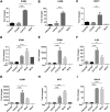
Effect of ToV-PLP deletion on innate immune gene expression levels. ST cells were infected with the parental virus EVG 08/NC_USA/2015, the cloned virus vEVG, or the PLP knockout mutant vPLP-KO at an MOI of 3. Mock-infected cells were used as a control. Total RNAs were then extracted from cell lysates to perform real-time RT-PCR. (A to C) Innate immune gene expression levels were measured at 10 hpi. (D to I) At 6 hpi, cells were stimulated with SeV (200 HA units/ml) for 4 h. Total RNAs were then extracted to perform real-time RT-PCR. Relative expression levels of innate immune genes were assessed by comparison with the values for the mock control. The experiment was repeated three times. Results are shown as mean values for each group. P values of <0.05 (*), <0.01 (**), <0.001 (***), and <0.0001 (****) are indicated.

Similar articles

Cited by

References

    1. Knowles NJ, Hovi T, Hyypiä T, King AMQ, Lindberg AM, Pallansch MA, Palmenberg AC, Simmonds P, Skern T, Stanway G, Yamashita T, Zell R. 2012. Virus taxonomy: classification and nomenclature of viruses, p 855–880. In King AMQ, ELefkowitz E, Adams MJ, Carstens EB (ed), Virus taxonomy. Classification and nomenclature of viruses. Ninth report of the International Committee on Taxonomy of Viruses Elsevier Academic Press, San Diego, CA.
    1. Boros Á, Nemes C, Pankovics P, Bíró H, Kapusinszky B, Delwart E, Reuter G. 2012. Characterization of a novel porcine enterovirus in wild boars in Hungary. Arch Virol 157:981–986. doi:10.1007/s00705-012-1255-5. - DOI - PMC - PubMed
    1. Boros Á, Pankovics P, Knowles NJ, Reuter G. 2012. Natural interspecies recombinant bovine/porcine enterovirus in sheep. J Gen Virol 93:1941–1951. doi:10.1099/vir.0.041335-0. - DOI - PubMed
    1. Moon H-J, Song D, Seon BH, Kim H-K, Park S-J, An D-J, Kim J-M, Kang B-K, Park B-K. 2012. Complete genome analysis of porcine enterovirus B isolated in Korea. J Virol 86:10250. doi:10.1128/JVI.01548-12. - DOI - PMC - PubMed
    1. Zoll J, Heus HA, van Kuppeveld FJ, Melchers WJ. 2009. The structure-function relationship of the enterovirus 3′-UTR. Virus Res 139:209–216. doi:10.1016/j.virusres.2008.07.014. - DOI - PubMed

Publication types