Exosomes as potential alternatives to stem cell therapy for intervertebral disc degeneration: in-vitro study on exosomes in interaction of nucleus pulposus cells and bone marrow mesenchymal stem cells
- PMID: 28486958
- PMCID: PMC5424403
- DOI: 10.1186/s13287-017-0563-9
Exosomes as potential alternatives to stem cell therapy for intervertebral disc degeneration: in-vitro study on exosomes in interaction of nucleus pulposus cells and bone marrow mesenchymal stem cells
Abstract
Background: The stem cell-based therapies for intervertebral disc degeneration have been widely studied. However, the mechanisms of mesenchymal stem cells interacting with intervertebral disc cells, such as nucleus pulposus cells (NPCs), remain unknown. Exosomes as a vital paracrine mechanism in cell-cell communication have been highly focused on. The purpose of this study was to detect the role of exosomes derived from bone marrow mesenchymal stem cells (BM-MSCs) and NPCs in their interaction with corresponding cells.
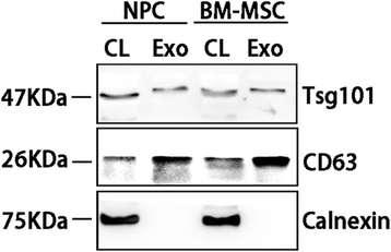
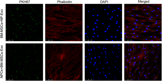
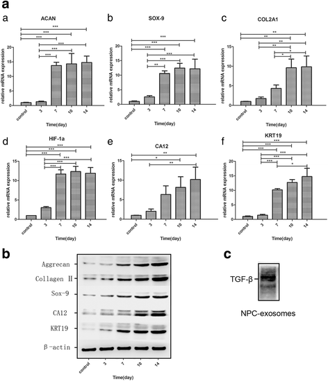
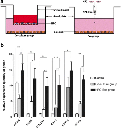
Methods: The exosomes secreted by BM-MSCs and NPCs were purified by differential centrifugation and identified by transmission electron microscope and immunoblot analysis of exosomal marker proteins. Fluorescence confocal microscopy was used to examine the uptake of exosomes by recipient cells. The effects of NPC exosomes on the migration and differentiation of BM-MSCs were determined by transwell migration assays and quantitative RT-PCR analysis of NPC phenotypic genes. Western blot analysis was performed to examine proteins such as aggrecan, sox-9, collagen II and hif-1α in the induced BM-MSCs. Proliferation and the gene expression profile of NPCs induced by BM-MSC exosomes were measured by Cell Counting Kit-8 and qRT-PCR analysis, respectively.
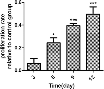
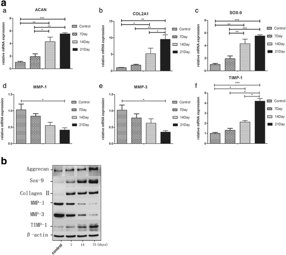
Results: Both the NPCs and BM-MSCs secreted exosomes, and these exosomes underwent uptake by the corresponding cells. NPC-derived exosomes promoted BM-MSC migration and induced BM-MSC differentiation to a nucleus pulposus-like phenotype. BM-MSC-derived exosomes promoted NPC proliferation and healthier extracellular matrix production in the degenerate NPCs.
Conclusion: Our study indicates that the exosomes act as an important vehicle in information exchange between BM-MSCs and NPCs. Given a variety of functions and multiple advantages, exosomes alone or loaded with specific genes and drugs would be an appropriate option in a cell-free therapy strategy for intervertebral disc degeneration.
Keywords: Differentiation; Exosomes; Intervertebral disc degeneration; Mesenchymal stem cell; Migration; Nucleus pulposus cell; Proliferation.
Figures


Similar articles
-
Effects of hypoxia on differentiation from human placenta-derived mesenchymal stem cells to nucleus pulposus-like cells.Spine J. 2014 Oct 1;14(10):2451-8. doi: 10.1016/j.spinee.2014.03.028. Epub 2014 Mar 21. Spine J. 2014. PMID: 24662208
-
Bone marrow mesenchymal stem cells slow intervertebral disc degeneration through the NF-κB pathway.Spine J. 2015 Mar 1;15(3):530-8. doi: 10.1016/j.spinee.2014.11.021. Epub 2014 Nov 29. Spine J. 2015. PMID: 25457469
-
Mesenchymal stem cells deliver exogenous miR-21 via exosomes to inhibit nucleus pulposus cell apoptosis and reduce intervertebral disc degeneration.J Cell Mol Med. 2018 Jan;22(1):261-276. doi: 10.1111/jcmm.13316. Epub 2017 Aug 14. J Cell Mol Med. 2018. PMID: 28805297 Free PMC article.
-
A Review: Methodologies to Promote the Differentiation of Mesenchymal Stem Cells for the Regeneration of Intervertebral Disc Cells Following Intervertebral Disc Degeneration.Cells. 2023 Aug 28;12(17):2161. doi: 10.3390/cells12172161. Cells. 2023. PMID: 37681893 Free PMC article. Review.
-
Mesenchymal stromal/stem cells and their exosomes application in the treatment of intervertebral disc disease: A promising frontier.Int Immunopharmacol. 2022 Apr;105:108537. doi: 10.1016/j.intimp.2022.108537. Epub 2022 Jan 29. Int Immunopharmacol. 2022. PMID: 35101851 Review.
Cited by
-
Extracellular vesicles produced by human-induced pluripotent stem cell-derived endothelial cells can prevent arterial stenosis in mice via autophagy regulation.Front Cardiovasc Med. 2022 Oct 17;9:922790. doi: 10.3389/fcvm.2022.922790. eCollection 2022. Front Cardiovasc Med. 2022. PMID: 36324745 Free PMC article.
-
MiR-200b in heme oxygenase-1-modified bone marrow mesenchymal stem cell-derived exosomes alleviates inflammatory injury of intestinal epithelial cells by targeting high mobility group box 3.Cell Death Dis. 2020 Jun 25;11(6):480. doi: 10.1038/s41419-020-2685-8. Cell Death Dis. 2020. PMID: 32587254 Free PMC article.
-
Umbilical cord mesenchymal stem cell (UC-MSC) transplantations for cerebral palsy.Am J Transl Res. 2018 Mar 15;10(3):901-906. eCollection 2018. Am J Transl Res. 2018. PMID: 29636880 Free PMC article.
-
Exosomes Derived from Bone Marrow Mesenchymal Stem Cells Prevent Acidic pH-Induced Damage in Human Nucleus Pulposus Cells.Med Sci Monit. 2020 May 21;26:e922928. doi: 10.12659/MSM.922928. Med Sci Monit. 2020. PMID: 32436493 Free PMC article.
-
A comparative study of mesenchymal stem cell transplantation and NTG-101 molecular therapy to treat degenerative disc disease.Sci Rep. 2021 Jul 20;11(1):14804. doi: 10.1038/s41598-021-94173-w. Sci Rep. 2021. PMID: 34285277 Free PMC article.
References
MeSH terms
Substances
LinkOut - more resources
Full Text Sources
Other Literature Sources
Research Materials
Miscellaneous

