. 2017 Jun;49(6):941-945.
doi: 10.1038/ng.3858. Epub 2017 May 1.

DUX-family transcription factors regulate zygotic genome activation in placental mammals

Affiliations

DUX-family transcription factors regulate zygotic genome activation in placental mammals

Alberto De Iaco et al. Nat Genet. 2017 Jun.

Abstract

In animal embryos, transcription is mostly silent for several cell divisions, until the release of the first major wave of embryonic transcripts through so-called zygotic genome activation (ZGA). Maternally provided ZGA-triggering factors have been identified in Drosophila melanogaster and Danio rerio, but their mammalian homologs are still undefined. Here, we provide evidence that the DUX family of transcription factors is essential to this process in mice and potentially in humans. First, human DUX4 and mouse Dux are both expressed before ZGA in their respective species. Second, both orthologous proteins bind the promoters of ZGA-associated genes and activate their transcription. Third, Dux knockout in mouse embryonic stem cells (mESCs) prevents the cells from cycling through a 2-cell-like state. Finally, zygotic depletion of Dux leads to impaired early embryonic development and defective ZGA. We conclude that DUX-family proteins are key inducers of zygotic genome activation in placental mammals.


Conflict of interest statement

Conflict of interest

The authors declare that they have no conflict of interest.

Figures

Figure 1
Figure 1. DUX4 promotes transcription of genes expressed during early ZGA
(A) Comparative expression during early human embryonic development of DUX4 (red) and the top 100 genes upregulated upon DUX4 overexpression in human primary myoblasts (blue, full line average, dashed lines 95% confidence interval around the mean). Oo, oocyte; Zy, zygote; 2C, 4C, 8C, corresponding n-cell stage; Mo, morula; Bl, blastocyst. (B) Cluster of genes differentially expressed during early embryonic development were selected from the previously identified subsets of genes (Supplementary Figure 1A) based on high expression at 4C (upper panels) and 8C (lower panels). Blue and dotted line delineate mean and 95% confidence, respectively. (C) Expression of genes from each cluster illustrated in (B) when DUX4 is ectopically expressed in human primary myoblasts. Lower parts of the panels depict the fold change expression of genes within these clusters, all randomly distributed along the y-axes, with kernel density plotted in the upper part.
Figure 2
Figure 2. DUX4 binds TSSs of genes expressed during early ZGA and activates their expression in hESCs.
(A) Average coverage normalized for sequencing depth of ChIP-seq signal of DUX4 (blue) when overexpressed in hESCs in a window of 5 kb from the annotated TSS of genes belonging to the 2-4C and 2-8C clusters from Figure 1B. Total input is represented in gray (line, average; shade, standard error of the mean). B) Fraction of genes belonging to each cluster from Figure 1B with a DUX4 peak within 5 kb of their annotated TSS. Fisher’s exact test was performed to compare maternal vs. 2-4C and 2-8C (p= 3.54e-61 and p= 2.23e-13 respectively) (C) Average coverage of ChIP-seq signal of DUX4 (blue) when overexpressed in hESCs within 5 kb of TFE of transcripts specifically upregulated at oocyte-to-4C and 4C-to-8C transitions. Total input is represented in gray (line, average; shade, standard error of the mean). (D) Fraction of TFE from oocyte-to-4C (n=32) and 4C-to-8C (n=128) transitions that have a DUX4 peak overlapping with their 5' end. Fisher’s exact test was performed to compare 4C-to-8C vs. oocyte-to-4C TFEs (p= 4.48e-17). (E) Comparative expression in hESCs of three genes activated at ZGA (ZSCAN4, MBD3L2 and DUXA) and two control housekeeping genes (ACTB and TBP) 24 hours after transfection with plasmids expressing LacZ (green squares) or DUX4 (blue circles). Expression was normalized to ACTB. Horizontal lines represent the mean. *** p ≤ 0.001, unpaired t-test.
Figure 3
Figure 3. Dux is necessary for formation of 2C-like mESCs.
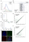
(A) Comparative expression of the two alternative transcripts of Dux, Dux (pink) and Gm4981 (orange), with genes (blue) and transposable elements (MERVL; green) specifically expressed during murine ZGA. Full lines represent the average and dashed lines the 95% confidence interval around the mean (B) Single-cell RNA-sequencing comparison between mESCs sorted for expression of both tomato and GFP reporters driven by MERVL and Zscan4 promoters, respectively (revelators of 2C-like cells), and the double negative population. Average gene expression was quantified and fold change between positive and negative cells was plotted. Dots are randomly distributed along the y-axes. The upper plot represents the kernel density estimate of middle-2C stage (blue line) and the rest of the genes (gray line). The Dux macrosatellite repeat was deleted in mESCs carrying a MERVL-GFP reporter by CRISPR/Cas9-mediated excision. (C) Fraction of GFP+ cells in WT or Dux-deleted cells. (D) RNA sequencing analysis of WT and Dux KO mESC clones. The dot plot displays the average gene expression of three independent clones from each cell type. (E) GFP expression in Dux KO (blue circles) and WT (green squares) mESC clones carrying an integrated MERVL-GFP reporter, and transiently expressing LacZ, DUX4, Dux or Gm4981 transgenes. (F) RNA sequencing analysis of Dux KO mESC clones transiently expressing Dux or control. The dot plot displays the average gene expression of two independent clones from each cell type. (G) Dux KO mESCs carrying an integrated MERVL-GFP reporter and transiently expressing a HA-tagged form of Dux were stained for HA and immunofluorescence was detected by confocal microscopy. DAPI, blue; GFP, green; HA, red. Horizontal bars in (C) and (E) represent the mean. *** p ≤ 0.001, unpaired t-test.
Figure 4
Figure 4. TRIM28 regulates formation of 2C-like mESCs by repressing Dux expression
(A) RNA sequencing analysis of WT and Trim28 KO mESCs. Average gene expression was quantified and fold change between KO and WT cells plotted. Dots are randomly distributed along the y-axes. The upper plot represents the kernel density estimate of genes specifically expressed in 2C-like mESCs (green line) and the rest of the genes (gray line). (B) WT (blue circles) and Dux KO (green squares) mESC clones carrying an integrated MERVL-GFP reporter were transduced with lentiviral vectors encoding for shRNAs targeting Trim28 or a control. 4 days later GFP expression was quantified. Horizontal lines represent the mean. *** p ≤ 0.001, unpaired t-test. (C) RNA sequencing of Trim28-depleted or control Dux KO mESC clones. The dot plot represents the average gene expression of three independent KO clones transduced with lentiviral vectors encoding for a control or a Trim28-specific shRNA. (D) Average coverage of ChIP-seq signal of Trim28 (top plot; blue lines; two replicates) and H3K9me3 (bottom plot; two replicates) in control (red lines) and Trim28 KD mESCs (green line) around the Dux gene. Total input is represented in gray. ChIP-seq reads were mapped on the genome, before focusing the analysis on a 500bp window around the main Dux gene. H3K9me3 peaks over the Dux macrosatellite repeat were only called in the control KD mESCs (Sicer; false discovery rate 0.05)
Figure 5
Figure 5. Dux is necessary for mouse early embryonic development
(A) Schematic of the Dux loss-of-function experiment in mouse pre-implantation embryos. Zygotes were first injected in the pronucleus with plasmids encoding for the Cas9 nuclease and sgRNAs targeting the flanking region of the Dux macrosatellite repeat or a non-targeting sgRNA, then were either (B) monitored for their ability to differentiate ex vivo or (C) collected at 2C-stage for mRNA quantification. (B) Average percent of embryos reaching the morula/blastocyst stages (white) or failing to differentiate (delayed/dead embryos, black; defective morula/blastocyst, grey) 4 days after pronuclear injection. The plot represents an average from 3 independent experiments with 16 to 23 embryos for each condition. Fisher’s exact test was performed to compare the embryonic stage of control against Dux KO (p= 1.54e-10) (C) Comparative expression of Dux, early ZGA genes (Zscan4, Sp110, B020004J07Rik, Dub1, Tdpoz4, Eif1a, Tcstv3, Cml2), 2C-restricted TE (MERVL, the LTR and int regions of which are detected with MT2_mm and MERVL-int primers, respectively), a gene (Mpo), the expression of which decreases at ZGA, 2 genes (Actb, Zbed3) stably expressed during pre-implantation embryonic development and a control TE (IAPEz) in 15 2C stage embryos (5 from each of 3 independent experiments) 15-24 hours after pronuclear injection with plasmids expressing Cas9 and control or Dux-specific sgRNAs. Boxes depict the 25 and 75 percentiles, line in the boxes represents the median. Expression was normalized to Actb. * p ≤ 0.05 ** p ≤ 0.01, *** p ≤ 0.001, Wilcoxon test.

Similar articles

Cited by

References

    1. Lee MT, Bonneau AR, Giraldez AJ. Zygotic genome activation during the maternal-to-zygotic transition. Annual review of cell and developmental biology. 2014;30:581–613. doi: 10.1146/annurev-cellbio-100913-013027. - DOI - PMC - PubMed
    1. Liang HL, et al. The zinc-finger protein Zelda is a key activator of the early zygotic genome in Drosophila. Nature. 2008;456:400–403. doi: 10.1038/nature07388. - DOI - PMC - PubMed
    1. Lee MT, et al. Nanog, Pou5f1 and SoxB1 activate zygotic gene expression during the maternal-to-zygotic transition. Nature. 2013;503:360–364. doi: 10.1038/nature12632. - DOI - PMC - PubMed
    1. Leidenroth A, et al. Evolution of DUX gene macrosatellites in placental mammals. Chromosoma. 2012;121:489–497. doi: 10.1007/s00412-012-0380-y. - DOI - PubMed
    1. Clapp J, et al. Evolutionary conservation of a coding function for D4Z4, the tandem DNA repeat mutated in facioscapulohumeral muscular dystrophy. American journal of human genetics. 2007;81:264–279. doi: 10.1086/519311. - DOI - PMC - PubMed