Abnormal Rab11-Rab8-vesicles cluster in enterocytes of patients with microvillus inclusion disease
- PMID: 28407399
- PMCID: PMC5693299
- DOI: 10.1111/tra.12486
Abnormal Rab11-Rab8-vesicles cluster in enterocytes of patients with microvillus inclusion disease
Abstract
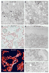
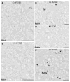
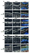
Microvillus inclusion disease (MVID) is a congenital enteropathy characterized by accumulation of vesiculo-tubular endomembranes in the subapical cytoplasm of enterocytes, historically termed "secretory granules." However, neither their identity nor pathophysiological significance is well defined. Using immunoelectron microscopy and tomography, we studied biopsies from MVID patients (3× Myosin 5b mutations and 1× Syntaxin3 mutation) and compared them to controls and genome-edited CaCo2 cell models, harboring relevant mutations. Duodenal biopsies from 2 patients with novel Myosin 5b mutations and typical clinical symptoms showed unusual ultrastructural phenotypes: aberrant subapical vesicles and tubules were prominent in the enterocytes, though other histological hallmarks of MVID were almost absent (ectopic intra-/intercellular microvilli, brush border atrophy). We identified these enigmatic vesiculo-tubular organelles as Rab11-Rab8-positive recycling compartments of altered size, shape and location harboring the apical SNARE Syntaxin3, apical transporters sodium-hydrogen exchanger 3 (NHE3) and cystic fibrosis transmembrane conductance regulator. Our data strongly indicate that in MVID disrupted trafficking between cargo vesicles and the apical plasma membrane is the primary cause of a defect of epithelial polarity and subsequent facultative loss of brush border integrity, leading to malabsorption. Furthermore, they support the notion that mislocalization of transporters, such as NHE3 substantially contributes to the reported sodium loss diarrhea.
Keywords: Myo5b; NHE3; Rab Small GTPases; Rab11a; Rab8a; Stx3; congenital diarrheal disorder; electron tomography; hereditary enteropathy; immunoelectron microscopy.
© 2017 John Wiley & Sons A/S. Published by John Wiley & Sons Ltd.
Figures


Similar articles
-
Loss of MYO5B Leads to Reductions in Na+ Absorption With Maintenance of CFTR-Dependent Cl- Secretion in Enterocytes.Gastroenterology. 2018 Dec;155(6):1883-1897.e10. doi: 10.1053/j.gastro.2018.08.025. Epub 2018 Aug 23. Gastroenterology. 2018. PMID: 30144427 Free PMC article.
-
Identification of intestinal ion transport defects in microvillus inclusion disease.Am J Physiol Gastrointest Liver Physiol. 2016 Jul 1;311(1):G142-55. doi: 10.1152/ajpgi.00041.2016. Epub 2016 May 26. Am J Physiol Gastrointest Liver Physiol. 2016. PMID: 27229121 Free PMC article.
-
Myosin Vb uncoupling from RAB8A and RAB11A elicits microvillus inclusion disease.J Clin Invest. 2014 Jul;124(7):2947-62. doi: 10.1172/JCI71651. Epub 2014 Jun 2. J Clin Invest. 2014. PMID: 24892806 Free PMC article.
-
An overview and online registry of microvillus inclusion disease patients and their MYO5B mutations.Hum Mutat. 2013 Dec;34(12):1597-605. doi: 10.1002/humu.22440. Epub 2013 Oct 16. Hum Mutat. 2013. PMID: 24014347 Review.
-
Trafficking Ion Transporters to the Apical Membrane of Polarized Intestinal Enterocytes.Cold Spring Harb Perspect Biol. 2018 Jan 2;10(1):a027979. doi: 10.1101/cshperspect.a027979. Cold Spring Harb Perspect Biol. 2018. PMID: 28264818 Free PMC article. Review.
Cited by
-
Advanced Microscopy for Liver and Gut Ultrastructural Pathology in Patients with MVID and PFIC Caused by MYO5B Mutations.J Clin Med. 2021 Apr 28;10(9):1901. doi: 10.3390/jcm10091901. J Clin Med. 2021. PMID: 33924896 Free PMC article.
-
Intestinal epithelial cell polarity defects in disease: lessons from microvillus inclusion disease.Dis Model Mech. 2018 Feb 13;11(2):dmm031088. doi: 10.1242/dmm.031088. Dis Model Mech. 2018. PMID: 29590640 Free PMC article. Review.
-
The Endosomal Recycling Pathway-At the Crossroads of the Cell.Int J Mol Sci. 2020 Aug 23;21(17):6074. doi: 10.3390/ijms21176074. Int J Mol Sci. 2020. PMID: 32842549 Free PMC article. Review.
-
Recent advances in understanding and managing malabsorption: focus on microvillus inclusion disease.F1000Res. 2019 Dec 5;8:F1000 Faculty Rev-2061. doi: 10.12688/f1000research.20762.1. eCollection 2019. F1000Res. 2019. PMID: 31824659 Free PMC article. Review.
-
Loss of MYO5B Leads to Reductions in Na+ Absorption With Maintenance of CFTR-Dependent Cl- Secretion in Enterocytes.Gastroenterology. 2018 Dec;155(6):1883-1897.e10. doi: 10.1053/j.gastro.2018.08.025. Epub 2018 Aug 23. Gastroenterology. 2018. PMID: 30144427 Free PMC article.
References
-
- Davidson GP, Cutz E, Hamilton JR, Gall DG. Familial enteropathy: a syndrome of protracted diarrhea from birth, failure to thrive, and hypoplastic villus atrophy. Gastroenterology. 1978;75(5):783–790. - PubMed
-
- Cutz E, Rhoads JM, Drumm B, Sherman PM, Durie PR, Forstner GG. Microvillus inclusion disease: an inherited defect of brush-border assembly and differentiation. N Engl J Med. 1989;320(10):646–651. - PubMed
-
- Phillips AD, Schmitz J. Familial microvillous atrophy: a clinicopathological survey of 23 cases. J Pediatr Gastroenterol Nutr. 1992;14(4):380–396. - PubMed
-
- Muller T, Hess MW, Schiefermeier N, Pfaller K, Ebner HL, Heinz-Erian P, Ponstingl H, Partsch J, Rollinghoff B, Kohler H, Berger T, Lenhartz H, Schlenck B, Houwen RJ, Taylor CJ, et al. MYO5B mutations cause microvillus inclusion disease and disrupt epithelial cell polarity. Nat Genet. 2008;40(10):1163–1165. - PubMed
Publication types
MeSH terms
Substances
Supplementary concepts
Grants and funding
LinkOut - more resources
Full Text Sources
Other Literature Sources
Medical

