. 2017 Apr 12;13(4):e1006273.
doi: 10.1371/journal.ppat.1006273. eCollection 2017 Apr.

A derivative of platelet-derived growth factor receptor alpha binds to the trimer of human cytomegalovirus and inhibits entry into fibroblasts and endothelial cells

Affiliations

A derivative of platelet-derived growth factor receptor alpha binds to the trimer of human cytomegalovirus and inhibits entry into fibroblasts and endothelial cells

Cora Stegmann et al. PLoS Pathog. .

Abstract

Human cytomegalovirus (HCMV) is a widely distributed herpesvirus that causes significant morbidity in immunocompromised hosts. Inhibitors of viral DNA replication are available, but adverse effects limit their use. Alternative antiviral strategies may include inhibition of entry. We show that soluble derivatives of the platelet-derived growth factor receptor alpha (PDGFR-alpha), a putative receptor of HCMV, can inhibit HCMV infection of various cell types. A PDGFR-alpha-Fc fusion protein binds to and neutralizes cell-free virus particles at an EC50 of 10-30 ng/ml. Treatment of particles reduced both attachment to and fusion with cells. In line with the latter, PDGFR-alpha-Fc was also effective when applied postattachment. A peptide scan of the extracellular domain of PDGFR-alpha identified a 40mer peptide that inhibits infection at an EC50 of 1-2 nmol/ml. Both, peptide and fusion protein, were effective against various HCMV strains and are hence promising candidates for the development of novel anti-HCMV therapies.


Conflict of interest statement

The authors have declared that no competing interests exist.

Figures
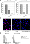
Fig 1
Fig 1. Effect of siRNA-mediated depletion of PDGFR-alpha on infection efficiency in fibroblasts and endothelial cells.
Fibroblasts (A) and endothelial cells (B) were transfected with siRNAs targeting PDGFR-alpha. Non-targeting (NT) siRNAs and siRNAs against the viral immediate early (IE) proteins were included as controls. Two days after transfection, cells were infected with HCMV strain TB40/E, and the next day viral IE antigens were detected by indirect immunofluorescence for visualization of infected cells. The number of IE antigen-positive cells was counted and compared to the NT control. Error bars in (A) and (B) represent the standard error of the mean (SEM). Significant differences as compared to NT control are indicated by asterisks. C: Examples of HCMV IE antigen expression (red fluorescence) in HFFs after treatment with the respective siRNAs. Nuclei of non-infected cells appear blue due to counterstaining with DAPI. D: Fluorescence-activated cell sorting (FACS) for detection of PDGFR-alpha on the surface of fibroblasts and endothelial cells 2 d after treatment with PDGFR-alpha siRNA or non-targeting (NT) siRNA. NT siRNA represents PDGFR-alpha levels without specific knockdown. Isotype control antibody was included as a negative control for staining.
Fig 2
Fig 2. Inhibitory effect of soluble PDGFR-alpha-Fc chimeras on HCMV infection.
Virus preparations of strain TB40/E were pretreated for two hours with PDGFR-alpha-Fc at indicated concentrations and then added to fibroblasts (HFFs) and endothelial cells (HECs). One day after infection, cells were fixed and stained for viral IE antigens by indirect immunofluorescence. The ratio of IE antigen-positive cells per total cell number was calculated to represent the degree of infection. The graphs show the inhibition of infection in treated cultures as compared to untreated controls. Error bars represent the standard error of the mean (SEM).
Fig 3
Fig 3. The inhibitory effect of soluble PDGFR-alpha is specific and affects various HCMV strains.
(A) PDGFR-alpha-Fc, PDGFR-beta-Fc and EGFR-Fc were compared regarding their inhibitory potential on infection of fibroblasts (HFFs) and endothelial cells (HECs) by HCMV strain TB40/E. Virus preparations were pretreated for 2 h with the respective growth factor receptor at indicated concentrations and then added to cell cultures for 2 h, followed by a medium exchange and incubation overnight. Cells were fixed and stained for viral IE antigens. The percentage of infection was calculated as the ratio of IE antigen-positive cells / total cell number. (B) The potential of PDGFR-alpha-Fc to inhibit fibroblast infection with various HCMV strains was tested using a collection of strains representing the known glycoprotein variants. The virus preparations were either preincubated with medium (no drug) or medium containing 250 ng/ml PDGFR-alpha-Fc. Error bars in (A) and (B) represent the standard error of the mean (SEM). The significant difference of VR1814 as compared to the other strains is indicated by an asterisk.
Fig 4
Fig 4. Binding of soluble growth factor receptor-Fc chimeras to HCMV particles.
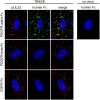
Virus preparations of strain TB40/E were pretreated for two hours with 500 ng/ml PDGFR-alpha-Fc, PDGFR-beta-Fc or EGFR-Fc and then incubated with fibroblasts for 90 min on ice. Cells were fixed and stained for the viral structural protein pUL32 (red) and for the Fc-fusion part (green). Nuclei were counterstained with DAPI.
Fig 5
Fig 5. Quantification of PDGFR-alpha-Fc binding to HCMV particles.
Virus preparations of strain TB40/E were preincubated with serial dilutions of PDGFR-alpha-Fc. Viruses were attached to fibroblasts by incubation on ice for 90 min followed by acetone fixation. Binding of PDGFR-alpha-Fc was assessed by direct immunofluorescence of the Fc-fusion part and quantification of signal intensities (maximum grey values per particle). In parallel, fibroblasts were incubated with the same mixtures at 37°C and stained for viral immediate-early antigens at one day postinfection to determine the fraction of infected cells. The intensity of the staining with PDGFR-alpha-Fc was measured for 100 particles in each condition. The same data set is provided with a linear scale and a logarithmic scale. Error bars indicate the error of the median.
Fig 6
Fig 6. Effect of soluble PDGFR-alpha on adsorption and penetration of HCMV.
Virus preparations of the dual fluorescent strain TB40-BACKL7-UL32EGFP-UL100mCherry were preincubated with 100 ng/ml soluble Fc-chimeras for two hours and subsequently used to infect fibroblasts (HFFs) and endothelial cells (HECs) at 37°C for two hours. (A) Adsorption was assessed by counting the total number of bound virus particles per cell (pUL32-EGFP signals regardless of the pUL100-Cherry signals). Each dot represents one cell, mean values are indicated by a horizontal line, and error bars represent the standard error of the mean (SEM). Significant differences as compared to the untreated controls are indicated by asterisks. (B) The fraction of penetrated particles was determined by counting the percentage of particles lacking the envelope (particles without the pUL100-mCherry signal). Bars indicate the mean values of 15 cells per condition and error bars represent the standard error of the mean (SEM). One representative experiment out of three is shown. The significant difference between PDGFR-alpha-Fc-treated HFFs and the untreated control is indicated by an asterisk. C: Examples of microscopic images taken in HFFs. Enveloped virus particles appear yellow due to an overlap of pUL32-EGFP signals and pUL100-Cherry signals. Penetrated non-enveloped particles are only EGFP-positive. Nuclei were stained with DAPI.
Fig 7
Fig 7. Postadsorption-inhibitory effect of soluble PDGFR-alpha-Fc.
Virus preparations were adsorbed to fibroblasts on ice for 60 min and subsequently incubated with 200 ng/ml PDGFR-alpha-Fc for two hours on ice. Cells were then either directly shifted to 37°C or first treated with the chemical fusogen PEG. After an overnight incubation, cells were fixed and stained for viral IE antigens. (A) For each condition, the fraction of infected cells (immediate-early antigen positive cells/total cells) was compared to the untreated control. Mean values of 3 independent experiments are shown. Error bars indicate the standard error of the mean (SEM). The significant difference between PDGFR-alpha-Fc-treated cells and the untreated control is indicated by an asterisk. (B) Examples of HCMV IE antigen expression (red fluorescence) after treatment with PDGFR-alpha-Fc alone or PDGFR-alpha-Fc and PEG. Nuclei were counterstained with DAPI.
Fig 8
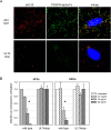
Fig 8. PDGFR-alpha-Fc binds HCMV particles only in the presence of the gH/gL/pUL74 complex.
(A) Virus preparations of strain TB40-BAC4 (wild type) or TB40-BAC4-UL74stop were pretreated with 500 ng/ml PDGFR-alpha-Fc for two hours at 37°C. Fibroblasts (HFFs) were then incubated with pretreated virus on ice followed by immunofluorescence staining for the viral structural protein pUL32 (red) and the Fc-fusion part of PDGFR-alpha-Fc (green). (B) Virus preparations were preincubated with variable concentrations of PDGFR-alpha-Fc for 2h before infection of HFFs and endothelial cells (HECs). One day postinfection, cells were fixed and stained by immunofluorescence for viral immediate early antigens. For each condition, the fraction of infected cells (immediate-early antigen positive cells/total cells) was compared to the untreated control. Mean values of 3 independent experiments are shown. Error bars indicate the standard error of the mean (SEM). Significant differences as compared to the untreated controls are indicated by asterisks.
Fig 9
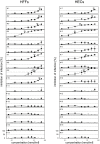
Fig 9. Inhibitory effect of PDGFR-alpha-derived peptides.
The luciferase reporter virus TB40-BAC4-IE-Gluc was preincubated with 40mer peptides derived from the extracellular domain of PDGFR-alpha at concentrations from 0.05–50 nmol/ml for 2 h before infection of fibroblasts (HFFs) and endothelial cells (HECs). Untreated virus was used as a control for uninhibited infection. One day postinfection, supernatants were analyzed for luciferase activity indicating the degree of infection. The extent of inhibition was calculated as 1 - (luminescence with peptide / luminescence without peptide) and is given as percentage. Error bars indicate the standard error of the mean (SEM) of four data sets per peptide. The number of the peptide (#1-#17) indicates its position in the protein, starting from the N-terminus. Arrowheads indicate the concentration at which 50% of infection are inhibited.

Similar articles

Cited by

References

    1. Ariza-Heredia EJ, Nesher L, Chemaly RF. Cytomegalovirus diseases after hematopoietic stem cell transplantation: a mini-review. Cancer Lett. 2014;342(1):1–8. Epub 2013/09/18. 10.1016/j.canlet.2013.09.004 - DOI - PubMed
    1. Gwee A, Curtis N, Connell TG, Garland S, Daley AJ. Ganciclovir for the treatment of congenital cytomegalovirus: what are the side effects? Pediatr Infect Dis J. 2014;33(1):115. Epub 2013/12/19. - PubMed
    1. Rasmussen LE, Nelson RM, Kelsall DC, Merigan TC. Murine monoclonal antibody to a single protein neutralizes the infectivity of human cytomegalovirus. Proc Natl Acad Sci USA. 1984;81(3):876–80. - PMC - PubMed
    1. Kropff B, Landini MP, Mach M. An ELISA using recombinant proteins for the detection of neutralizing antibodies against human cytomegalovirus. J Med Virol. 1993;39(3):187–95. Epub 1993/03/01. - PubMed
    1. Gerna G, Sarasini A, Patrone M, Percivalle E, Fiorina L, Campanini G, et al. Human cytomegalovirus serum neutralizing antibodies block virus infection of endothelial/epithelial cells, but not fibroblasts, early during primary infection. Journal of General Virology. 2008;89(4):853–65. - PubMed

MeSH terms

Substances

Grants and funding

This work was supported by grants from the Wilhelm-Sander-Foundation (Project 2013.002.1) to CSi and from the Medical Faculty of Ulm University (Baustein L.SBN.0094) to DL. The funders had no role in study design, data collection and analysis, decision to publish, or preparation of the manuscript.