. 2017 Apr 12;13(4):e1006281.
doi: 10.1371/journal.ppat.1006281. eCollection 2017 Apr.

Human cytomegalovirus glycoprotein complex gH/gL/gO uses PDGFR-α as a key for entry

Affiliations

Human cytomegalovirus glycoprotein complex gH/gL/gO uses PDGFR-α as a key for entry

Yiquan Wu et al. PLoS Pathog. .

Abstract

Herpesvirus gH/gL envelope glycoprotein complexes are key players in virus entry as ligands for host cell receptors and by promoting fusion of viral envelopes with cellular membranes. Human cytomegalovirus (HCMV) has two alternative gH/gL complexes, gH/gL/gO and gH/gL/UL128,130,131A which both shape the HCMV tropism. By studying binding of HCMV particles to fibroblasts, we could for the first time show that virion gH/gL/gO binds to platelet-derived growth factor-α (PDGFR-α) on the surface of fibroblasts and that gH/gL/gO either directly or indirectly recruits gB to this complex. PDGFR-α functions as an entry receptor for HCMV expressing gH/gL/gO, but not for HCMV mutants lacking the gH/gL/gO complex. PDGFR-α-dependent entry is not dependent on activation of PDGFR-α. We could also show that the gH/gL/gO-PDGFR-α interaction starts the predominant entry pathway for infection of fibroblasts with free virus. Cell-associated virus spread is either driven by gH/gL/gO interacting with PDGFR-α or by the gH/gL/UL128,130,131A complex. PDGFR-α-positive cells may thus be preferred first target cells for infections with free virus which might have implications for the design of future HCMV vaccines or anti-HCMV drugs.


Conflict of interest statement

The authors have declared that no competing interests exist.

Figures

Fig 1
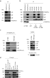
Fig 1. Anti-gH antibodies co-precipitate PDGFR-α, gO and gB from lysates of HFF co-incubated with TB40-UL131Astop virions.
HFF surface proteins were biotinylated and lysates of HFF (1), lysates of HFF mixed with lysates of TB40-UL131Astop virions (2) and lysates of HFF co-incubated with TB40-UL131stop virions (3) were subjected to anti-gH immunoprecipitation. The precipitates were analyzed by Western blot using antibodies directed against PDGFR-α, gO, gH and gB.
Fig 2
Fig 2. Co-precipitation of PDGFR-α and gH is dependent on gO.
(a) Lysates of TB40-UL131Astop virions were co-incubated with PDGFR-Fc fusion proteins and then the Fc fusion proteins precipitated with protein A sepharose. (b) Lysates of wt TB40 virions were treated as described under (a). Additionally, lysates of HFF (1), lysates of HFF mixed with lysates of wt TB40 virions (2) and lysates of HFF co-incubated with wt TB40 virions (3) were subjected to anti-gH and anti-gB immunoprecipitation. Negatively stained gB bands in gB precipitations are due to abundant protein. (c) Left panel: Lysates of wt TB40 and TB40-ΔgO virions were co-incubated with PDGFR-α-Fc fusion protein and the Fc fusion proteins precipitated with protein A sepharose. Right panel: Lysates of wt TB40 and TB40-ΔgO virions adapted for equal amounts of MCP protein. UL128 was additionally precipitated from the lysates with a rabbit antiserum specific for HCMV UL131A. (d) Lysates of HFF mixed with lysates of virions, lysates of virions, or an HFF control lysate were subjected to anti-gH immunoprecipitation. The immunoprecipitates were analyzed by Western blot using antibodies directed against PDGFR-α, gO, gH and gB and the lysates under (c) additionally with antibodies directed against MCP and UL128.
Fig 3
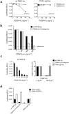
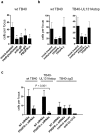
Fig 3. PDGFR-α-dependent and gO-dependent entry are congruent.
HFF (a) to (c) or 293 cells (d) were infected on 96 well plates. (a) Before infection, wt TB40-luc or TB40-ΔgO-luc virus were pre-incubated with the indicated amounts of PDGFR-α-Fc or as a control PDGFR-β-Fc for one hour at 4°C. Cells were infected with the virion-PDGFR-Fc mixtures using centrifugal enhancement, washed three times with PBS and then incubated for 24 hours before infection was determined by a luciferase assay. (b) The experiment was performed as described under (a) with the difference that infections with wt TB40-luc and TB40-UL131stop-luc virus were compared and that cells were infected with a 50fold higher m.o.i. (c) Before infection, cells were pre-incubated with the indicated amounts of PDGF-AA for one hour at 4°C. Cells were then infected in the presence of PDGF-AA. After infection, free virus and cell surface-bound virus were inactivated by washing the cells three times with PBS, pH 3.0. 24 hours after infection cells were analyzed by a luciferase assay. The left panel shows inhibition of wt TB40-luc infection in the presence of increasing concentrations of PDGF-AA and the right panel shows inhibition of wt TB40-luc, TB40-UL131stop-luc, and TB40-ΔgO-luc viruses after infection of HFF pre-incubated with 0 ng ml-1 PDGF-AA (set to 100%) and with 100 ng ml-1 PDGF-AA. (d) 293 cells were transfected with pCMV-PDGFR-α or a control vector and 2 days later infected with wt TB40-luc, TB40-UL131Astop-luc or TB40-ΔgO-luc viruses. 24 hours after infection, cells were analyzed by a luciferase assay. (a), (c), and (d) show means +/- SD of three independent experiments done in triplicates. (b) and the left panel of (c) show one representative experiment done in triplicates.
Fig 4
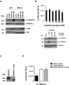
Fig 4. HCMV infection of fibroblasts is not dependent on phosphorylation of PDGFR-α.
(a) Serum-starved HFF or MRC-5 were co-incubated with wt TB40 (m.o.i of 10 in 0.05% DMEM), PDGF-AA (100 ng ml-1), PDGF-BB (100 ng ml-1) or 0.05% DMEM (mock) for 1 hour at 4°C and then shifted to 37°C for 20 min. p-PDGFR-α or PDGFR-α and p-Akt or Akt were detected by Western blot analysis of total cell lysates. (b) HFF were pre-incubated with different concentrations of imatinib mesylate for 1 hour at 37°C and then infected with wt TB40-luc (m.o.i. of 1) for 90 min. Free virus and cell surface-bound virus were inactivated by washing the cells three times with PBS, pH 3.0. After 24 hours in medium containing the respective amounts of imatinib mesylate, a luciferase assay was performed to determine infection of cells. One representative experiment done in triplicates is shown. To control for the activity of imatinib mesylate, HFF were in parallel pre-treated with the indicated concentrations of imatinib mesylate for 1 hour at 37°C and then stimulated with PDGF-BB (100 ng ml-1) or mock-treated for 20 min. p742- and p762-PDGFR-α or PDGFR-α were detected by Western blot analysis of total cell lysates. (c,d) 293 cells were transfected with pCMV-PDGFR-α, pCMV-PDGFR-α(1–558), or a control vector. (c) Total cell extracts of pCMV-PDGFR-α- or pCMV-PDGFR-α(1–558)-transfected cells were analyzed for PDGFR-α expression by Western blot 24 hours after transfection using an antibody recognizing the N-terminus of human PDGFR-α (2D2-1A11). (d) 48 hours after transfection 293 cells were infected with wt TB40-luc. 24 hours after infection, cells were analyzed by a luciferase assay. Shown are means +/- SD of three independent experiments done in triplicates.
Fig 5
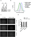
Fig 5. Silencing of PDGFR-α reduces infection of fibroblasts with gH/gL/gO-positive HCMV.
HFF were transfected with PDGFR-α siRNAs, non-targeting (NT) siRNAs or mock-transfected. (a) 72 and 108 hours after transfection, total cell lysates were analyzed by Western blot for the expression of PDGFR-α or GAPDH. (b) 72 hours after transfection, cell surfaces of siRNA-transfected cells were stained with an anti-PDGFR-α antibody (35248) and a secondary Fluor 488-labelled anti-mouse antibody and analyzed by FACS. (c) 48 hours after transfection, cells were infected with wt TB40, TB40-UL131Astop and TB40-ΔgO viruses and 24 hours post infection stained for HCMV IE1 by indirect immunofluorescence. (d) HFF were infected as described under (c) and the percentage of IE1-positive nuclei was determined. Infection of cells transfected with NT siRNAs (set to 100%) and infection of PDGFR-α-silenced HFF is shown. Shown are means +/- SD from two independent experiments done in triplicates.
Fig 6
Fig 6. Silencing of PDGFR-α reduces cell-associated spread of gH/gL/gO-positive HCMV.
(a) Confluent monolayers of HFF were infected with wt TB40 virus at a very low m.o.i. After infection, cells were either overlaid with methylcellulose or medium containing anti-gB antibodies (SM5-1, 2μg ml-1), anti-gH antibodies (14-4B), PDGFR-α-Fc (2 μg ml-1), or no inhibitor (mock-treated). (b) HFF infected with wt TB40 or TB40-UL131Astop virus were mixed with uninfected cells. After adherence, cells were either overlaid with methylcellulose or fresh medium was added containing 2 μg ml-1 PDGFR-α-Fc, 2 μg ml-1 PDGFR-β-Fc, or no inhibitor (mock-treated). (a,b) 5 days later, cells were stained for HCMV IE1 by indirect immunofluorescence and cells per focus counted. For each treatment, at least 10 (a) or 20 (b) foci were counted and depicted as means +/- SD. Shown are representative experiments. Asterisks under (a) represent P<0.001 values determined by comparing foci in mock-treated monolayers with foci in monolayers overlaid with methylcellulose, co-incubated with antibodies, or co-incubated with PDGFR-α-Fc (Mann-Whitney Rank Sum test). (c) NT siRNA or PDGFR-α siRNA-transfected HFF 48 hours after transfection were mixed with HFF infected with wt TB40, TB40-UL131Astop, or TB40-ΔgO virus. After adherence, cells were overlaid with methylcellulose or methylcellulose containing anti-UL131A rabbit antiserum (1:40) or a control rabbit antiserum (1:40). 5 days later, cells were analyzed as described under (a). The P value shown (Mann-Whitney Rank Sum test) was determined by comparing cell spread of wt TB40 virus with spread of TB40-UL131Astop virus in PDGFR-α-silenced HFF.

Similar articles

Cited by

References

    1. Boppana SB, Britt WJ. Synopsis of clinical aspects of human cytomegalovirus disease In: Reddehase MJ, editors. Cytomegaloviruses: From Molecular Pathogenesis to Intervention. Norfolk, UK: Caister Academic Press; 2013; pp 1–25.
    1. Plachter B, Sinzger C, Jahn G. Cell types involved in replication and distribution of human cytomegalovirus. Adv Virus Res. 1996; 46:195–261. - PubMed
    1. Compton T, Nowlin DM, Cooper NR. Initiation of human cytomegalovirus infection requires initial interaction with cell surface heparan sulfate. Virology. 1993; 193:834–841. 10.1006/viro.1993.1192 - DOI - PubMed
    1. Boyle KA, Compton T. Receptor-binding properties of a soluble form of human cytomegalovirus glycoprotein B. J Virol. 1998; 72:1826–1833. - PMC - PubMed
    1. Kari B, Gehrz R. A human cytomegalovirus glycoprotein complex designated gC-II is a major heparin-binding component of the envelope. J Virol. 1992; 66:1761–1764. - PMC - PubMed

MeSH terms

Substances

Grants and funding

This work was supported by grants from the Deutsche Forschungsgemeinschaft given to BA (AD 131/3-2 and AD 131/4-1). YW was funded by a PhD grant from the China Scholarship Council. The funders had no role in study design, data collection and analysis, decision to publish, or preparation of the manuscript.