. 2018 Jan 1;28(1):62-77.
doi: 10.1089/ars.2016.6871. Epub 2017 May 1.

A Peroxidase Peroxiredoxin 1-Specific Redox Regulation of the Novel FOXO3 microRNA Target let-7

Affiliations

A Peroxidase Peroxiredoxin 1-Specific Redox Regulation of the Novel FOXO3 microRNA Target let-7

Barbara L Hopkins et al. Antioxid Redox Signal. .

Abstract

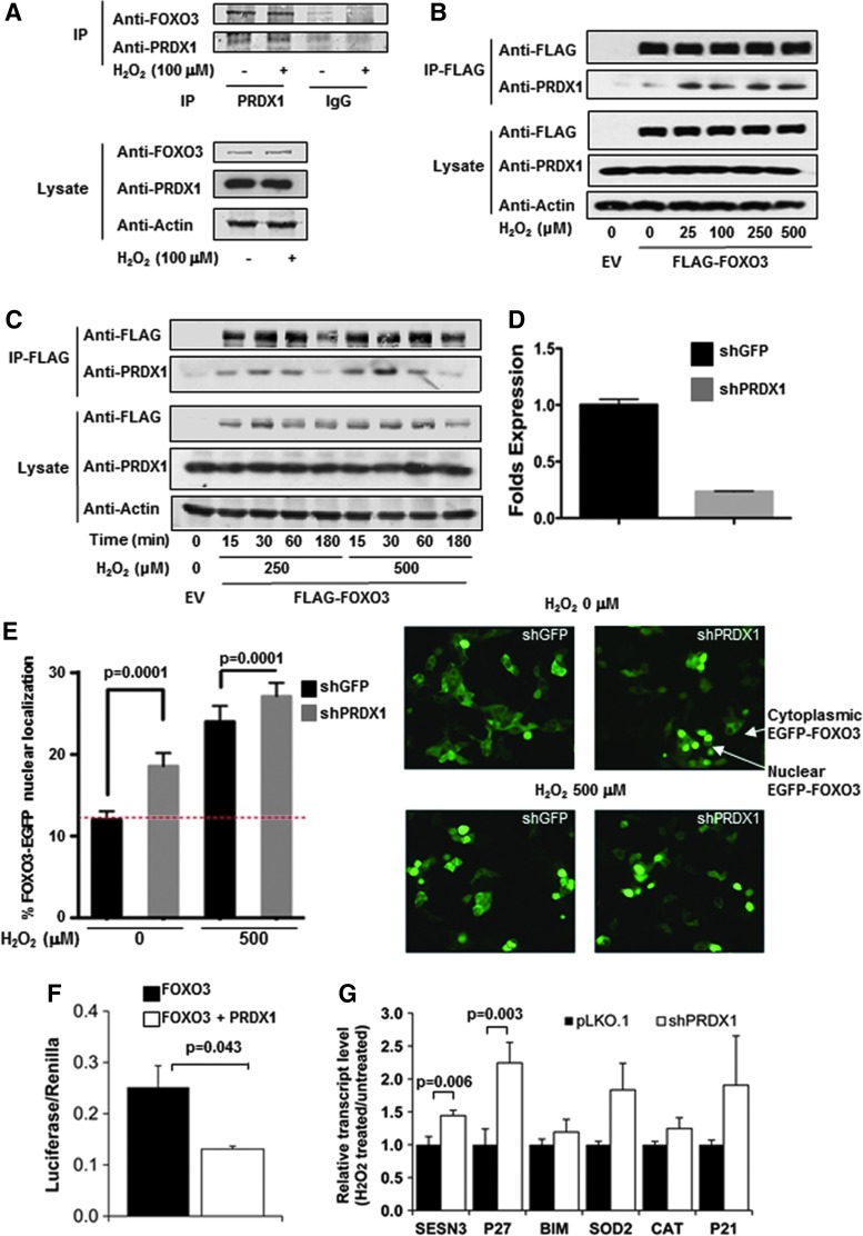
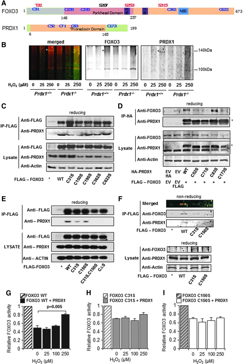
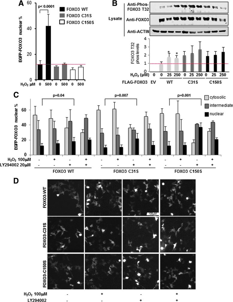
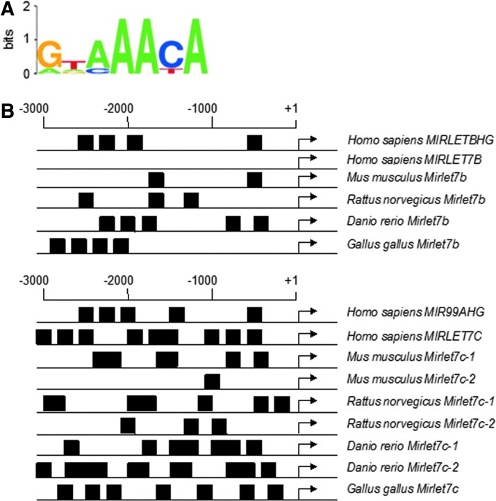
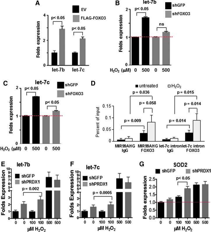
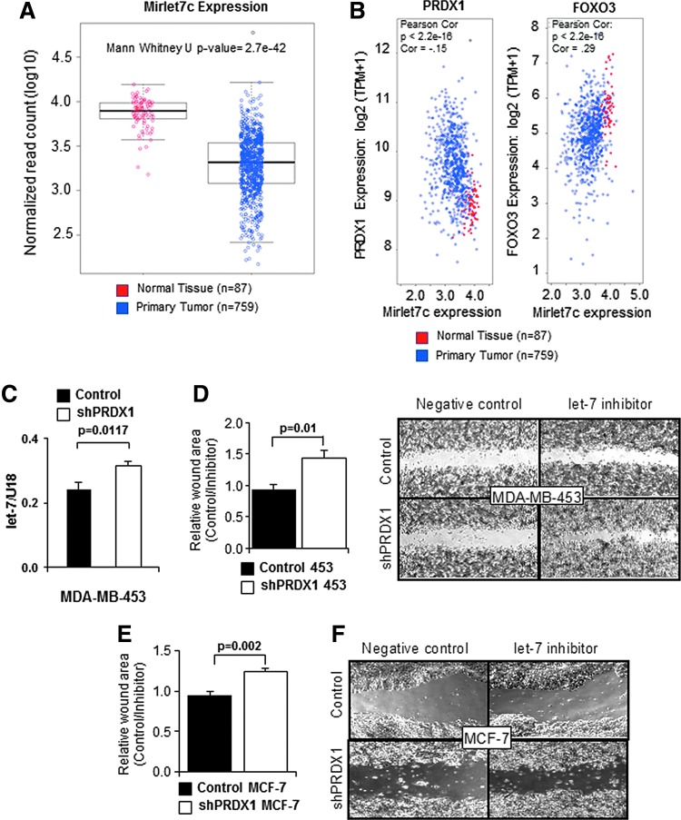
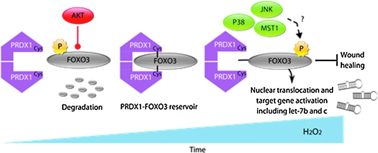
Precision in redox signaling is attained through posttranslational protein modifications such as oxidation of protein thiols. The peroxidase peroxiredoxin 1 (PRDX1) regulates signal transduction through changes in thiol oxidation of its cysteines. We demonstrate here that PRDX1 is a binding partner for the tumor suppressive transcription factor FOXO3 that directly regulates the FOXO3 stress response. Heightened oxidative stress evokes formation of disulfide-bound heterotrimers linking dimeric PRDX1 to monomeric FOXO3. Absence of PRDX1 enhances FOXO3 nuclear localization and transcription that are dependent on the presence of Cys31 or Cys150 within FOXO3. Notably, FOXO3-T32 phosphorylation is constitutively enhanced in these mutants, but nuclear translocation of mutant FOXO3 is restored with PI3K inhibition. Here we show that on H2O2 exposure, transcription of tumor suppressive miRNAs let-7b and let-7c is regulated by FOXO3 or PRDX1 expression levels and that let-7c is a novel target for FOXO3. Conjointly, inhibition of let-7 microRNAs increases let-7-phenotypes in PRDX1-deficient breast cancer cells. Altogether, these data ascertain the existence of an H2O2-sensitive PRDX1-FOXO3 signaling axis that fine tunes FOXO3 activity toward the transcription of gene targets in response to oxidative stress. Antioxid. Redox Signal. 28, 62-77.

Keywords: FOXO3; PRDX1; breast cancer; let-7; oxidative stress; tumor suppressor.


Conflict of interest statement

No competing financial interests exist.

Figures

<b>FIG. 1.</b>
FIG. 1.
PRDX1 binds to FOXO3 in an H2O2 dose-dependent manner and regulates FOXO3 nuclear localization. (A) Immunoprecipitation of precleared lysate with PRDX1 or IgG antibodies found PRDX1 bound FOXO3. 293T cells underwent serum starvation for 30 min and were then treated with the 0 or 100 μM H2O2 for an additional 30 min. (B and C) 293T cells were transfected with pcDNA3-FLAG-FOXO3A or EV and treated with increasing concentrations of H2O2 for the indicated times. Before lysis, cells were washed with 20 mM NEM in phosphate-buffered saline to block lysis-induced disulfide bond formation. FLAG-labeled proteins were immunoprecipitated and detected by immunoblot with FLAG and PRDX1 antibodies. (D and E) The percentage of 293T cells displaying nuclear FOXO3-EGFP localization was enhanced with reduction of PRDX1; 150 or more cells analyzed per sample, p < 0.0001 (t-test). Cells infected with shPRDX1A or control lentivirus for 48 h, followed by transfection with FOXO3-EGFP for 24 h. H2O2 was added during the last 30 min. (F) HA-PRDX reduced FLAG-FOXO3 activity when transiently cotransfected into MEF and analyzed utilizing a dual-luciferase reporter assay. Values (mean + SE) were normalized to vehicle treatment. *p < 0.05, t-test (N = 3). (G) qPCR gene transcription of SESN3 and P27 was increased in PRDX1-deficient 293T cells (white bars) treated with 250 μM H2O2 compared to pLKO.1 control cells (black bars) after 16 h. Values (mean + SE) were normalized to vehicle treatment. *p < 0.05, t-test (N = 3). MEF, murine embryonic fibroblast; NEM, N-ethylmaleimide; SE, standard error.
<b>FIG. 2.</b>
FIG. 2.
PRDX1 and FOXO3 form disulfide bonds. (A) Domain structures of FOXO3 and PRDX1 proteins. (B) A PRDX1-FOXO3 complex was detected under nonreducing conditions in Prdx1+/+, but not Prdx1−/− MEFs, when treated with the indicated concentrations of H2O2 for 30 min by immunoblot with two-color IR antibody detection. (C) Anti-FLAG IP of 293T cells transfected with EV or FLAG-FOXO3 (WT or single C-to-S mutants) displayed reduced PRDX1 binding to FOXO3 C31S or C150S when treated with H2O2 for 30 min by immunoblot. (D) Anti-HA IP of 293T cells transfected with EV or HA-PRDX1 (WT or C-to-S mutants) showed reduced FOXO3 binding with PRDX1 C52S, C71S, or C173S mutants when treated with H2O2 for 30 min (*HA-PRDX1). (E) Mutation of FOXO3 cysteines reduced PRDX1 binding when treated with 25 μM H2O2 for 30 min. Anti-FLAG IP of 293T cells transfected with EV or FLAG-FOXO3 (WT, C31S, C150S, C31, 150S double mutant, or ΔCys mutants) was detected by immunoblot. (F) A PRDX1-FOXO3 complex was detected in FLAG-FOXO3 WT, but not C31S or C150S mutant Anti-FLAG samples under nonreducing conditions in 293T cells cotransfected with FLAG-FOXO3 constructs and HA-PRDX1 treated with 25 μM H2O2 for 30 min. (G–I) The ability of PRDX1 to reduce FOXO3 activity was inhibited with H2O2 treatment in 293T cells in FOXO3 WT, but not C31S or C150S mutants. Cells were transiently transfected with FOXO3 and PRDX1 constructs in a dual-luciferase assay treated with 0–250 μM H2O2. Values (mean + SE) were normalized to FOXO3 vehicle treatment. *p < 0.05, t-test (N = 3). NES, nuclear export sequence; NLS, nuclear localization signal.
<b>FIG. 3.</b>
FIG. 3.
Interdisulfides between PRDX1 and FOXO3 modulate FOXO3 subcellular localization and FOXO3 phosphorylation. (A) Thirty minutes of H2O2 treatment enhanced nuclear EGFP-FOXO3 content in WT, but not C31 or C150 mutant constructs, 48 h following transfection in 293T cells. Percentage of cells displaying nuclear FOXO3-EGFP localization is indicated. (B) T32 phosphorylation of FOXO3 C31S or C150S was heightened under basal conditions compared to FOXO3 WT. 293T cells transfected with FLAG-FOXO3 (WT, C31, or C150 mutants) were treated with H2O2 and detected by immunoblot for FOXO3 Phospho-T32, FOXO3, and Actin. Phospho-T32 signals were normalized to total FOXO3. Values represent mean + SD normalized to WT 0 μM H2O2. *p < 0.05 (N = 3). (C) PI3K inhibition with 20 μM LY294002 enhanced nuclear FOXO3-EGFP WT, C1S, or C2S mutants in transiently transfected 293T cells after 24 h. Values represent mean + SE (N = 3) with 150–400 cells counted per sample by fluorescence microscopy. Experiment was repeated twice. (D) Representative pictures of (C).
<b>FIG. 4.</b>
FIG. 4.
FOXO3 consensus binding sequence found in let-7b and let-7c promoter region. (A) Putative FOXO3 binding sequence (10). (B) Putative FOXO binding motifs in forward and reverse are located in the 5′ UTR of let-7b and -7c in several species. The consensus FOXO binding sequences in the WebLogo (black boxes) are indicated. Numbering is relative to the transcription start site obtained through the University of California Santa Cruz Genome Browser. UTR, untranslated region.
<b>FIG. 5.</b>
FIG. 5.
FOXO3 regulates expression of let-7 miRNAs under H2O2. (A) Overexpression of FLAG-FOXO3 enhanced let-7b and c transcription in Hela cells 48 h after transfection. Cells were harvested, lysed, and analyzed for miRNA expression by TaqMan assays, using the delta CT method with mir-30c as the internal standard. (B, C) FOXO3-deficient HeLa cells were nonresponsive to H2O2 treatment. Thirty hours following transfection of shGFP or shFOXO3, cells were treated with or without H2O2 for 18 h. Expression profiles of let-7b and let-7c were assessed by individual TaqMan assays with U18 as the internal standard (N = 3). (D) ChIP assays indicate transfected FLAG-FOXO3 binds to the MIR99AHG and intronic let-7c promoter regions and is enhanced following 100 μM H2O2 treatment for 30 min in 293T cells. Quantification of immunoprecipitated DNA was performed in triplicate by quantitative PCR and evaluated by the delta CT method. Values of each immunoprecipitated sample are expressed as a percentage relative to their respective input (no antibody). (E–G) let-7b and c transcription was increased in PRDX1-deficient cells. HeLa cells were transfected with either a shGFP or shPRDX1 construct for 56 h, followed by treatment with or without H2O2 for 18 h. HeLa cells were analyzed for gene expression by TaqMan expression assays using the delta CT method with U18 as the internal standard (F, G) or β-actin as the internal standard (H).
<b>FIG. 6.</b>
FIG. 6.
Lack of PRDX1 enhances levels and function of let-7 miRNAs under H2O2. (A) Mirlet7 expression in normal and breast cancer tissue from TCGA. (B) Expression of FOXO3 and PRDX1 from TCGA breast cancer cases and normal tissue was compared to Mirlet7c expression. (C) EV or shPRDX1 MDA-MB-453 cells were harvested, lysed, and analyzed for miRNA expression by TaqMan assays using the delta CT method with U18 as the internal standard (N = 3). (D–E) Wound healing was increased following let-7 inhibition in PRDX1-deficient breast cancer cells. 1 × 106 shPRDX1 MDA-MB-453 or MCF-7 (white bar) cells or control (black bar) cells were transfected with a let-7 miRNA inhibitor in a 12-well plate and wound healing was assessed after 48 h (MDA-MB-453) or 24 h (MCF-7) in the presence of mitomycin C (0.5 μg/ml). Wound area of let-7 inhibitor-treated cells was normalized to control miRNA inhibitor-treated cells (mean + SE) N = 4. Right side, representative photographs of wound healing assays in MDA-MB-453 or MCF-7 cells. TCGA, The Cancer Genome Atlas.
<b>FIG. 7.</b>
FIG. 7.
Model: stepwise oxidation of PRDX1 regulates FOXO3 under oxidative stress. PRDX1 and FOXO3 interact in an oxidative stress-dependent way. This involves the catalytic/peroxidatic cysteine and C71 of PRDX1 and C31 and C150 of FOXO3. Our data strongly suggest disulfide bonding between PRDX1 and FOXO3 involving PRDX1 C52, C71, and C173 and FOXO3 C31 and C150 regulating the AKT-induced phosphorylation of T32 on FOXO3 and 14-3-3 binding and dissociation. Nuclear localization and 14-3-3 dissociation of FOXO3 may be promoted by monoubiquitination or phosphorylation by over-oxidized PRDX1 activating MST1 or oxidative stress activation of JNK or p38. This results in expression in let-7b and let-7c and inhibition of migration.

Similar articles

Cited by

References

    1. Bailey CK, Mittal MK, Misra S, and Chaudhuri G. High motility of triple-negative breast cancer cells is due to repression of plakoglobin gene by metastasis modulator protein SLUG. J Biol Chem 287: 19472–19486, 2012 - PMC - PubMed
    1. Belguise K, Guo S, and Sonenshein GE. Activation of FOXO3a by the green tea polyphenol epigallocatechin-3-gallate induces estrogen receptor alpha expression reversing invasive phenotype of breast cancer cells. Cancer Res 67: 5763–5770, 2007 - PubMed
    1. Blake DC, Jr, Mikse OR, Freeman WM, and Herzog CR. FOXO3a elicits a pro-apoptotic transcription program and cellular response to human lung carcinogen nicotine-derived nitrosaminoketone (NNK). Lung Cancer 67: 37–47 - PubMed
    1. Borniquel S, Garcia-Quintans N, Valle I, Olmos Y, Wild B, Martinez-Granero F, Soria E, Lamas S, and Monsalve M. Inactivation of Foxo3a and subsequent downregulation of PGC-1 alpha mediate nitric oxide-induced endothelial cell migration. Mol Cell Biol 30: 4035–4044, 2010 - PMC - PubMed
    1. Boyerinas B, Park SM, Hau A, Murmann AE, and Peter ME. The role of let-7 in cell differentiation and cancer. Endocr Relat Cancer 17: F19–F36, 2010 - PubMed

LinkOut - more resources