

β Cell Aging Markers Have Heterogeneous Distribution and Are Induced by Insulin Resistance
- PMID: 28380379
- PMCID: PMC5471618
- DOI: 10.1016/j.cmet.2017.03.015
β Cell Aging Markers Have Heterogeneous Distribution and Are Induced by Insulin Resistance
Abstract
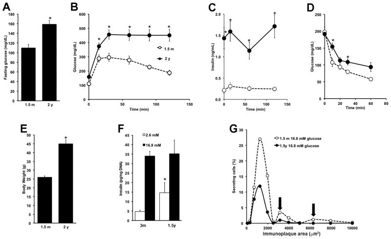
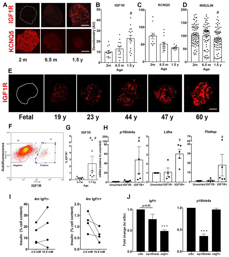
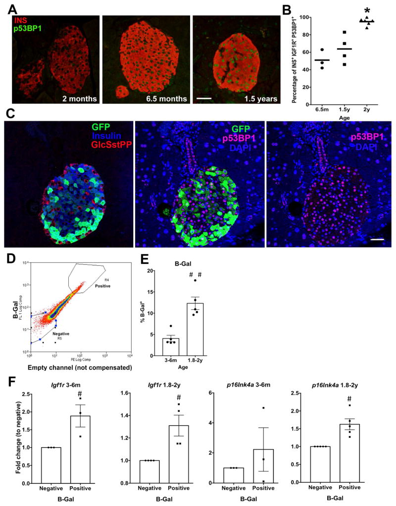
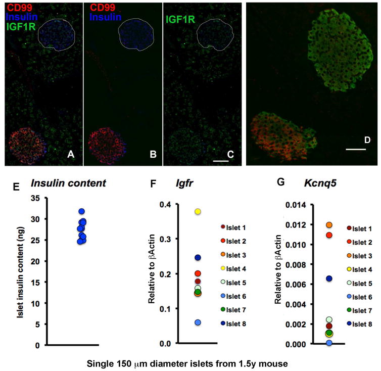
We hypothesized that the known heterogeneity of pancreatic β cells was due to subpopulations of β cells at different stages of their life cycle with different functional capacities and that further changes occur with metabolic stress and aging. We identified new markers of aging in β cells, including IGF1R. In β cells IGF1R expression correlated with age, dysfunction, and expression of known age markers p16ink4a, p53BP1, and senescence-associated β-galactosidase. The new markers showed striking heterogeneity both within and between islets in both mouse and human pancreas. Acute induction of insulin resistance with an insulin receptor antagonist or chronic ER stress resulted in increased expression of aging markers, providing insight into how metabolic stress might accelerate dysfunction and decline of β cells. These novel findings about β cell and islet heterogeneity, and how they change with age, open up an entirely new set of questions about the pathogenesis of type 2 diabetes.
Keywords: aging markers; beta-cell heterogeneity; islets.
Copyright © 2017 Elsevier Inc. All rights reserved.
Figures


Similar articles
-
p16(Ink4a)-induced senescence of pancreatic beta cells enhances insulin secretion.Nat Med. 2016 Apr;22(4):412-20. doi: 10.1038/nm.4054. Epub 2016 Mar 7. Nat Med. 2016. PMID: 26950362 Free PMC article.
-
Decreased IGF1R attenuates senescence and improves function in pancreatic β-cells.Front Endocrinol (Lausanne). 2023 Jun 27;14:1203534. doi: 10.3389/fendo.2023.1203534. eCollection 2023. Front Endocrinol (Lausanne). 2023. PMID: 37441495 Free PMC article.
-
Effects of ageing and senescence on pancreatic β-cell function.Diabetes Obes Metab. 2016 Sep;18 Suppl 1:58-62. doi: 10.1111/dom.12719. Diabetes Obes Metab. 2016. PMID: 27615132 Review.
-
Skeletal muscle insulin resistance in zebrafish induces alterations in β-cell number and glucose tolerance in an age- and diet-dependent manner.Am J Physiol Endocrinol Metab. 2015 Apr 15;308(8):E662-9. doi: 10.1152/ajpendo.00441.2014. Epub 2015 Feb 10. Am J Physiol Endocrinol Metab. 2015. PMID: 25670827 Free PMC article.
-
Investigation of Pancreatic-beta Cells Role in the Biological Process of Ageing.Endocr Metab Immune Disord Drug Targets. 2024;24(3):348-362. doi: 10.2174/1871530323666230822095932. Endocr Metab Immune Disord Drug Targets. 2024. PMID: 37608675 Review.
Cited by
-
Pancreas Optical Clearing and 3-D Microscopy in Health and Diabetes.Front Endocrinol (Lausanne). 2021 Apr 26;12:644826. doi: 10.3389/fendo.2021.644826. eCollection 2021. Front Endocrinol (Lausanne). 2021. PMID: 33981285 Free PMC article. Review.
-
Phlda3 regulates beta cell survival during stress.Sci Rep. 2019 Sep 6;9(1):12827. doi: 10.1038/s41598-019-49289-5. Sci Rep. 2019. PMID: 31492921 Free PMC article.
-
β-Cell DNA Damage Response Promotes Islet Inflammation in Type 1 Diabetes.Diabetes. 2018 Nov;67(11):2305-2318. doi: 10.2337/db17-1006. Epub 2018 Aug 27. Diabetes. 2018. PMID: 30150306 Free PMC article.
-
Increased glycolysis affects β-cell function and identity in aging and diabetes.Mol Metab. 2022 Jan;55:101414. doi: 10.1016/j.molmet.2021.101414. Epub 2021 Dec 3. Mol Metab. 2022. PMID: 34871777 Free PMC article.
-
Opportunities and Challenges in Building a Spatiotemporal Multi-scale Model of the Human Pancreatic β Cell.Cell. 2018 Mar 22;173(1):11-19. doi: 10.1016/j.cell.2018.03.014. Cell. 2018. PMID: 29570991 Free PMC article. Review.
References
-
- Bader E, MA, Gegg M, Moruzzi N, Gerdes J, Roscioni SS, Bakhti M, Brandl E, Irmler M, Beckers J, Aichler M, Feuchtinger A, Leitzinger C, Zischka H, Wang-Sattler R, Jastroch M, Tschöp M, Machicao F, Staiger H, Haering H-U, Chmelova H, Chouinard JA, Oskolkov N, Korsgren O, Speier S, Lickert H. Identification of proliferative and mature beta cells in the islet of Langerhans. Nature. 2016;535:430–434. - PubMed
MeSH terms
Substances
Grants and funding
LinkOut - more resources
Full Text Sources
Other Literature Sources
Molecular Biology Databases
Miscellaneous
