. 2017 Apr 7:6:e24146.
doi: 10.7554/eLife.24146.

La-related protein 1 (LARP1) binds the mRNA cap, blocking eIF4F assembly on TOP mRNAs

Affiliations

La-related protein 1 (LARP1) binds the mRNA cap, blocking eIF4F assembly on TOP mRNAs

Roni M Lahr et al. Elife. .

Abstract

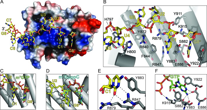
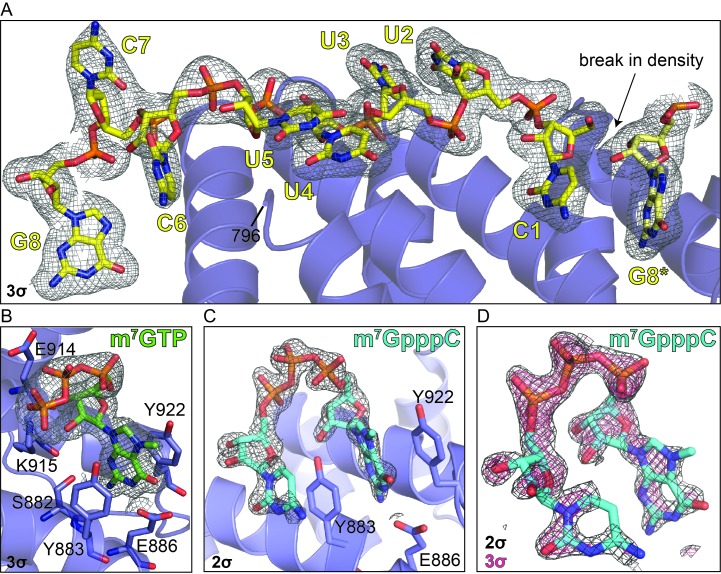
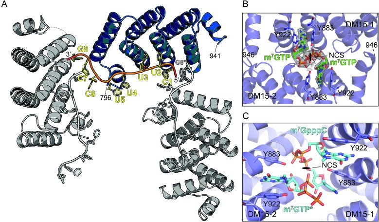
The 5'terminal oligopyrimidine (5'TOP) motif is a cis-regulatory RNA element located immediately downstream of the 7-methylguanosine [m7G] cap of TOP mRNAs, which encode ribosomal proteins and translation factors. In eukaryotes, this motif coordinates the synchronous and stoichiometric expression of the protein components of the translation machinery. La-related protein 1 (LARP1) binds TOP mRNAs, regulating their stability and translation. We present crystal structures of the human LARP1 DM15 region in complex with a 5'TOP motif, a cap analog (m7GTP), and a capped cytidine (m7GpppC), resolved to 2.6, 1.8 and 1.7 Å, respectively. Our binding, competition, and immunoprecipitation data corroborate and elaborate on the mechanism of 5'TOP motif binding by LARP1. We show that LARP1 directly binds the cap and adjacent 5'TOP motif of TOP mRNAs, effectively impeding access of eIF4E to the cap and preventing eIF4F assembly. Thus, LARP1 is a specialized TOP mRNA cap-binding protein that controls ribosome biogenesis.

Keywords: 5' cap; E. coli; La-related protein 1; RNA binding protein; TOP mRNAs; X-ray crystallography; biochemistry; biophysics; eIF4E; human; structural biology.


Conflict of interest statement

The authors declare that no competing interests exist.

Figures

Figure 1.
Figure 1.. The LARP1 DM15 region recognizes the 7-methylguanosine cap and invariant 5’cytidine of TOP mRNAs.
(A) Protein surface representation is colored according to electrostatic potential (−74 kEV, red; 74 kEV, blue). (B) Zoomed view of the DM15 RNA binding site. (C) Superimposition of DM15 bound to RNA and bound to cap analog, m7GTP. (D) Superimposition of DM15 bound to RNA and bound to m7GpppC. (E–F) Zoomed views of the specific recognition of C1 (E) and m7GTP (F). Potential hydrogen bonds indicated by dotted lines. DOI: http://dx.doi.org/10.7554/eLife.24146.002
Figure 1—figure supplement 1.
Figure 1—figure supplement 1.. Electron density reveals RNA, cap analog, and m7GpppC bind in the same location on the conserved surface of the DM15 region of LARP1.
Composite omit maps carved around the (A) RNA (3σ), (B) m7GTP cap analog (3σ), and (C) m7GpppC dinucleotide (2σ). (D) Composite omit map carved around the m7GpppC dinucleotide at 2σ (grey) and 3σ (magenta) for comparison. DOI: http://dx.doi.org/10.7554/eLife.24146.003
Figure 1—figure supplement 2.
Figure 1—figure supplement 2.. The DM15 region of LARP1 recognizes a guanosine.
(A) Three neighboring unit cells from the DM15-RNA co-crystal are shown. The protein monomer colored in blue interacts with two molecules of RNA: one from its unit cell and one originating in the unit cell on the right. This places a guanosine nucleotide 5’ to the TOP motif in the binding site of the blue DM15 region. (B) Two non-crystallographic symmetry mates from the DM15-m7GTP co-crystal show the guanine of the cap analog binds the same place as the G8* residue binds in the DM15-RNA co-crystal (A). (C) Two non-crystallographic symmetry mates from the DM15-m7GpppC co-crystal structure reveal that the m7G and C moieties bind in the same place as the G8* and C1 residues in the DM15-RNA co-crystal, respectively. In the other non-crystallographic symmetry-mate shown, only m7GTP is modeled because only one dinucleotide can bind per asymmetric unit since the inverted triphosphate linkage sits on the NCS 2-fold axis (see Figure 1—figure supplement 5). DOI: http://dx.doi.org/10.7554/eLife.24146.004
Figure 1—figure supplement 3.
Figure 1—figure supplement 3.. The amino acids of the DM15 region that directly bind G8*, C1, and m7GTP are nearly 100% conserved.
Frequency plots were generated from amino acid sequences of LARP1 from H. sapiens (NP_056130.2), M. musculus (NP_082727.1), D. rerio (XP_001920902.3), X. laevis (NP_001089363.1), D. melanogaster (NP_524998.1), C. elegans (NP_001040867.2), A. thaliana (NP_001190354.1), O. sativa (XP_015621184.1), B. dendrobatidis (XP_006676827.1), and M. brevicollis (XP_001744631.1). Asterisks indicate amino acids shown in Figure 1. Numbering is based on the DM15 region construct cloned from LARP1 isoform 2 (NP_056130.2). DOI: http://dx.doi.org/10.7554/eLife.24146.005
Figure 1—figure supplement 4.
Figure 1—figure supplement 4.. The DM15 region of LARP1 has evolved a unique fold to support a canonical cap-binding pocket.
The amino acids responsible for cap recognition in each of the proteins are shown as white sticks and the cap analog is shown as green sticks. PDBs used to generate each panel: eIF4E (1EJ1), VP23 (1AV6), VP39 D182A (4DCG), CBP20 (1H2T), LARP1 DM15 (5V4R), DXO (4J7N), PARN (3CTR), DCP1-DCP2 (5KQ4), DCPS (1XMM). DOI: http://dx.doi.org/10.7554/eLife.24146.006
Figure 1—figure supplement 5.
Figure 1—figure supplement 5.. The m7GpppC-DM15 co-crystal non-crystallographic symmetry reveals one ordered dinucleotide and one partially ordered dinucleotide.
The composite omit map shown is contoured at 1σ and reveals an ordered m7GpppC dinucleotide bound to the DM15 NCS-mate on the right. The DM15 NCS-mate on the left binds the m7G in the G-binding pocket, while the triphosphate moiety is ordered and facing away from the C-binding pocket (m7GTP*). Presumably, the density for the cytidine is mostly disordered: based on the path of the m7G triphosphate moiety (dotted line and 3'C label), the cytidine is most likely in the solvent channel; density for the Watson-Crick face of a cytidine is visible in the C-binding site in this copy of DM15, but is presumably of very low occupancy. Based on this data and the data shown in Figure 1—figure supplement 2B, only one inverted triphosphate per asymmetric unit can be accommodated in the anion binding site that overlaps with the non-crystallographic 2-fold axis. DOI: http://dx.doi.org/10.7554/eLife.24146.007
Figure 2.
Figure 2.. The LARP1 DM15 region recognizes capped TOP sequences and outcompetes eIF4E for their binding.
(A–C) Electrophoretic mobility shift assays using the indicated RNA. (D) Quantitation of 3 replicate EMSAs; error bars represent standard deviation. (E) Competition assays analyzed by native gel electrophoresis using labeled m7Gppp-RPS6 as substrate with the indicated protein concentrations. DOI: http://dx.doi.org/10.7554/eLife.24146.009
Figure 2—figure supplement 1.
Figure 2—figure supplement 1.. Mutation of amino acids that stabilize the cap and the RNA decreases the affinity of the DM15 region for capped TOP RNA.
(A–C) EMSA of capped RPS6 42-mer 5’UTR with each indicated point mutant. (D) Quantification of three replicates of each indicated EMSA. ND, not determined. DOI: http://dx.doi.org/10.7554/eLife.24146.011
Figure 2—figure supplement 2.
Figure 2—figure supplement 2.. Capping another TOP sequence enhances the affinity of DM15 for the RNA.
(A) EMSA of DM15 with an uncapped RNA representing the first 42 nucleotides of the 5’UTR of RPL32. (B) EMSA of DM15 with the capped version of the RNA from panel (A). DOI: http://dx.doi.org/10.7554/eLife.24146.012
Figure 2—figure supplement 3.
Figure 2—figure supplement 3.. The interaction between the DM15 region of LARP1 and the cap moiety is specific.
Competition assay challenging DM15 bound to labeled capped non-TOP RNA with capped (lanes 3–5) or uncapped (lanes 6–8) cold competitor RNA. DOI: http://dx.doi.org/10.7554/eLife.24146.013
Figure 2—figure supplement 4.
Figure 2—figure supplement 4.. Cap analog, m7GTP, and m7GpppC stabilize the protein fold of the DM15 region of LARP1.
Quantification of thermal shift assays conducted in the presence of GTP, m7GTP, ATP, m7GpppG, or m7GpppC; n = 3 for all assays; error bars show 1 standard deviation. DOI: http://dx.doi.org/10.7554/eLife.24146.014
Figure 2—figure supplement 5.
Figure 2—figure supplement 5.. eIF4E binds non-TOP mRNAs and outcompetes the DM15 region of LARP1 for their binding.
(A–B) EMSAs with the indicated substrates and increasing concentrations of recombinant human eIF4E. (C) Quantitation of EMSAs, n = 3. Error bars indicate standard deviation. (D–E) Competition assays for the capped non-TOP substrate. DOI: http://dx.doi.org/10.7554/eLife.24146.015
Figure 3.
Figure 3.. LARP1 prevents eIF4F assembly on 5’TOP mRNAs.
(A and B). Extracts of HEK293T cells that were transfected with empty vector (EV), FLAG-tagged wild type LARP1 (WT) or FLAG-LARP1 double-mutant (R840E/Y883A), were immunoprecipitated with anti-eIF4G antibody. Inputs were analyzed by Western blot (A) and eIF4G-IPs were analyzed for TOP mRNA abundance by RT-ddPCR (B). Data were normalized to input mRNA levels. Three biological replicates were performed and error bars denote propagated standard deviation. DOI: http://dx.doi.org/10.7554/eLife.24146.016

Similar articles

Cited by

References

    1. Adams PD, Afonine PV, Bunkóczi G, Chen VB, Davis IW, Echols N, Headd JJ, Hung LW, Kapral GJ, Grosse-Kunstleve RW, McCoy AJ, Moriarty NW, Oeffner R, Read RJ, Richardson DC, Richardson JS, Terwilliger TC, Zwart PH. PHENIX: a comprehensive Python-based system for macromolecular structure solution. Acta Crystallographica. Section D, Biological Crystallography. 2010;66:213–221. doi: 10.1107/S0907444909052925. - DOI - PMC - PubMed
    1. Altschul SF, Madden TL, Schäffer AA, Zhang J, Zhang Z, Miller W, Lipman DJ. Gapped BLAST and PSI-BLAST: a new generation of protein database search programs. Nucleic Acids Research. 1997;25:3389–3402. doi: 10.1093/nar/25.17.3389. - DOI - PMC - PubMed
    1. Aoki K, Adachi S, Homoto M, Kusano H, Koike K, Natsume T. LARP1 specifically recognizes the 3' terminus of poly(A) mRNA. FEBS Letters. 2013;587:2173–2178. doi: 10.1016/j.febslet.2013.05.035. - DOI - PubMed
    1. Avni D, Shama S, Loreni F, Meyuhas O. Vertebrate mRNAs with a 5'-terminal pyrimidine tract are candidates for translational repression in quiescent cells: characterization of the translational cis-regulatory element. Molecular and Cellular Biology. 1994;14:3822–3833. doi: 10.1128/MCB.14.6.3822. - DOI - PMC - PubMed
    1. Biberman Y, Meyuhas O. TOP mRNAs are translationally inhibited by a titratable repressor in both wheat germ extract and reticulocyte lysate. FEBS Letters. 1999;456:357–360. doi: 10.1016/S0014-5793(99)00983-7. - DOI - PubMed

Publication types

MeSH terms