. 2017 Apr 4:7:45489.
doi: 10.1038/srep45489.

A Gate Hinge Controls the Epithelial Calcium Channel TRPV5

Affiliations

A Gate Hinge Controls the Epithelial Calcium Channel TRPV5

Jenny van der Wijst et al. Sci Rep. .

Abstract

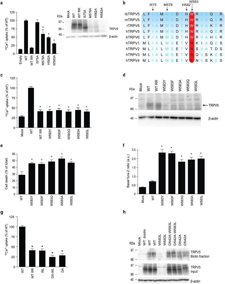
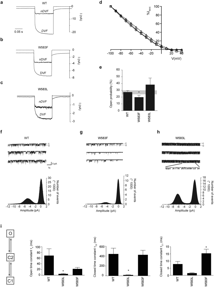
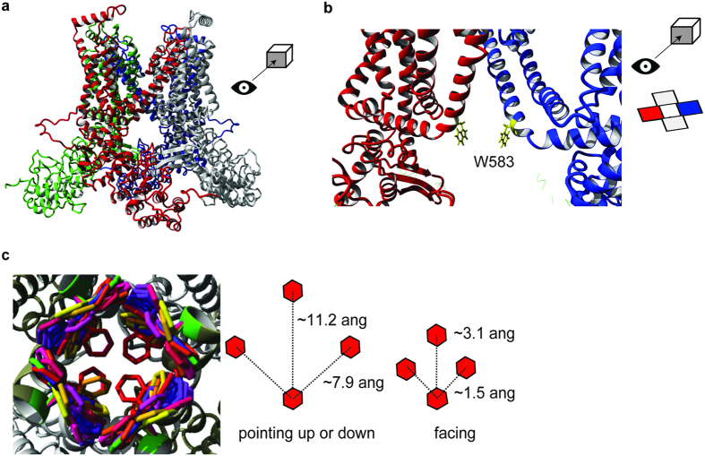
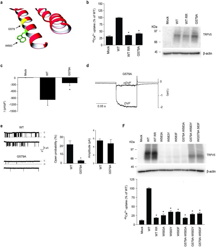
TRPV5 is unique within the large TRP channel family for displaying a high Ca2+ selectivity together with Ca2+-dependent inactivation. Our study aims to uncover novel insights into channel gating through in-depth structure-function analysis. We identify an exceptional tryptophan (W583) at the terminus of the intracellular pore that is unique for TRPV5 (and TRPV6). A combination of site-directed mutagenesis, biochemical and electrophysiological analysis, together with homology modeling, demonstrates that W583 is part of the gate for Ca2+ permeation. The W583 mutants show increased cell death due to profoundly enhanced Ca2+ influx, resulting from altered channel function. A glycine residue above W583 might act as flexible linker to rearrange the tryptophan gate. Furthermore, we hypothesize functional crosstalk between the pore region and carboxy terminus, involved in Ca2+-calmodulin-mediated inactivation. This study proposes a unique channel gating mechanism and delivers detailed molecular insight into the Ca2+ permeation pathway that can be extrapolated to other Ca2+-selective channels.


Conflict of interest statement

The authors declare no competing financial interests.

Figures

Figure 1
Figure 1. Effect of W583 mutations on TRPV5 function.
(a) Left panel depicts a 45Ca2+ uptake assay of HEK293 cells transfected with either wild type (WT) TRPV5, I575A, M578A, H582A, or W583A. Data is shown as percentage of WT (left panel) (N = 9, three independent experiments performed in triplicate). Ruthenium red (RR), 10 μM, is used as control to inhibit the TRPV5-mediated Ca2+ uptake. The right panel shows a representative immunoblot using HA antibody for TRPV5 expression, and β-actin as loading control. (b) Multiple sequence alignment of the region surrounding W583 in different species of TRPV5 and amongst other TRPV family members. Light blue letters represent conserved amino acids and the red box indicates the conservation of W583. (c) 45Ca2+ uptake assay of HEK293 cells transfected with either wild type (WT) TRPV5 or the indicated mutants, depicted as percentage of WT (upper panel) (N = 9, three independent experiments performed in triplicate). Ruthenium red (RR) is used as negative control. (d) Representative immunoblot of cell lysates of the respective Ca2+ uptake experiments with HA antibody for TRPV5 and β-actin as loading control. (e) Quantification of cell death by counting the number of trypan blue stained (death) HEK293 cells upon transfecting wild type (WT) TRPV5 or the indicated mutants, depicted as percentage of the total cell amount (N = 6, three independent experiments performed in duplicate). (f) Basal intracellular Ca2+ levels are shown as fura-2 ratio in arbitrary units (a.u.) for HEK293 cells expressing either wild type (WT) TRPV5 or the indicated mutants (N = 30–40, total from two independent experiments). Values are shown as mean ± SEM. Asterisk indicates statistical significance (p < 0.05) compared to WT. (g) 45Ca2+ uptake assay of HEK293 cells transfected with wild type (WT) TRPV5 or the indicated mutants, depicted as percentage of WT and corrected for protein expression levels (upper panel) (N = 9, three independent experiments performed in triplicate). Ruthenium red (RR) is used as a negative control. (h) Cell surface biotinylation of HEK293 cells transfected with wild type (WT) TRPV5 or indicated mutants. The biotin fraction represents the TRPV5 present at the plasma membrane (top panel) and input demonstrates TRPV5 expression in total cell lysates (bottom panel). Representative immunoblot of three independent experiments is shown.
Figure 2
Figure 2. Channel activity is altered in the TRPV5 W583 mutants.
(ac) Representative traces of the whole-cell Na+ currents at the voltage step to −100 mV of HEK293 cells expressing either wild type (WT) TRPV5 or the indicated mutants (W583L and W583F) in nominally DVF (nDVF) and divalent-free (DVF) solutions. (d) Representative current-voltage relationship of wild type (WT) TRPV5 (circle), W583F (triangle), and W583L (square) measured from a voltage step protocol of −100 to + 40 mV in the whole cell configuration in DVF solution and current is depicted as percentage of the maximal current at −100 mV (%Imax). (e) Histogram showing the average open probability at −80 mV for wild type (WT) TRPV5, W583F, and W583L. Values are shown as mean ± SEM (N > 7 per condition). Asterisk indicates statistical significance (p < 0.05) compared to WT. (fh) Cell-attached single channel recordings were measured during a 10 s step to −80 mV (holding potential 0 mV) in wild type (WT) TRPV5 (f), W583F (g), and W583L (h). Bottom panels show the amplitude histograms with a Gaussian fit function corresponding to the closed and open states in the upper panels. (i) The dwell time constants (τ) are extracted via a three-state model of one open (o) and two closed states (C1 and C2). Average τ values of wild type (WT) TRPV5, W583F, and W583L were derived by two-exponential fit of the model-based distribution of dwell times, obtained from single channel recordings (N > 7). Values are shown as mean ± SEM. Asterisk indicates statistical significance (p < 0.05) compared to WT.
Figure 3
Figure 3. Homology model of TRPV5.
(a) The tetrameric TRPV5 structure is depicted in front view. Each monomer is color-coded. (b) The detailed front view of the pore region highlights the side chains of W583 (yellow) in two monomers. The side chains are sticking towards the permeation pathway. (c) The possible rotameric positions for the side chain of W583 are shown in different colors (left panel). Overall, three main rotameric positions are detected with the side chains either pointing upwards, downwards or pointing towards each other. The distance between the W583 side chains is based on the homology model and is depicted for the main rotameric positions (right panel).
Figure 4
Figure 4. Analysis of the G579A mutation on TRPV5 activity.
(a) Zoomed image of the α-helix containing W583 and G579. (b) 45Ca2+ uptake assay of HEK293 cells transfected with either wild type (WT) TRPV5 or the G579A mutant. Ruthenium red (RR) is used as control for TRPV5-mediated uptake. The protein expression level is shown in the representative immunoblot (N = 9, three independent experiments performed in triplicate). (c) Averaged Na+ current densities of mock, wild type (WT) TRPV5 or G579A transfected HEK293 cells are presented at −80 mV in DVF solution (N = 8–10). (d) Representative traces of the whole-cell Na+ currents of HEK293 cells expressing TRPV5 G579A in nDVF and DVF solutions, in response to a voltage step protocol (−100 to +40 mV). (e) The open probability at −80 mV is assessed using cell-attached patch clamp in cells expressing the G579A mutant. Typical single-channel behavior is shown for wild type (WT) TRPV5 and G579A. The average open probability (middle panel) and amplitude (right panel) are determined (N > 7 per condition). Values are shown as mean ± SEM. Asterisk indicates statistical significance (p < 0.05) compared to WT. (f) Cell lysates of wild type (WT) TRPV5 and the indicated single (W583A, W583Y, W583F) and double (W583A-G579A, W583Y-G579A, W583F-G579A) mutants were immunoblotted with HA antibody, and using β-actin as loading control. A representative immunoblot is shown in the upper panel. The lower panel shows a 45Ca2+ uptake assay of HEK293 cells transfected with either wild type (WT) TRPV5 or the indicated mutants (N = 9, three independent experiments performed in triplicate). Ruthenium red (RR) is used as control for TRPV5-mediated uptake. Values are shown as mean +/− SEM. Asterisk indicates statistical significance (p < 0.05) compared to WT.
Figure 5
Figure 5. Rescue the TRPV5 CaM binding mutants by the G579A mutation.
(a) 45Ca2+ uptake assay of HEK293 cells transfected with either wild type (WT) TRPV5 or the indicated mutants (G579A, W583A, S698X, W702A, G579-W583A, G579A-S698X, and G579A-W702A), depicted as percentage of WT (upper panel) (N = 9, three independent experiments performed in triplicate). Ruthenium red (RR) is used as a negative control. (b) Cell lysates of the respective experiments were immunoblotted with HA antibody, and using β-actin as loading control. A representative immunoblot is shown. (c) The basal intracellular Ca2+ levels are shown as fura-2 ratio in arbitrary units (a.u.) for HEK293 cells expressing either wild type (WT) TRPV5 or the indicated mutants (N = 11–66, total from two independent experiments). Values are shown as mean ± SEM. Asterisk indicates statistical significance (p < 0.05) compared to WT. (d) CaM binding assay of HEK293 cells transfected with wild type (WT) or indicated mutants of TRPV5. Samples were analyzed by immunoblotting with HA antibody. The CaM beads fraction represents the TRPV5 bound to the CaM agarose beads (top panel) and input demonstrates TRPV5 expression in total cell lysates (bottom panel). Representative immunoblot of three independent experiments is depicted.

Similar articles

Cited by

References

    1. Nilius B. & Voets T. TRP channels: a TR(I)P through a world of multifunctional cation channels. Pflugers Arch. 451, 1–10 (2005). - PubMed
    1. Ramsey I. S., Delling M. & Clapham D. E. An introduction to TRP channels. Annu. Rev. Physiol. 68, 619–647 (2006). - PubMed
    1. Venkatachalam K. & Montell C. TRP channels. Annu. Rev. Biochem. 76, 387–417 (2007). - PMC - PubMed
    1. Stock L., Souza C. & Treptow W. Structural basis for activation of voltage-gated cation channels. Biochemistry 52, 1501–1513 (2013). - PubMed
    1. Hoenderop J. G. J. & Bindels R. J. M. Calciotropic and magnesiotropic TRP channels. Physiology (Bethesda) 23, 32–40 (2008). - PubMed

Publication types

LinkOut - more resources