Case Reports
. 2017 Jun 1;26(11):2118-2132.
doi: 10.1093/hmg/ddx107.

Localized TWIST1 and TWIST2 basic domain substitutions cause four distinct human diseases that can be modeled in Caenorhabditis elegans

Affiliations
Case Reports

Localized TWIST1 and TWIST2 basic domain substitutions cause four distinct human diseases that can be modeled in Caenorhabditis elegans

Sharon Kim et al. Hum Mol Genet. .

Abstract

Twist transcription factors, members of the basic helix-loop-helix family, play crucial roles in mesoderm development in all animals. Humans have two paralogous genes, TWIST1 and TWIST2, and mutations in each gene have been identified in specific craniofacial disorders. Here, we describe a new clinical entity, Sweeney-Cox syndrome, associated with distinct de novo amino acid substitutions (p.Glu117Val and p.Glu117Gly) at a highly conserved glutamic acid residue located in the basic DNA binding domain of TWIST1, in two subjects with frontonasal dysplasia and additional malformations. Although about one hundred different TWIST1 mutations have been reported in patients with the dominant haploinsufficiency Saethre-Chotzen syndrome (typically associated with craniosynostosis), substitutions uniquely affecting the Glu117 codon were not observed previously. Recently, subjects with Barber-Say and Ablepharon-Macrostomia syndromes were found to harbor heterozygous missense substitutions in the paralogous glutamic acid residue in TWIST2 (p.Glu75Ala, p.Glu75Gln and p.Glu75Lys). To study systematically the effects of these substitutions in individual cells of the developing mesoderm, we engineered all five disease-associated alleles into the equivalent Glu29 residue encoded by hlh-8, the single Twist homolog present in Caenorhabditis elegans. This allelic series revealed that different substitutions exhibit graded severity, in terms of both gene expression and cellular phenotype, which we incorporate into a model explaining the various human disease phenotypes. The genetic analysis favors a predominantly dominant-negative mechanism for the action of amino acid substitutions at this highly conserved glutamic acid residue and illustrates the value of systematic mutagenesis of C. elegans for focused investigation of human disease processes.


Figures

Figure 1
Figure 1
Craniofacial appearance of individuals with p.Glu117 substitutions in TWIST1. (A–C) Subject 1 with de novo TWIST1 c.350A > T variant, leading to the substitution p.Glu117Val. In (A, B) (aged 4 months), note the hypertelorism, prominent forehead, upper eyelid colobomas, deficient bony orbits with pseudoproptosis, hypoplastic alae nasi, short columella and philtrum and small low-set dysplastic cupped ears. (C) CT of skull aged 4 months. Note severely hypoplastic frontal bones separated by patent metopic suture (arrowhead), massive enlargement of anterior fontanelle (bounded by arrows), patent coronal and sagittal sutures, hypertelorism and hypoplasia of zygomatic bones, mandible and maxilla. (D–F) Subject 2 with a de novo TWIST1 c.350A > G variant, leading to the substitution p.Glu117Gly (D aged 6 days, E and F aged 15 weeks, postoperative tarsorraphies). Note that this individual is more severely affected but with a similar combination of hair configuration in widow’s peak, hypertelorism, upper eyelid colobomas, very broad nasal bridge with a broad and flattened nasal tip and a low columella and small crumpled ears.
Figure 2
Figure 2
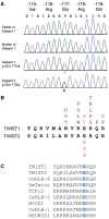
Identification of TWIST1 mutations altering the Glu117 codon. (A) Dideoxy-sequencing traces of TWIST1 in Subject 1 and unaffected parents (above) and Subject 2 (below). The heterozygous substitutions A > T and A > G are indicated in the two probands. (B) Amino acid sequences (bold black lettering) of basic DNA binding domains of human TWIST1 and TWIST2. Above the TWIST1 sequence are shown the positions of amino acid substitutions observed in SCS (unbolded black lettering; public data available from Human Gene Mutation Database, plus an unpublished c.347G > C (p.Arg116Pro) mutation identified by the Oxford Genetics Laboratories) and SwCoS (red lettering). Below the TWIST2 sequence are shown the positions of amino acid substitutions observed in AMS and BSS (red lettering). (C) Amino acid sequence alignment of basic domains from various bHLH proteins; the glutamic acid (blue in all the sequences) is invariant throughout various bHLH human homologs as well as in D. melanogaster (Dm) Twist and C. elegans (Ce) bHLH proteins.
Figure 3
Figure 3
Phenotypes of C. elegans hlh-8 CRISPR/Cas9 alleles. (A) Table indicates all of the phenotypes examined for the disease-associated Glu29† and Arg28† alleles and the related control strains. In experiments for which the data in the subsequent figures are summarized (starting with the row ‘Early M lineage’ and all subsequent rows), the + and – marks indicate the percent of the population with the WT phenotype [e.g. for the Early M lineage—two cells in the dorsal/ventral planes or robust expression of the reporter genes (with bright or medium GFP signal):  +++, 80–100%;  ++, 60–80%; +, 40–60%; +/–, 20–40%; –, 0–20%]; nd, not determined. (B–G) Micrographs representing the major phenotypes in the mutant animals (E–G) compared with WT (B–D). (B, E) The central region of adult hermaphrodites showing the egg-laying defective (Egl) phenotype with two representative embryos (white arrowheads) that are in an early stage of embryogenesis in WT (B) compared with embryos that are just prior to hatching in Egl mutants (E). WT hermaphrodites already would have laid these later stage embryos. (C, F) The central region of L4 larvae showing the constipated (Con) phenotype with the intestinal lumen (white arrow) that is expanded in the constipated mutant (F) compared with the narrow lumen in the WT animal (C). (D, G) The posterior region of adult hermaphrodites showing the deformed anal region (Dar) phenotype with the anal opening (open arrow) surrounded by a smooth, tapered tail in WT (D) and a wide, deformed tail in the mutant (G). In (A), n values for Egl, Con, and development time are all >30 animals per genotype. Subsequent n values and data can be found in the following figures: Average Brood Size (Supplementary Material, Fig. S3), Early M lineage (Fig. 4A), Late M lineage (two sex myoblasts) (Fig. 4B), Late M lineage (SM descendants) (Fig. 4C), arg-1 VM expression (Fig. 5G), arg-1 Ent expression (Fig. 5G), egl-15 expression (Fig. 5J), E29†/+ nonRet (Fig. 6A) Note that the nonRet phenotype in heterozygotes (E29†/+) was analyzed in a egl-15::gfp sensitized background., E29†/+ arg-1 VMs (Fig. 6C), E29†/+ arg-1 Ents (Supplementary Material, Fig. S4), E29†/+ egl-15 (Fig. 6B). Ent refers to the enteric muscles collectively: intestinal muscles (Ints), anal sphincter (Sph) and anal depressor (Dep) (see Supplementary Material, Figs S4, S5).
Figure 4
Figure 4
hlh-8 homozygous Glu29† mutant animals display distinct defects in the M (postembryonic mesodermal) lineage. (A) Animals were scored for the first division of the M cell in L1 larvae—in WT animals, one daughter cell is on the dorsal side and one is on the ventral side of the animal (D/V). Some mutant animals have both cells on the dorsal (D) or ventral (V) side. (B) L3 larvae were scored for the number of sex myoblasts (SMs), the range being from 1 to 6 or more SMs (6+). (C) L4 larvae at the same stage of vulval development were scored for the presence of SM descendants (SMd). When the SMs divide, the resulting cells are reduced in size such that they are approximately a quarter of the size of WT SM cells. Animals that have six or more of these small cells (indicative of at least three SM divisions) are considered to have SMd (blue bars). Animals that still contained the large blast cells at this stage that would be either undivided or underdivided are also indicated (red bars). Some animals exhibit both phenotypes so the two categories are >100% (e.g. fs2728); an animal with four SMs may have many smaller SMd cells and still have a few large undivided SMs. Here and in subsequent figures, the n values of animals examined with each genotype are indicated in parentheses. Error bars on each graph are standard error of the proportion.
Figure 5
Figure 5
hlh-8 homozygous Glu29† mutant animals disrupt target gene expression. GFP reporters [arg-1::gfp (A–F) and egl-15::gfp (H, I)] were crossed into the Glu29† mutants and the expression patterns were scored for each cell type: head mesodermal cell (HMC), vulval muscles (VMs), intestinal muscles (Ints), anal sphincter (Sph) and anal depressor (Dep or mu anal). For the egl-15::gfp expression, animals were categorized as having bright green (I), dim green or no green VMs (see images in Fig. 6). The percentage of the animals in the population with each expression pattern is graphed (G, J). A–F and H–I are modified from (46). White arrow in (F) indicates the anal opening. Error bars on each graph are standard error of the proportion.
Figure 6
Figure 6
hlh-8 Glu29†/+ heterozygotes retain embryos and are distinctly dominant for target gene expression in the vulval muscles. (A) Heterozygous animals were scored for the number of pharyngeal GFP-expressing embryos in the uterus indicating the hermaphrodite is retaining embryos. Since the pharynx develops during a stage of embryogenesis after embryos are typically laid on plates and no longer present in the uterus, hermaphrodites with green embryos in utero are retaining embryos (Ret phenotype). (B, C) Heterozygous animals were examined for vulval muscle expression of egl-15::gfp (B) and arg-1::gfp (C). Animals were scored as having bright (very bright gfp expression, comparable to the pharyngeal gfp expression in the heterozygous animals), medium (clearly defined vulval muscles but not overly bright), dim (can discern outline of the vulval muscles but can barely see the expression) or no GFP expression. (Images of representative egl-15::gfp expression taken with the same exposure time (50 ms) for each category are shown beneath (B and C). Error bars on each graph are standard error of the proportion.
Figure 7
Figure 7
Clinical phenotypes associated with TWIST1 and TWIST2 disease alleles. The clinical manifestations of altered TWIST1 (left) and TWIST2 (right) function fall on a continuum of decreasing total protein activity. In all cases, except for the mouse knockout (TWIST1) and Setleis syndrome (TWIST2), phenotypes are associated with heterozygous mutations. Since SCS alleles behave dominantly (due to haploinsufficiency), missense alleles such as Arg116† are grouped with deletion alleles at ∼50% protein activity. For TWIST2 heterozygotes, 50% protein activity yields a normal phenotype. Relative protein activities for substitutions at Glu117 (TWIST1) or Glu75 (TWIST2) are based on the experimental observations in heterozygous worms presented in Figure 6 and summarized in Figure 3. See Discussion for further explanation.

Similar articles

Cited by

References

    1. Ledent V., Paquet O., Vervoort M. (2002) Phylogenetic analysis of the human basic helix-loop-helix proteins. Genome Biol., 3, RESEARCH0030.. - PMC - PubMed
    1. Simpson P. (1983) Maternal-zygotic gene interactions during formation of the dorsoventral pattern in Drosophila embryos. Genetics, 105, 615–632. - PMC - PubMed
    1. Harfe B.D., Vaz Gomes A., Kenyon C., Liu J., Krause M., Fire A. (1998) Analysis of a Caenorhabditis elegans Twist homolog identifies conserved and divergent aspects of mesodermal patterning. Genes Dev., 12, 2623–2635. - PMC - PubMed
    1. Franco H.L., Casasnovas J., Rodríguez-Medina J.R., Cadilla C.L. (2011) Redundant or separate entities? Roles of Twist1 and Twist2 as molecular switches during gene transcription. Nucleic Acids Res., 39, 1177–1186. - PMC - PubMed
    1. Tukel T., Šošić D., Al-Gazali L.I., Erazo M., Casasnovas J., Franco H.L., Richardson J.A., Olson E.N., Cadilla C.L., Desnick R.J. (2010) Homozygous nonsense mutations in TWIST2 cause Setleis syndrome. Am. J. Hum. Genet., 87, 289–296. - PMC - PubMed

Publication types

MeSH terms

Supplementary concepts