. 2017 Mar 16:8:286.
doi: 10.3389/fimmu.2017.00286. eCollection 2017.

Exosomes Isolated from Ascites of T-Cell Lymphoma-Bearing Mice Expressing Surface CD24 and HSP-90 Induce a Tumor-Specific Immune Response

Affiliations

Exosomes Isolated from Ascites of T-Cell Lymphoma-Bearing Mice Expressing Surface CD24 and HSP-90 Induce a Tumor-Specific Immune Response

Florencia Menay et al. Front Immunol. .

Abstract

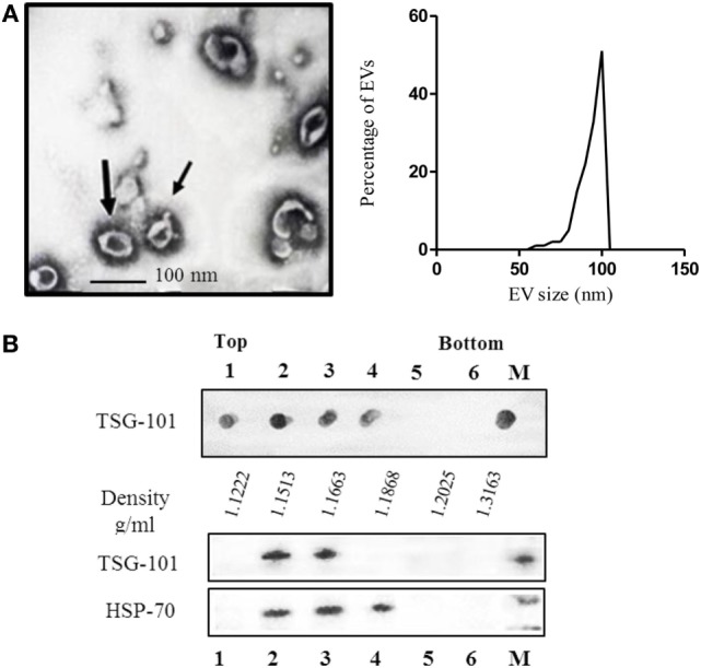
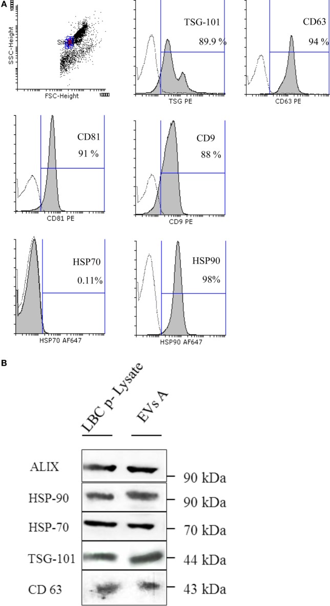
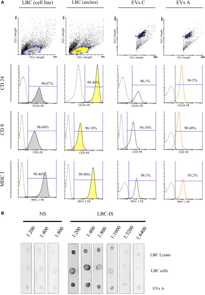
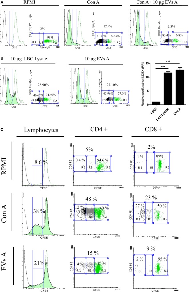
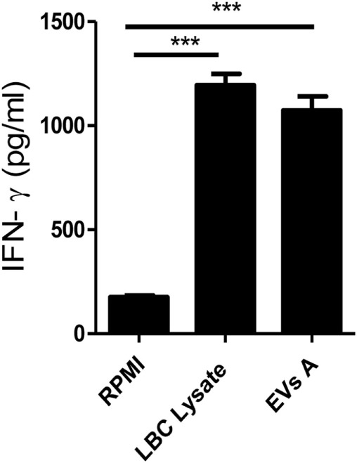
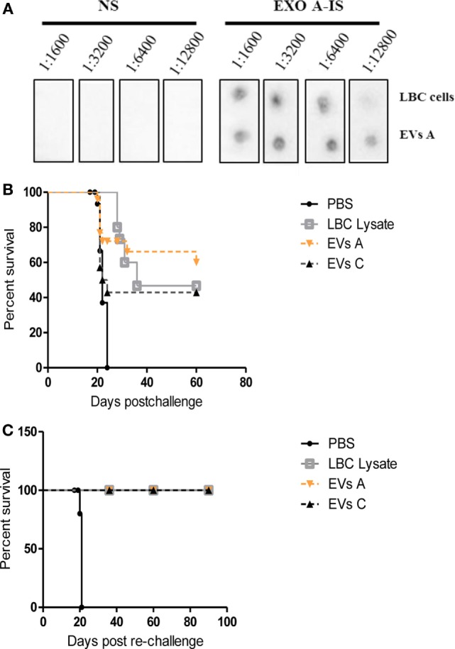
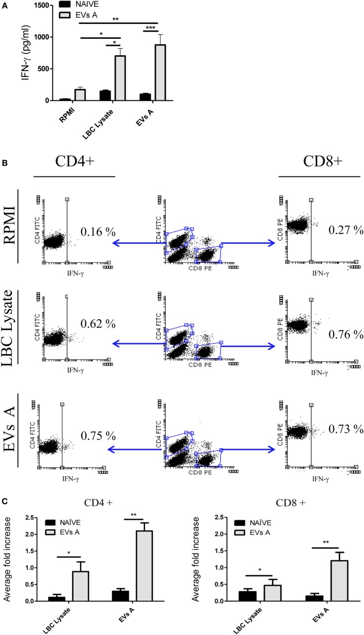
Extracellular vesicles (EVs), including endosome-derived nanovesicles (exosomes), are involved in cell-cell communication. Through transfer of their molecular contents, extracellular nanovesicles can alter the function of recipient cells. Due to these characteristics, EVs have shown potential as a new alternative for cancer immunotherapy. Tumor exosomes isolated from malignant ascites can activate dendritic cells, thereby priming the immune system to recognize and kill cancer cells. However, a suppressive role on tumor immune response has also been reported, suggesting that the neoplastic stage of carcinogenesis and the microenvironment where tumor cells grow may influence the amount of EVs released by the cell. This neoplastic stage and microenvironment may also impact EVs' components such as proteins and miRNA, determining their biological behavior. Most T-cell lymphomas have an aggressive clinical course and poor prognosis. Consequently, complementary alternative therapies are needed to improve the survival rates achieved with conventional treatments. In this work, we have characterized EVs isolated from ascites of mice bearing a very aggressive murine T-cell lymphoma and have studied their immunogenic properties. Small EVs were isolated by differential centrifugation, ultrafiltration, and ultracentrifugation at 100,000 × g on a sucrose cushion. The EVs were defined as exosomes by their morphology and size analyzed by electron microscopy, their floating density on a sucrose gradient, as well as their expression of endosome marker proteins ALIX, TSG-101; the tetraspanins CD63, CD9, and CD81. In addition, they contain tumor antigens, the marker for malignancy CD24, the heat shock protein HSP-70, and an unusual surface expression of HSP-90 was demonstrated. The administration of EVs isolated from ascites (EVs A) into naïve-syngeneic mice induced both humoral and cellular immune responses that allowed the rejection of subsequent tumor challenges. However, the immunization had no effect on a non-related mammary adenocarcinoma, demonstrating that the immune response elicited was specific and also it induced immune memory. In vitro analysis demonstrated that T-cells from EVs A-immunized mice secrete IFN-γ in response to tumor stimulation. Furthermore, tumor-specific CD4+ and CD8+ IFN-γ secreting cells could be efficiently expanded from mice immunized with EVs A, showing that a T helper 1 response is involved in tumor rejection. Our findings confirm exosomes as promising defined acellular tumor antigens for the development of an antitumor vaccine.

Keywords: T-cell lymphoma; ascites; exosomes; immune response; tumor vaccine.


Figures

Figure 1
Figure 1
(A) Electron microscopy of purified extracellular vesicles (EVs) isolated from ascitic fluid samples, demonstrating the typical shape and size (60–110 nm) of exosomes, and graphic of the size-distribution of the vesicles measured with ImageJ software of at least eight micro-photos. (B) Characterization of EVs isolated by sucrose density gradient. The sample was loaded on top of a step sucrose gradient, and six fractions were collected and analyzed for exosomal proteins. Aliquots of fractions collected from the top of the gradient were analyzed by dot blot for TSG-101 and western blotting with antibodies against HSP-70 and TSG-101. M = pellet from 100,000 × g ultracentrifugation not submitted to a sucrose gradient sedimentation.
Figure 2
Figure 2
(A) Flow cytometry detection of surface molecules on extracellular vesicles (EVs). EVs were incubated with aldehyde–sulfate latex beads and stained with monoclonal antibodies or specific isotype-matched control antibodies. Analysis was performed on singlet gate of a forward scatter versus side scatter dot plot. Filled histograms represent exosome–bead complexes stained with specific monoclonal antibodies, the unfilled histogram represents isotype control antibodies. (B) Immunoblot protein analyses of EVs. EVs A (EVs isolated from ascites) and the LBC-cell lysate (LBC p-Lysate) were separated on a SDS-PAGE and electroblotted onto a nitrocellulose membrane. Samples were incubated with antibodies specific for heat shock proteins (HSP-70 and HSP-90) and specific markers for exosome as ALIX, TSG-101, and CD63.
Figure 3
Figure 3
Flow cytometry analysis of extracellular vesicles (EVs) and LBC cells. (A) Samples were incubated with antibodies specific for CD24, CD8, and MHC I. Analysis was performed on singlet gate of a forward scatter versus side scatter dot plot. Filled histograms represent LBC cells stained with specific monoclonal antibodies, the unfilled histograms represent EVs–bead complexes, and dot line represents irrelevant-isotype-control antibodies. Graphs correspond to one representative of four independent experiments. EVs C, EVs isolated from LBC cell line. (B) Dot blot characterization of tumor-associated antigens in EVs A and LBC cells. Dot blot analysis of 1 μg LBC-cell lysates, 10,000 LBC cells (LBC), and 1 μg of LBC EVs. EVs proteins and LBC lysates proteins were separated on a SDS-PAGE and electroblotted to nitrocellulose. Blots were probed with sera from naïve (NS) or LBC lysate-immunized (LBC-IS) mice.
Figure 4
Figure 4
Evaluation of the immune properties of EVs A in vitro. (A) Immunosuppressive capacity of EVs A. Splenocytes obtained from naïve mice were stained with carboxyfluorescein succinimidyl ester (CFSE) and incubated with 1 μg of concanavalin A (CON A) or 1 μg CON A + 10 μg EXO A in RPMI plus 5% fetal-bovine serum for 5 day. (B) Stimulatory capacity of EVs A. Splenocytes obtained from mice sensitized in vivo with LBC lysate were labeled with 5 μM CFSE and cultured for 5 days with 10 μg EVs A or 10 μg of LBC lysate (green-filled histogram). Flow cytometry histograms corresponding to non-stimulated splenocytes stained with CFSE used as negative control (green line histogram), unstained non-stimulated splenocytes (black line histogram). RPI, relative proliferative index, is defined as the ratio between the percentages of stimulated cells by the percentage of control cells. The percentage of proliferating cells was taken from G0 (not included) to the beginning of autofluorescence. The numbers in the graph correspond to the percentage of the gated region: R2, unstimulated CFSE stained cells; R1, autofluorescence; and R3, proliferating cells. Graphs correspond to one representative of two independent experiments. (C) Proliferation of CD4+ and CD8+ lymphocytes. Splenocytes obtained from mice sensitized in vivo with LBC lysate were labeled with 5 μM CFSE and cultured for 5 days with 10 μg EVs A or 0.5 μg CON A. Flow cytometry histograms corresponding to non-stimulated splenocytes stained with CFSE used as negative control (green line histogram), unstained non-stimulated splenocytes (black line histogram). The percentage of proliferating cells was taken from G0 (not included) to the beginning of autofluorescence. Graphs correspond to one representative of two independent experiments. Lymphocyte gate was determined by forward and side scatter. CFSE dye dilution was analyzed gating on CD4 and CD8 cell fractions.
Figure 5
Figure 5
IFN-γ production. Spleen cells obtained from mice previously immunized with LBC lysate were incubated for 5 days at 37°C. Supernatants were then collected and IFN-γ levels were measured in culture supernatants by enzyme-linked immunosorbent assays. Error bars indicate SD of duplicate samples from a representative of two independent experiments. Asterisks indicate significant differences (***p < 0.001).
Figure 6
Figure 6
Protective immune response against LBC tumor induced by EVs A immunization. (A) Serum-specific antibodies against LBC cells. One microgram of exosomes or 10,000 LBC cells were spotted onto a nitrocellulose membrane. Membranes were blocked and incubated overnight at 4°C with different dilutions (from 1:1,600 to 1:12,800) of sera from naïve mice (NS) or from mice immunized with LBC lysate (LBC-IS). (B) Survival curves of mice immunized with EVs A. Immunocompetent naïve BALB/c mice were inoculated i.p. twice with 10 μg/mouse of EVs A with an interval of 7 days or with LBC lysate or EVs C. Seven days after the last immunization, mice were challenged with 1.0 × 106 LBC tumor cells i.p. Phosphate-buffered saline (PBS) corresponds to the non-immunized control group, inoculated with PBS and challenged with the LBC tumor cells. N = 12/group. Graphs correspond to one representative of two independent experiments. (C) Survival curves of mice after tumor rechallenge. Mice vaccinated with EVs A or LBC lysate that had rejected the first tumor challenge were rechallenged with 1 × 106 LBC tumor cells i.p. 30 days after the first tumor injection. N = 6/group. The curves corresponding to EVs A, EVs C, and LBC lysate overlap.
Figure 7
Figure 7
Immune response induced in EVs A-immunized mice. (A) IFN-γ levels in the supernatant of splenocytes from mice immunized with EVs. Spleen cells obtained from naïve mice or mice previously immunized with EVs A were incubated for 48 h at 37°C. Supernatants were then collected, and IFN-γ levels were measured in culture supernatants by enzyme-linked immunosorbent assays. Error bars indicate SD of duplicate replicates from a representative of two independent experiments. Asterisks indicate significant differences (*p < 0.05, **p < 0.01, and ***p < 0.001). (B) Intracellular staining for IFN-γ in lymphocytes from mice immunized with EVs A. Spleen cells obtained from naïve or mice previously immunized with EVs A were incubated for 48 h at 37°C. Double-color surface staining was first performed with conjugated mAbs fluorescein isothiocyanate -anti-CD4 and PE-anti-CD8, and then cells were permeabilized with saponin and stained with allophycocyanin-anti-IFN-γ. To analyze intracellular cytokine expression by CD4+ and CD8+ lymphocytes, a gate was first drawn around the lymphocytes in a dot plot of forward scatter versus SSC. A second gate was drawn around the CD4+ or CD8+ lymphocytes in the CD4 versus CD8 dot plot. Non-stimulated splenocytes were used as negative control (RPMI). Splenocytes cultured 6 h with PMA at 50 ng/ml and ionomycin at 1 μM were used and as positive control (data not shown). (C) Average fold increase in IFN-γ-producing cells. This index is defined as the ratio between % stimulated IFN-γ-producing cells and % IFN-γ-producing cells of unstimulated splenocytes. The % IFN-γ-producing cells used for the calculation was previously subtracted by the % of unstimulated control. Error bars indicate SD of duplicate samples from a representative of two independent experiments. Asterisks indicate significant differences (*p < 0.05, **p < 0.01, and ***p < 0.001). N = 6/group.

Similar articles

Cited by

References

    1. El Andaloussi S, Mäger I, Breakefield XO, Wood MJ. Extracellular vesicles: biology and emerging therapeutic opportunities. Nat Rev Drug Discov (2013) 12:347–57.10.1038/nrd3978 - DOI - PubMed
    1. Colombo M, Raposo G, Théry C. Biogenesis, secretion, and intercellular interactions of exosomes and other extracellular vesicles. Annu Rev Cell Dev Biol (2014) 30:255–89.10.1146/annurev-cellbio-101512-122326 - DOI - PubMed
    1. Kowal J, Arras G, Colombo M, Jouve M, Morath JP, Primdal-Bengtson B, et al. Proteomic comparison defines novel markers to characterize heterogeneous populations of extracellular vesicle subtypes. Proc Natl Acad Sci U S A (2016) 113:E968–77.10.1073/pnas.1521230113 - DOI - PMC - PubMed
    1. De Toro J, Herschlik L, Waldner C, Mongini C. Emerging roles of exosomes in normal and pathological conditions: new insights for diagnosis and therapeutic applications. Front Immunol (2015) 6:203.10.3389/fimmu.2015.00203 - DOI - PMC - PubMed
    1. Hanahan D, Weinberg RA. Hallmarks of cancer: the next generation. Cell (2011) 144:646–74.10.1016/j.cell.2011.02.013 - DOI - PubMed