. 2017 Feb 15;31(4):422-437.
doi: 10.1101/gad.290155.116. Epub 2017 Mar 9.

A microRNA family exerts maternal control on sex determination in C. elegans

Affiliations

A microRNA family exerts maternal control on sex determination in C. elegans

Katherine McJunkin et al. Genes Dev. .

Abstract

Gene expression in early animal embryogenesis is in large part controlled post-transcriptionally. Maternally contributed microRNAs may therefore play important roles in early development. We elucidated a major biological role of the nematode mir-35 family of maternally contributed essential microRNAs. We show that this microRNA family regulates the sex determination pathway at multiple levels, acting both upstream of and downstream from her-1 to prevent aberrantly activated male developmental programs in hermaphrodite embryos. Both of the predicted target genes that act downstream from the mir-35 family in this process, suppressor-26 (sup-26) and NHL (NCL-1, HT2A, and LIN-41 repeat) domain-containing-2 (nhl-2), encode RNA-binding proteins, thus delineating a previously unknown post-transcriptional regulatory subnetwork within the well-studied sex determination pathway of Caenorhabditis elegans Repression of nhl-2 by the mir-35 family is required for not only proper sex determination but also viability, showing that a single microRNA target site can be essential. Since sex determination in C. elegans requires zygotic gene expression to read the sex chromosome karyotype, early embryos must remain gender-naïve; our findings show that the mir-35 family microRNAs act in the early embryo to function as a developmental timer that preserves naïveté and prevents premature deleterious developmental decisions.

Keywords: embryonic development; maternal control; microRNAs; mir-35–41; mir-35–42; sex determination.


Figures

Figure 1.
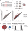
Figure 1.
Many genes are up-regulated in mir-35–41(nDf50) mutants but are not predicted mir-35 family targets. (A) Sequences of mature miRNAs comprising the mir-35 family. mir-35–41 are processed from a single transcript, while mir-42 is located in a separate cluster. Red text indicates the seed sequence. (B) mir-35–41(nDf50) is a temperature-sensitive mutant in which embryonic lethality is low at 20°C and highly penetrant at 25°C. Gene expression in embryos raised at each temperature was profiled using microarrays. (C) Many more genes are differentially expressed in mir-35–41(nDf50) embryos at 25°C than at 20°C. More genes are up-regulated than are down-regulated. (D) The genes that are differentially expressed at 20°C are largely a subset of those observed at 25°C. (E) The sets of differentially expressed genes at 20°C and at 25°C overlap much more than expected by chance. (***) P-value < 1 × 10−100; (**) P-value < 1 × 10⁻⁵, hypergeometric test. (F) The genes differentially expressed in mir-35–41(nDf50) embryos do not contain more mir-35 family seed matches than expected in a randomly chosen gene set.
Figure 2.
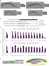
Figure 2.
Gene expression changes in mir-35–41(nDf50) mutants are similar to those in sex determination mutants. (A) Comparison of gene expression changes in mir-35–41(nDf50) embryos and sdc-2(lf) embryos, each normalized to wild type. (B) The set of genes differentially regulated in mir-35–41(nDf50) and sdc-2(lf) overlaps more than expected by chance. (***) P-value < 1 × 10−80; (*) P-value < 1 × 10⁻⁵, hypergeometric test. (C) Genetic model of C. elegans sex determination and dosage compensation. (DF) Quantitative RT–PCR (qRT–PCR) of transcripts indicated on the X-axis in embryos of the genotypes indicated above the graph. The mean and SEM of three biological replicates are shown. (D) Both genotypes were normalized to xol-1(y9). (E,F) All genotypes were normalized to wild-type XX embryos.
Figure 3.
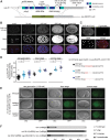
Figure 3.
mir-35–41 are required for proper sex determination in hermaphrodites. (A) Differential interference contrast (DIC) micrographs of wild-type and her-1(gf) and mir-35–41(nDf50) single- and double-mutant adults on the second day of adulthood. (B) Quantification of sex determination phenotypes; darker-colored bars indicate more severe masculinization. (C) The requirement for mir-35–41 in sex determination is dose-dependent, since heterozygotes display enhanced masculinization. Removing both copies of zygotic mir-35–41 does not strongly further enhance masculinization, indicating a strong requirement for the maternal contribution of mir-35–41. (+*) mIn1 balancer chromosome.
Figure 4.
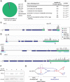
Figure 4.
The mir-35 family target genes sup-26 and nhl-2 are required for the sex determination phenotypes of mir-35–41(nDf50). (A) Representative DIC micrographs of adults on the second day of adulthood. mir-35–41(nDf50);sup-26(n1091);her-1(n695gf) and mir-35–41(nDf50);nhl-2(ok818);her-1(n695gf) adults are slightly smaller than age-matched her-1(n695gf) or wild type (not shown). (B) Quantification of sex determination phenotypes; darker-colored bars indicate more severe masculinization. (C,D) qRT–PCR in embryos raised at 20°C. The mean and SEM of three biological replicates are shown. All genotypes were normalized to wild-type XX embryos. (E, left) Summary of genetic interactions of mir-35–41(nDf50) with sup-26(lf) or nhl-2(lf). (Right) Genetic model highlighting pathways whose disruption in mir-35–41(nDf50) mutants underlies each aspect of the mutant sex determination phenotype. Whether the ΨGE phenotype contributes to the synTra phenotype is yet to be determined.
Figure 5.
Figure 5.
Sup-26 is a direct target gene of the mir-35 family. (A, top) Gene model of sup-26 depicting exon structure, protein domains, mutant alleles, and CRISPR–Cas9 editing sites. (Bottom) Schematic of homologous recombination strategy to generate sup-26(GFP_null). (B) Maximum projection of confocal stacks showing immunofluorescence of SUP-26::Flag. (C) Single confocal image of SUP-26::Flag costaining with PGL-1, a core component of P granules, in an ∼48-cell stage embryo (prior to the division of the germline primordium from a single cell [P4] into two cells [Z2 and Z3]). (D) Quantification of fluorescence in embryos containing the indicated mutations at the mir-35 family seed match in the 3′ UTR of the sup-26(GFP_null) locus. (*) P < 0.0001, Student's t-test. (E) Representative epifluorescence images of embryos with wild-type or mutated mir-35 family seed match in the sup-26(GFP_null) 3′ UTR. (F) The role of sup-26 in sex determination is dose-dependent. sup-26(n1091) heterozygosity strongly suppresses the mir-35–41(nDf50);her-1(gf) synTra phenotype. (+) qC1 balancer chromosome.
Figure 6.
Figure 6.
SUP-26 binds numerous 3′ UTRs, including those of sup-26 and nhl-2. (A) Functional annotation of significant SUP-26::Flag CLIP peaks. (B) Top significantly enriched gene ontology terms among transcripts bound by SUP-26::Flag. (CE) Example CLIP traces showing SUP-26::Flag binding to the 3′ UTR of transcripts implicated in sex determination and DC (tra-2 and dpy-30) (C), the 5′ UTR and 3′ UTR of sup-26 and the poly(A)-binding protein 2 (pab-2) (D), and the 3′ UTR of nhl-2 (E). The broken line indicates that the gene model continues outside the frame. (F) Western blot showing up-regulation of NHL-2 in sup-26(GFP_null) mutants. Two biological replicates are shown. (G) Simplified working model of the mir-35–41 regulatory network. mir-35–41 likely acts in early development in both XX and XO embryos to repress sup-26 and nhl-2 prior to sex determination. sup-26 and nhl-2 promote male development and may indirectly regulate HER-1 by affecting DCC activity (not shown).
Figure 7.
Figure 7.
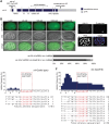
The mir-35 family seed match in nhl-2 cannot be mutated in a wild-type nhl-2 background. (A) Gene model of nhl-2 depicting exon structure, protein domains, mutant alleles, and CRISPR–Cas9 editing sites. (B) Epifluorescence and corresponding DIC images of embryos expressing GFP::NHL-2. (C) Maximum projection of a confocal stack of NHL-2 costaining with a P-granule-specific antibody (K76) in a ∼48-cell stage embryo (when the germline primordium is a single cell [P4]). (D) nhl-2(ok818) heterozygosity weakly suppresses the synTra phenotype. All animals of both genotypes contain one copy of the qC1 balancer chromosome. (E,F) Representation of CRISPR/Cas9 alleles generated when targeting the mir-35 family seed match in the nhl-2 3′ UTR in a nhl-2 wild-type background (E) or nhl-2(null) background (F). (Top) Graphs indicate the frequency of genomic mutation at each nucleotide in and around the mir-35 family seed match in lines selected for visible co-CRISPR editing events. Arrowheads denote potential sites of Cas9 cleavage corresponding to injected single-guide RNAs (sgRNAs). (Bottom) The number at the right of the sequence indicates the number of alleles isolated bearing the indicated mutation. Events marked with double lines and “2” were homozygous. Arrowheads indicate that deletion extends out of frame.

Similar articles

Cited by

References

    1. Alvarez-Saavedra E, Horvitz HR. 2010. Many families of C. elegans microRNAs are not essential for development or viability. Curr Biol 20: 367–373. - PMC - PubMed
    1. Bazzini AA, Lee MT, Giraldez AJ. 2012. Ribosome profiling shows that miR-430 reduces translation before causing mRNA decay in zebrafish. Science 336: 233–237. - PMC - PubMed
    1. Chiang HR, Schoenfeld LW, Ruby JG, Auyeung VC, Spies N, Baek D, Johnston WK, Russ C, Luo S, Babiarz JE, et al. 2010. Mammalian microRNAs: experimental evaluation of novel and previously annotated genes. Genes Dev 24: 992–1009. - PMC - PubMed
    1. Chuck G, Meeley R, Irish E, Sakai H, Hake S. 2007. The maize tasselseed4 microRNA controls sex determination and meristem cell fate by targeting Tasselseed6/indeterminate spikelet1. Nat Genet 39: 1517–1521. - PubMed
    1. Duerr JS. 2013. Antibody staining in C. elegans using ‘freeze-cracking’. J Vis Exp 10.3791/50664. - DOI - PMC - PubMed

Publication types

MeSH terms