

A mature macrophage is a principal HIV-1 cellular reservoir in humanized mice after treatment with long acting antiretroviral therapy
- PMID: 28279181
- PMCID: PMC5345240
- DOI: 10.1186/s12977-017-0344-7
A mature macrophage is a principal HIV-1 cellular reservoir in humanized mice after treatment with long acting antiretroviral therapy
Abstract
Background: Despite improved clinical outcomes seen following antiretroviral therapy (ART), resting CD4+ T cells continue to harbor latent human immunodeficiency virus type one (HIV-1). However, such cells are not likely the solitary viral reservoir and as such defining where and how others harbor virus is imperative for eradication measures. To such ends, we used HIV-1ADA-infected NOD.Cg-Prkdc scid Il2rg tm1Wjl /SzJ mice reconstituted with a human immune system to explore two long-acting ART regimens investigating their abilities to affect viral cell infection and latency. At 6 weeks of infection animals were divided into four groups. One received long-acting (LA) cabotegravir (CAB) and rilpivirine (RVP) (2ART), a second received LA CAB, lamivudine, abacavir and RVP (4ART), a third were left untreated and a fourth served as an uninfected control. After 4 weeks of LA ART treatment, blood, spleen and bone marrow (BM) cells were collected then phenotypically characterized. CD4+ T cell subsets, macrophages and hematopoietic progenitor cells were analyzed for HIV-1 nucleic acids by droplet digital PCR.
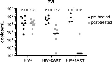
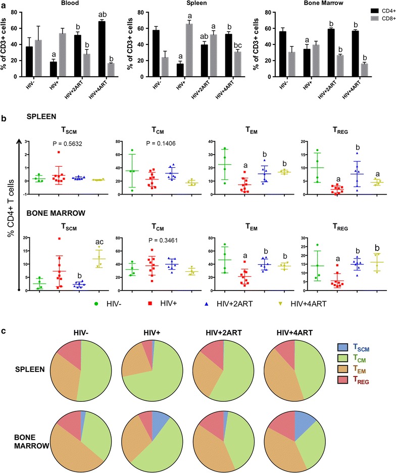
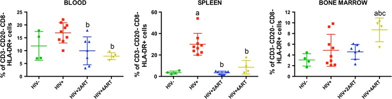
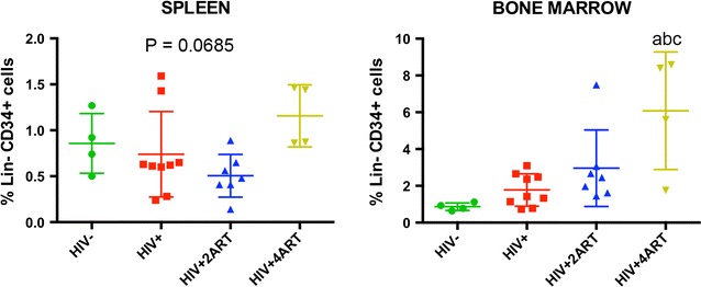
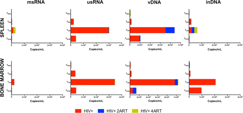
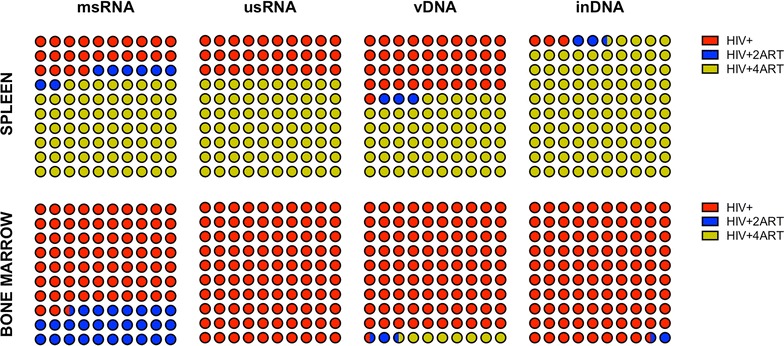
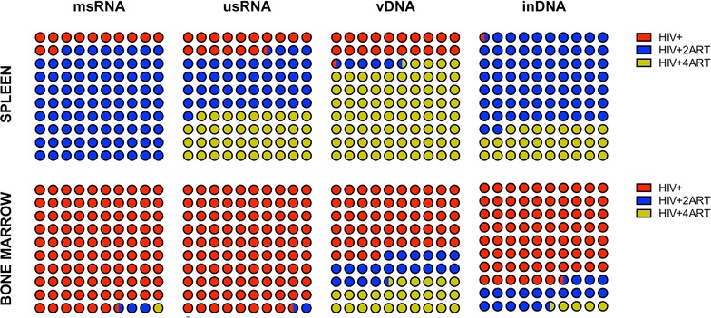
Results: Plasma viral loads were reduced by two log10 or to undetectable levels in the 2 and 4ART regimens, respectively. Numbers and distributions of CD4+ memory and regulatory T cells, macrophages and hematopoietic progenitor cells were significantly altered by HIV-1 infection and by both ART regimens. ART reduced viral DNA and RNA in all cell and tissue compartments. While memory cells were the dominant T cell reservoir, integrated HIV-1 DNA was also detected in the BM and spleen macrophages in both regimen-treated mice.
Conclusion: Despite vigorous ART regimens, HIV-1 DNA and RNA were easily detected in mature macrophages supporting their potential role as an infectious viral reservoir.
Keywords: Antiretroviral therapy; HIV-1; Humanized mice; Monocyte–macrophage; T effector cells; Viral reservoirs.
Figures
Similar articles
-
Lack of concordance between residual viremia and viral variants driving de novo infection of CD4(+) T cells on ART.Retrovirology. 2016 Aug 2;13(1):51. doi: 10.1186/s12977-016-0282-9. Retrovirology. 2016. PMID: 27484989 Free PMC article.
-
HIV-1 infection, response to treatment and establishment of viral latency in a novel humanized T cell-only mouse (TOM) model.Retrovirology. 2013 Oct 24;10:121. doi: 10.1186/1742-4690-10-121. Retrovirology. 2013. PMID: 24156277 Free PMC article.
-
Reservoirs for HIV-1: mechanisms for viral persistence in the presence of antiviral immune responses and antiretroviral therapy.Annu Rev Immunol. 2000;18:665-708. doi: 10.1146/annurev.immunol.18.1.665. Annu Rev Immunol. 2000. PMID: 10837072 Review.
-
Peripheral T Follicular Helper Cells Are the Major HIV Reservoir within Central Memory CD4 T Cells in Peripheral Blood from Chronically HIV-Infected Individuals on Combination Antiretroviral Therapy.J Virol. 2015 Dec 16;90(6):2718-28. doi: 10.1128/JVI.02883-15. J Virol. 2015. PMID: 26676775 Free PMC article. Clinical Trial.
-
HIV-1 Reservoirs During Suppressive Therapy.Trends Microbiol. 2016 May;24(5):345-355. doi: 10.1016/j.tim.2016.01.006. Epub 2016 Feb 12. Trends Microbiol. 2016. PMID: 26875617 Free PMC article. Review.
Cited by
-
HIV-associated neurocognitive disorder: key implications of the microbiota-gut-brain axis.Front Microbiol. 2024 Aug 2;15:1428239. doi: 10.3389/fmicb.2024.1428239. eCollection 2024. Front Microbiol. 2024. PMID: 39155987 Free PMC article. Review.
-
Persistent Viral Reservoirs in Lymphoid Tissues in SIV-Infected Rhesus Macaques of Chinese-Origin on Suppressive Antiretroviral Therapy.Viruses. 2019 Jan 27;11(2):105. doi: 10.3390/v11020105. Viruses. 2019. PMID: 30691203 Free PMC article.
-
Recent Updates on Mouse Models for Human Immunodeficiency, Influenza, and Dengue Viral Infections.Viruses. 2019 Mar 13;11(3):252. doi: 10.3390/v11030252. Viruses. 2019. PMID: 30871179 Free PMC article. Review.
-
Immune Activations and Viral Tissue Compartmentalization During Progressive HIV-1 Infection of Humanized Mice.Front Immunol. 2019 Feb 28;10:340. doi: 10.3389/fimmu.2019.00340. eCollection 2019. Front Immunol. 2019. PMID: 30873181 Free PMC article.
-
Tobacco and Antiretrovirals Modulate Transporter, Metabolic Enzyme, and Antioxidant Enzyme Expression and Function in Polarized Macrophages.Curr HIV Res. 2018;16(5):354-363. doi: 10.2174/1570162X17666190130114531. Curr HIV Res. 2018. PMID: 30706821 Free PMC article.
References
-
- Cooper ER, Charurat M, Mofenson L, Hanson IC, Pitt J, Diaz C, et al. Combination antiretroviral strategies for the treatment of pregnant HIV-1-infected women and prevention of perinatal HIV-1 transmission. J Acquir Immune Defic Syndr. 2002;29:484–494. doi: 10.1097/00042560-200204150-00009. - DOI - PubMed
Publication types
MeSH terms
Substances
Grants and funding
- R01 NS036126/NS/NINDS NIH HHS/United States
- P01 NS043985/NS/NINDS NIH HHS/United States
- R21 DA041018/DA/NIDA NIH HHS/United States
- P01 DA028555/DA/NIDA NIH HHS/United States
- P30 MH062261/MH/NIMH NIH HHS/United States
- P01 NS031492/NS/NINDS NIH HHS/United States
- P01 MH064570/MH/NIMH NIH HHS/United States
- R01 MH104147/MH/NIMH NIH HHS/United States
- R01 AG043540/AG/NIA NIH HHS/United States
- P30 GM103509/GM/NIGMS NIH HHS/United States
- P30 AI078498/AI/NIAID NIH HHS/United States
- R01 NS034239/NS/NINDS NIH HHS/United States
- R24 OD018546/OD/NIH HHS/United States
LinkOut - more resources
Full Text Sources
Other Literature Sources
Medical
Research Materials
