Interaction between HCMV pUL83 and human AIM2 disrupts the activation of the AIM2 inflammasome
- PMID: 28219398
- PMCID: PMC5319029
- DOI: 10.1186/s12985-016-0673-5
Interaction between HCMV pUL83 and human AIM2 disrupts the activation of the AIM2 inflammasome
Abstract
Background: AIM2, a cytosolic DNA sensor, plays an important role during infection caused by pathogens with double-stranded DNA; however, its role in human cytomegalovirus (HCMV) infection remains unclear. Previously, we showed an increase in AIM2 protein levels during the early stage of HCMV infection and a decrease 24 h post infection. Because HCMV has developed a variety of strategies to evade host immunity, we speculated that this decline might be attributed to a viral immune escape mechanism. The tegument protein pUL83 is an important immune evasion protein and several studies have reported that pUL83 binds to specific cellular proteins, such as AIM2-like receptor IFI16, to affect their functions. To determine whether pUL83 contributes to the variation in AIM2 levels during HCMV infection, we investigated the pUL83/AIM2 interaction and its impact on the AIM2 inflammasome activation.
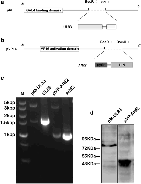
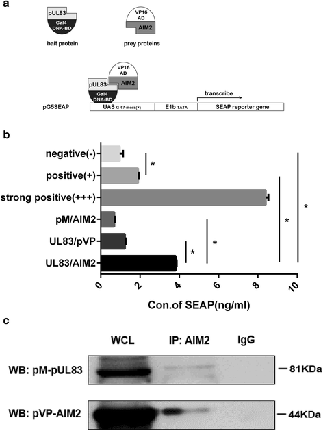
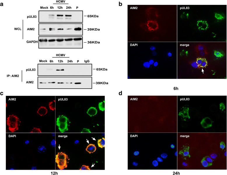
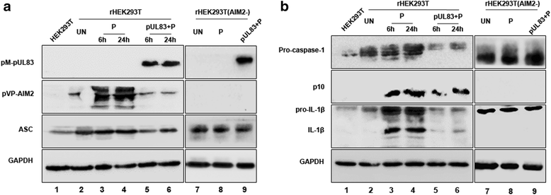
Methods: We constructed plasmids expressing recombinant pUL83 and AIM2 proteins for two-hybrid and chemiluminescence assays. Using co-immunoprecipitation and immunofluorescent co-localization, we confirmed the interaction of pUL83/AIM2 in THP-1-derived macrophages infected with HCMV AD169 strain. Furthermore, by investigating the expression and cleavage of inflammasome-associated proteins in recombinant HEK293T cells expressing AIM2, apoptosis-associated speck-like protein (ASC), pro-caspase-1 and pro-IL-1β, we evaluated the effect of pUL83 on the AIM2 inflammasome.
Results: An interaction between pUL83 and AIM2 was detected in macrophages infected with HCMV as well as in transfected HEK293T cells. Moreover, transfection of the pUL83 expression vector into recombinant HEK293T cells stimulated by poly(dA:dT) resulted in reduced expression and activation of AIM2 inflammasome-associated proteins, compared with the absence of pUL83.
Conclusions: Our data indicate that pUL83 interacts with AIM2 in the cytoplasm during the early stages of HCMV infection. The pUL83/AIM2 interaction deregulates the activation of AIM2 inflammasome. These findings reveal a new strategy of immune evasion developed by HCMV, which may facilitate latent infection.
Keywords: AIM2 inflammasome; HCMV; Immune evasion; pUL83.
Figures


Similar articles
-
Human cytomegalovirus triggers the assembly of AIM2 inflammasome in THP-1-derived macrophages.J Med Virol. 2017 Dec;89(12):2188-2195. doi: 10.1002/jmv.24846. Epub 2017 Aug 29. J Med Virol. 2017. PMID: 28480966
-
Regulatory Interaction between the Cellular Restriction Factor IFI16 and Viral pp65 (pUL83) Modulates Viral Gene Expression and IFI16 Protein Stability.J Virol. 2016 Aug 26;90(18):8238-50. doi: 10.1128/JVI.00923-16. Print 2016 Sep 15. J Virol. 2016. PMID: 27384655 Free PMC article.
-
Human cytomegalovirus pUL83 stimulates activity of the viral immediate-early promoter through its interaction with the cellular IFI16 protein.J Virol. 2010 Aug;84(15):7803-14. doi: 10.1128/JVI.00139-10. Epub 2010 May 26. J Virol. 2010. PMID: 20504932 Free PMC article.
-
The human cytomegalovirus tegument protein pp65 (pUL83): a key player in innate immune evasion.New Microbiol. 2018 Apr;41(2):87-94. Epub 2018 Jan 31. New Microbiol. 2018. PMID: 29384558 Review.
-
The absent in melanoma 2 (AIM2) inflammasome in microbial infection.Clin Chim Acta. 2019 Aug;495:100-108. doi: 10.1016/j.cca.2019.04.052. Epub 2019 Apr 5. Clin Chim Acta. 2019. PMID: 30959045 Review.
Cited by
-
The Trinity of cGAS, TLR9, and ALRs Guardians of the Cellular Galaxy Against Host-Derived Self-DNA.Front Immunol. 2021 Feb 11;11:624597. doi: 10.3389/fimmu.2020.624597. eCollection 2020. Front Immunol. 2021. PMID: 33643304 Free PMC article. Review.
-
The Interplay between Human Cytomegalovirus and Pathogen Recognition Receptor Signaling.Viruses. 2018 Sep 20;10(10):514. doi: 10.3390/v10100514. Viruses. 2018. PMID: 30241345 Free PMC article. Review.
-
Activation and Immune Regulation Mechanisms of PYHIN Family During Microbial Infection.Front Microbiol. 2022 Jan 25;12:809412. doi: 10.3389/fmicb.2021.809412. eCollection 2021. Front Microbiol. 2022. PMID: 35145495 Free PMC article. Review.
-
The Viral Tegument Protein pp65 Impairs Transcriptional Upregulation of IL-1β by Human Cytomegalovirus through Inhibition of NF-kB Activity.Viruses. 2018 Oct 16;10(10):567. doi: 10.3390/v10100567. Viruses. 2018. PMID: 30332797 Free PMC article.
-
DNA Sensing in the Innate Immune Response.Physiology (Bethesda). 2020 Mar 1;35(2):112-124. doi: 10.1152/physiol.00022.2019. Physiology (Bethesda). 2020. PMID: 32027562 Free PMC article. Review.
References
Publication types
MeSH terms
Substances
LinkOut - more resources
Full Text Sources
Other Literature Sources
Miscellaneous

