

RNA Sequencing Analysis Reveals Interactions between Breast Cancer or Melanoma Cells and the Tissue Microenvironment during Brain Metastasis
- PMID: 28210624
- PMCID: PMC5292181
- DOI: 10.1155/2017/8032910
RNA Sequencing Analysis Reveals Interactions between Breast Cancer or Melanoma Cells and the Tissue Microenvironment during Brain Metastasis
Abstract
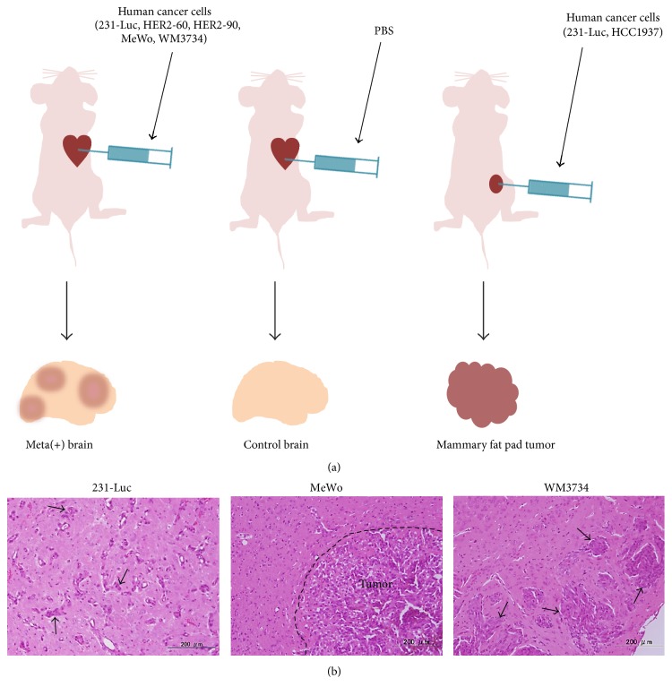
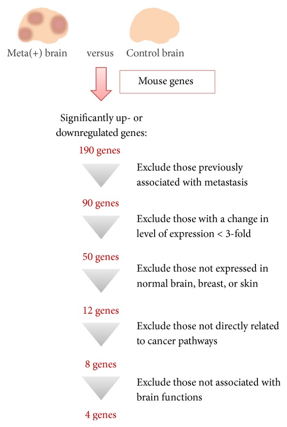
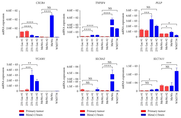
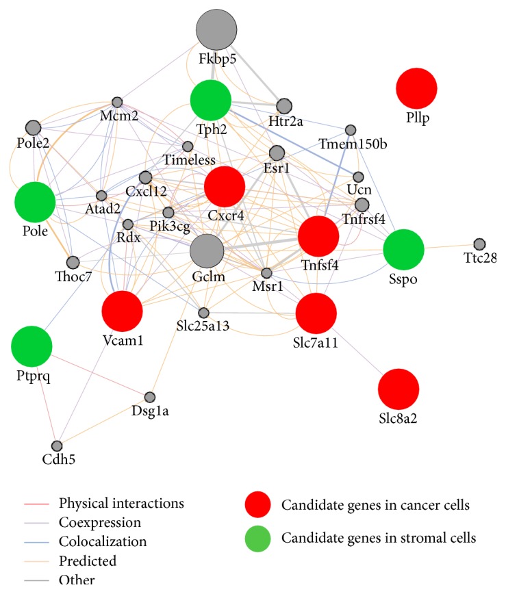
Metastasis is the main cause of treatment failure and death in cancer patients. Metastasis of tumor cells to the brain occurs frequently in individuals with breast cancer, non-small cell lung cancer, or melanoma. Despite recent advances in our understanding of the causes and in the treatment of primary tumors, the biological and molecular mechanisms underlying the metastasis of cancer cells to the brain have remained unclear. Metastasizing cancer cells interact with their microenvironment in the brain to establish metastases. We have now developed mouse models of brain metastasis based on intracardiac injection of human breast cancer or melanoma cell lines, and we have performed RNA sequencing analysis to identify genes in mouse brain tissue and the human cancer cells whose expression is associated specifically with metastasis. We found that the expressions of the mouse genes Tph2, Sspo, Ptprq, and Pole as well as those of the human genes CXCR4, PLLP, TNFSF4, VCAM1, SLC8A2, and SLC7A11 were upregulated in brain tissue harboring metastases. Further characterization of such genes that contribute to the establishment of brain metastases may provide a basis for the development of new therapeutic strategies and consequent improvement in the prognosis of cancer patients.
Conflict of interest statement
The authors declare that there are no competing interests regarding the publication of this paper.
Figures

Similar articles
-
αv-Integrin isoform expression in primary human tumors and brain metastases.Int J Cancer. 2013 Nov 15;133(10):2362-71. doi: 10.1002/ijc.28267. Epub 2013 Jun 10. Int J Cancer. 2013. PMID: 23661241
-
Cystatin C takes part in melanoma-microglia cross-talk: possible implications for brain metastasis.Clin Exp Metastasis. 2018 Aug;35(5-6):369-378. doi: 10.1007/s10585-018-9891-0. Epub 2018 May 2. Clin Exp Metastasis. 2018. PMID: 29722001 Free PMC article.
-
The metastatic microenvironment: Brain-derived soluble factors alter the malignant phenotype of cutaneous and brain-metastasizing melanoma cells.Int J Cancer. 2012 Dec 1;131(11):2509-18. doi: 10.1002/ijc.27552. Epub 2012 Apr 12. Int J Cancer. 2012. PMID: 22447293
-
Epigenomic landscape of melanoma progression to brain metastasis: unexplored therapeutic alternatives.Epigenomics. 2015;7(8):1303-11. doi: 10.2217/epi.15.77. Epub 2015 Dec 7. Epigenomics. 2015. PMID: 26638944 Review.
-
Cell-cell communication characteristics in breast cancer metastasis.Cell Commun Signal. 2024 Jan 19;22(1):55. doi: 10.1186/s12964-023-01418-4. Cell Commun Signal. 2024. PMID: 38243240 Free PMC article. Review.
Cited by
-
Cystine/cysteine metabolism regulates the progression and response to treatment of triple‑negative breast cancer (Review).Oncol Lett. 2024 Aug 30;28(5):521. doi: 10.3892/ol.2024.14654. eCollection 2024 Nov. Oncol Lett. 2024. PMID: 39268159 Free PMC article. Review.
-
Molecular and cellular mechanisms underlying brain metastasis of breast cancer.Cancer Metastasis Rev. 2020 Sep;39(3):711-720. doi: 10.1007/s10555-020-09881-y. Cancer Metastasis Rev. 2020. PMID: 32399646 Free PMC article. Review.
-
Lineage plasticity in cancer: a shared pathway of therapeutic resistance.Nat Rev Clin Oncol. 2020 Jun;17(6):360-371. doi: 10.1038/s41571-020-0340-z. Epub 2020 Mar 9. Nat Rev Clin Oncol. 2020. PMID: 32152485 Free PMC article. Review.
-
The progress of microenvironment-targeted therapies in brain metastases.Front Mol Biosci. 2023 Mar 28;10:1141994. doi: 10.3389/fmolb.2023.1141994. eCollection 2023. Front Mol Biosci. 2023. PMID: 37056723 Free PMC article. Review.
-
Comprehensive molecular biomarker identification in breast cancer brain metastases.J Transl Med. 2017 Dec 29;15(1):269. doi: 10.1186/s12967-017-1370-x. J Transl Med. 2017. PMID: 29287594 Free PMC article.
References
MeSH terms
Substances
LinkOut - more resources
Full Text Sources
Other Literature Sources
Medical
Miscellaneous
