

Autophagy, Metabolism, and Cancer
- PMID: 28209717
- PMCID: PMC5521269
- DOI: 10.1101/sqb.2016.81.030981
Autophagy, Metabolism, and Cancer
Abstract
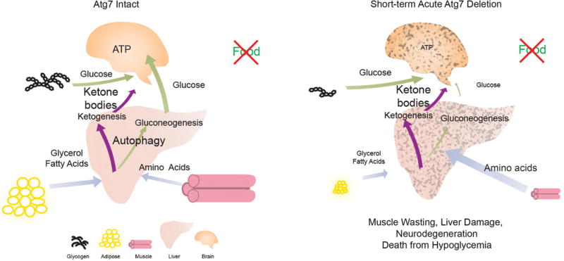
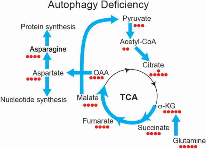
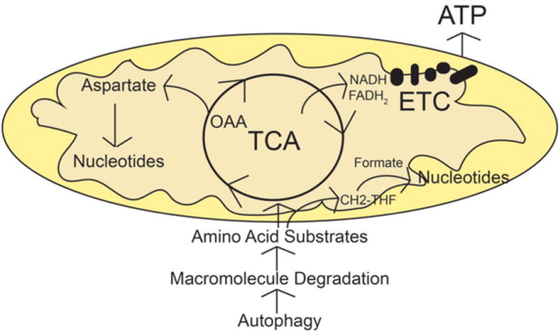
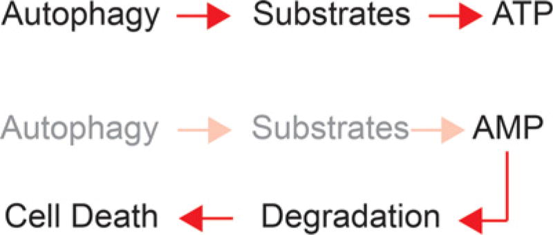
Macroautophagy (autophagy hereafter) is a process that collects cytoplasmic components, particularly mitochondria, and degrades them in lysosomes. In mammalian systems, basal autophagy levels are normally low but are profoundly stimulated by starvation and essential for survival. Cancer cells up-regulate autophagy and can be more autophagy-dependent than most normal tissues. Genetic deficiency in essential autophagy genes in tumors in many autochthonous mouse models for cancer reduces tumor growth. In K-rasG12D-driven non-small cell lung cancer (NSCLC) and other models, autophagy sustains metabolism and survival. The mechanism by which autophagy promotes tumorigenesis varies in different contexts, but evidence points to a critical role for autophagy in sustaining metabolism, thereby preventing p53 activation, energy crisis, growth arrest, apoptosis, senescence, and activation of the immune response. Autophagy in NSCLC preserves mitochondrial quality and regulates their abundance. By degrading macromolecules in lysosomes, autophagy provides mitochondria with substrates to prevent energy crisis and fatal nucleotide pool depletion in starvation. We review here how autophagy supports mammalian survival and how cancer cells usurp this survival mechanism to maintain mitochondrial metabolism for their own benefit. Insights from these studies provide the rationale and approach to target the autophagy survival pathway for cancer therapy.
© 2016 Guo and White; Published by Cold Spring Harbor Laboratory Press.
Conflict of interest statement
E.W. is on the Scientific Advisory Board of Forma Therapeutics.
Figures
Similar articles
-
Autophagy is required for mitochondrial function, lipid metabolism, growth, and fate of KRAS(G12D)-driven lung tumors.Autophagy. 2013 Oct;9(10):1636-8. doi: 10.4161/auto.26123. Epub 2013 Aug 15. Autophagy. 2013. PMID: 23959381 Free PMC article. Review.
-
Autophagy suppresses progression of K-ras-induced lung tumors to oncocytomas and maintains lipid homeostasis.Genes Dev. 2013 Jul 1;27(13):1447-61. doi: 10.1101/gad.219642.113. Genes Dev. 2013. PMID: 23824538 Free PMC article.
-
Autophagy provides metabolic substrates to maintain energy charge and nucleotide pools in Ras-driven lung cancer cells.Genes Dev. 2016 Aug 1;30(15):1704-17. doi: 10.1101/gad.283416.116. Epub 2016 Aug 11. Genes Dev. 2016. PMID: 27516533 Free PMC article.
-
Autophagy, Metabolism, and Cancer.Clin Cancer Res. 2015 Nov 15;21(22):5037-46. doi: 10.1158/1078-0432.CCR-15-0490. Clin Cancer Res. 2015. PMID: 26567363 Free PMC article. Review.
-
Activated Ras requires autophagy to maintain oxidative metabolism and tumorigenesis.Genes Dev. 2011 Mar 1;25(5):460-70. doi: 10.1101/gad.2016311. Epub 2011 Feb 11. Genes Dev. 2011. PMID: 21317241 Free PMC article.
Cited by
-
Autophagy modulates lipid metabolism to maintain metabolic flexibility for Lkb1-deficient Kras-driven lung tumorigenesis.Genes Dev. 2019 Feb 1;33(3-4):150-165. doi: 10.1101/gad.320481.118. Epub 2019 Jan 28. Genes Dev. 2019. PMID: 30692209 Free PMC article.
-
The role of programmed cell death in diabetic foot ulcers.Int Wound J. 2023 Sep 22;21(2):e14399. doi: 10.1111/iwj.14399. Online ahead of print. Int Wound J. 2023. PMID: 37736955 Free PMC article. Review.
-
Mitochondrial homeostasis is maintained in the absence of autophagy.Mol Cell Oncol. 2021 Sep 30;8(5):1984162. doi: 10.1080/23723556.2021.1984162. eCollection 2021. Mol Cell Oncol. 2021. PMID: 34859144 Free PMC article.
-
Potential of antiviral drug oseltamivir for the treatment of liver cancer.Int J Oncol. 2021 Dec;59(6):109. doi: 10.3892/ijo.2021.5289. Epub 2021 Dec 3. Int J Oncol. 2021. PMID: 34859259 Free PMC article.
-
Biological effects of selective COX-2 inhibitor NS398 on human glioblastoma cell lines.Cancer Cell Int. 2020 May 13;20:167. doi: 10.1186/s12935-020-01250-7. eCollection 2020. Cancer Cell Int. 2020. PMID: 32435158 Free PMC article.
References
-
- Degenhardt K, Chen G, Lindsten T, White E. BAX and BAK mediate p53-independent suppression of tumorigenesis. Cancer Cell. 2002;2:193–203. - PubMed
Publication types
MeSH terms
Grants and funding
LinkOut - more resources
Full Text Sources
Other Literature Sources
Medical
Research Materials
Miscellaneous