. 2017 Feb 14;8(1):e02294-16.
doi: 10.1128/mBio.02294-16.

Spatial and Temporal Analysis of Alphavirus Replication and Assembly in Mammalian and Mosquito Cells

Affiliations

Spatial and Temporal Analysis of Alphavirus Replication and Assembly in Mammalian and Mosquito Cells

Joyce Jose et al. mBio. .

Abstract

Sindbis virus (SINV [genus Alphavirus, family Togaviridae]) is an enveloped, mosquito-borne virus. Alphaviruses cause cytolytic infections in mammalian cells while establishing noncytopathic, persistent infections in mosquito cells. Mosquito vector adaptation of alphaviruses is a major factor in the transmission of epidemic strains of alphaviruses. Though extensive studies have been performed on infected mammalian cells, the morphological and structural elements of alphavirus replication and assembly remain poorly understood in mosquito cells. Here we used high-resolution live-cell imaging coupled with single-particle tracking and electron microscopy analyses to delineate steps in the alphavirus life cycle in both the mammalian host cell and insect vector cells. Use of dually labeled SINV in conjunction with cellular stains enabled us to simultaneously determine the spatial and temporal differences of alphavirus replication complexes (RCs) in mammalian and insect cells. We found that the nonstructural viral proteins and viral RNA in RCs exhibit distinct spatial organization in mosquito cytopathic vacuoles compared to replication organelles from mammalian cells. We show that SINV exploits filopodial extensions for virus dissemination in both cell types. Additionally, we propose a novel mechanism for replication complex formation around glycoprotein-containing vesicles in mosquito cells that produced internally released particles that were seen budding from the vesicles by live imaging. Finally, by characterizing mosquito cell lines that were persistently infected with fluorescent virus, we show that the replication and assembly machinery are highly modified, and this allows continuous production of alphaviruses at reduced levels.IMPORTANCE Reemerging mosquito-borne alphaviruses cause serious human epidemics worldwide. Several structural and imaging studies have helped to define the life cycle of alphaviruses in mammalian cells, but the mode of virus replication and assembly in the invertebrate vector and mechanisms producing two disease outcomes in two types of cells are yet to be identified. Using transmission electron microscopy and live-cell imaging with dual fluorescent protein-tagged SINV, we show that while insect and mammalian cells display similarities in entry and exit, they present distinct spatial and temporal organizations in virus replication and assembly. By characterizing acutely and persistently infected cells, we provide new insights into alphavirus replication and assembly in two distinct hosts, resulting in high-titer virus production in mammalian cells and continuous virus production at reduced levels in mosquito cells-presumably a prerequisite for alphavirus maintenance in nature.


Figures

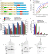
FIG 1
FIG 1
Construction and characterization of FP-tagged viruses. (A) FP-tagged SINV constructs generated for the study. Sequences encoding the fluorescent proteins eYFP and mCherry were cloned into pToto64, a cDNA clone of SINV, as fusion proteins of nsP2, nsP3, or E2. (B) One-step growth curve analysis of FP-tagged viruses from BHK cells. BHK cells were infected with WT or FP-tagged viruses at an MOI of 5, medium was changed every hour for 12 h, and the rate of virus release (PFU per milliliter per hour) was determined using standard plaque assays. (C) Quantitation of the number of virus particles released into the medium for WT and FP-tagged viruses at 4, 6, 8, 10 and 12 h p.i. The total number of genome RNA molecules was determined by qRT-PCR using a standard curve of a known amount of in vitro-transcribed SINV RNA molecules. The PFU in these samples were determined by standard plaque assays of the virus supernatant used collected at 4, 6, 8, 10, and 12 h p.i. from infected BHK cells. (D) Specific infectivity (particle/PFU ratio) of FP-tagged virus calculated from panel C. (E) Western analysis of the cytoplasmic extracts of FP-tagged SINV-infected BHK cell lysates to detect nonstructural protein processing. The blot was probed with anti-nsP2 and anti-nsP4 rabbit polyclonal antibodies. (F) Western analysis of the cytoplasmic extracts of FP-tagged SINV-infected BHK cell lysates. The blot was probed with anti-CP and anti-E2 rabbit polyclonal antibodies. (G) SDS-PAGE analysis of purified FP-tagged and WT virus showing the mCherry protein tagged to the E2 protein.
FIG 2
FIG 2
Replication and growth kinetic analyses of BHK and C6/36 cells infected with WT and mCherry-E2 viruses. Medium over 106 cells or lysates of 106 cells were used for the analysis. Virus-infected cells were lysed with repeated freeze-thaw cycles to recover cell-associated virus, and the numbers of infectious particles in the supernatant and cell-associated viruses were determined by standard plaque assays. Total RNA was extracted from 106 infected cells and culture supernatants, and the total number of viral genome RNA molecules present in the extracts was quantified using qRT-PCR. The number of RNA molecules from virus culture supernatant or cytoplasmic extracts and the PFU associated with the cells and supernatants were plotted for the WT from BHK cells (A), WT from C6/36 cells (B), mCherry-E2 from BHK cells (C), and mCherry-E2 from C6/36 cells (D).
FIG 3
FIG 3
IF analysis of BHK-15 cells infected with FP-tagged viruses showing the distribution of viral replication complexes and glycoprotein E2. Cells infected with FP-tagged SINV were subjected to IF analysis at 6 h p.i. using antibodies against dsRNA (A and C), nsP1 (B), or nsP4 (D and E), as indicated in the figure. The filamentous actin was detected using phalloidin (F).
FIG 4
FIG 4
IF analysis of C6/36 cells infected with FP-tagged viruses. C6/36 cells were infected with FP-tagged viruses and were subjected to IF analysis at 12 h p.i. using antibodies against dsRNA (A and C), nsP1 (B), or nsP4 (D and E), as indicated in the figure. The filamentous actin was detected using phalloidin (F).
FIG 5
FIG 5
TEM analyses of infected BHK cells. Shown are the results of TEM analysis of BHK cells infected with WT and mCherry-E2 virus. BHK cells were infected with WT (A, B, D, and E) or mCherry-E2 (C and F) virus at an MOI of 5 and fixed for TEM analysis at 6 h (A to C) and 12 h (D to F) p.i. Budding viruses (open white arrows), NCs (solid black arrows), and replication spherules (solid white arrows) are marked. Solid white arrowheads indicate CPV-I, and solid black arrowheads indicate CPV-II. Scale bars represent 200 nm. CPV-I does not contain any internally budded particles (A and E).
FIG 6
FIG 6
TEM analysis of mosquito cells infected with WT and mCherry-E2 virus. C6/36 cells were infected with WT (A and D) or mCherry-E2 virus (B, C, E, and F) at an MOI of 5 and fixed for TEM analysis at 12 h (A to C) and 24 h (D and E) p.i. Budding viruses (open white arrows), NCs (solid black arrows), and replication spherules (solid white arrows) are shown. Solid black arrowheads point to internally budded virions. Scale bars represent 200 nm. Large cytopathic vacuoles (diameter of 0.5 to 2 μm) containing replication spherules and internally budded virus particles (A and D) are seen. NCs are also seen at the cytoplasmic side of the vesicles.
FIG 7
FIG 7
Live imaging of FP-tagged viruses. (A) BHK cells infected with nsP3-eYFP/mCherry-E2 dually labeled virus showing the distribution of replication proteins and glycoproteins (representative image from Movie S1). (B) C6/36 cells infected with nsP3-eYFP/mCherry-E2 dually labeled virus showing the localization of nsP3 around large cytopathic vacuoles (representative image from Movie S2). (C) BHK cells transfected with RNA from nonbudding, dually labeled negative control with 400YAL402/AAA mutation in the E2 (representative image from Movie S3) and nonfusing dually labeled negative control (D) with G91D mutation in E1 fusion loop (representative image from Movie S4). (D) Magenta arrows indicate budding viruses, and cyan arrows indicate internalized viruses that are unable to fuse at the endosomes. (E) Live BHK cells infected with nsP2-eYFP/mCherry-E2 virus (green, nsP2-eYFP; red, mCherry-E2) stained with Hoechst stain (nucleus, blue). (F and G) Three-dimensional reconstruction of deconvoluted z-stack images of nsP2-eYFP/mCherry-E2 (F) and nsP3-eYFP/mCherry-E2 (G) virus-infected C6/36 cells. Glycoprotein vesicles are colocalizing with a network of green replication proteins; nsP2 (F [nsP2-eYFP/mCherry-E2]) and nsP3 (G [nsP3-eYFP/mCherry-E2]). (H) Three-dimensional reconstruction of deconvoluted z-stack images of nsP3-eYFP/mCherry-E2 dually labeled virus-infected live BHK and C6/36 (I) cells: green, nsP3-eYFP; red, mCherry-E2; blue, nucleus (Hoechst stain). (J) Live image representing Movie S5 of C6/36 cells infected with mCherry-E2 virus and stained with LysoTracker blue. Green arrows represent LysoTracker blue-positive vesicles, with red glycoprotein-containing endocytic vesicles appearing magenta. (K) Live image representing Movie S6 of C6/36 cells infected with mCherry-E2 virus at 24 h p.i. showing the release of virus from internal cytopathic vacuoles. Virus particles from large cytopathic vacuoles (indicated by green arrows), are transported to the PM and are secreted into the medium by exocytosis. The secreted virus particles stay associated with the filopodial extensions during release (indicated by magenta arrows).
FIG 8
FIG 8
Characterization of persistently infected C6/36 cells. (A) Live image of C6/36 cells persistently infected with mCherry-E2 (a representative image from Movie S7). (B) Three-dimensional reconstruction of deconvoluted z-stack images showing the presence of mCherry-E2 glycoprotein-containing large cytopathic vacuoles in C6/36 cells persistently infected FP-tagged virus. (C and D) Quantification of number of RNA molecules and PFU in C6/36 cells persistently infected with WT (C) or mCherry-E2 (D) virus. C6/36 cells persistently infected with WT virus were plated onto 35-mm culture dishes (106 cells/ml), and virus PFU and RNA were collected from cell culture supernatants and lysed cells at the indicated time points postplating. (E and F) TEM analysis of C6/36 cells persistently infected with WT (Ea to -c) and mCherry-E2 (Fd to -f) virus. Budding virus (open white arrows), budded virions (solid black arrowheads), NCs (solid black arrows), and replication spherules (solid white arrowheads) are shown. Scale bars represent 200 nm for panels Ea to -c and Fb and -c. For panel Fd, the scale bar represents 500 nm. Cytopathic vacuoles that contain internally budded particles are lined with NCs on the cytoplasmic side (Eb and Fd). Budded virus particles associate with the filopodial extension outside the cell similar to acute infection (Eb).
FIG 9
FIG 9
Models of the alphavirus life cycle and the virus-induced structures in mammalian (A) and insect (B) cells. Following attachment and receptor binding (steps 1 and 2), SINV is internalized by clathrin-mediated endocytosis (step 3). Low-pH-mediated fusion (step 4) in the late endosome releases nucleocapsid (NC), and after disassembly (step 5), nonstructural polyproteins are translated (step 6) from viral mRNA. Replication proteins and host proteins along with the viral RNA form replication complexes (step 7) that replicate and transcribe (step 8) viral RNA and induce spherule structures on endosomal and plasma membranes. Internalization of replication spherules from the plasma membrane via vesicles and subsequent fusion of these vesicles with lysosomes generate CPV-I structures. Structural polyprotein translated from the subgenomic RNA (step 9) is processed into capsid protein (CP), and envelope polyproteins that are translocated (step 10) to the ER, processed by signalase (step 11) and glycosylated and transported through the Golgi complex, where fuin cleavage removes E3 from E2 (step 12) to the PM via the secretory pathway. CP binds genome RNA to form NC (step 13) that binds the glycoprotein spikes on the PM and virus buds from PM (step 14). CPV-II structures that contain glycoprotein spikes and attached NCs originate from the Golgi complex and accumulate in the cells at the late stage of infection. In mosquito cells, replication spherules are present only on large internal vesicles that also contain NCs and internally budded virus particles. Glycoprotein-containing vesicles are internalized from the plasma membrane, and fusion of smaller internalized vesicles produces large cytopathic vacuoles. Virus particles bud into the large cytopathic vacuoles (step 15), which also contain replication spherules and nucleocapsid cores. Internally budded virions accumulate in large vesicles. Spherules on the plasma membrane and structurally distinct CPV-I and CPV-II structures are not observed in mosquito cells.

Similar articles

Cited by

References

    1. Raghow RS, Grace TD, Filshie BK, Bartley W, Dalgarno L. 1973. Ross River virus replication in cultured mosquito and mammalian cells: virus growth and correlated ultrastructural changes. J Gen Virol 21:109–122. doi:10.1099/0022-1317-21-1-109. - DOI - PubMed
    1. Strauss JH, Strauss EG. 1994. The alphaviruses: gene expression, replication, and evolution. Microbiol Rev 58:491–562. - PMC - PubMed
    1. Enserink M. 2014. Infectious diseases. Crippling virus set to conquer Western hemisphere. Science 344:678–679. doi:10.1126/science.344.6185.678. - DOI - PubMed
    1. Grimley PM, Berezesky IK, Friedman RM. 1968. Cytoplasmic structures associated with an arbovirus infection: loci of viral ribonucleic acid synthesis. J Virol 2:1326–1338. - PMC - PubMed
    1. Friedman RM, Levin JG, Grimley PM, Berezesky IK. 1972. Membrane-associated replication complex in arbovirus infection. J Virol 10:504–515. - PMC - PubMed

Publication types