. 2017 Apr 21;292(16):6821-6837.
doi: 10.1074/jbc.M116.770362. Epub 2017 Feb 14.

Molecular determinants of the N-terminal acetyltransferase Naa60 anchoring to the Golgi membrane

Affiliations

Molecular determinants of the N-terminal acetyltransferase Naa60 anchoring to the Golgi membrane

Henriette Aksnes et al. J Biol Chem. .

Abstract

Nα-Acetyltransferase 60 (Naa60 or NatF) was recently identified as an unconventional N-terminal acetyltransferase (NAT) because it localizes to organelles, in particular the Golgi apparatus, and has a preference for acetylating N termini of the transmembrane proteins. This knowledge challenged the prevailing view of N-terminal acetylation as a co-translational ribosome-associated process and suggested a new mechanistic functioning for the enzymes responsible for this increasingly recognized protein modification. Crystallography studies on Naa60 were unable to resolve the C-terminal tail of Naa60, which is responsible for the organellar localization. Here, we combined modeling, in vitro assays, and cellular localization studies to investigate the secondary structure and membrane interacting capacity of Naa60. The results show that Naa60 is a peripheral membrane protein. Two amphipathic helices within the Naa60 C terminus bind the membrane directly in a parallel position relative to the lipid bilayer via hydrophobic and electrostatic interactions. A peptide corresponding to the C terminus was unstructured in solution and only folded into an α-helical conformation in the presence of liposomes. Computational modeling and cellular mutational analysis revealed the hydrophobic face of two α-helices to be critical for membranous localization. Furthermore, we found a strong and specific binding preference of Naa60 toward membranes containing the phosphatidylinositol PI(4)P, thus possibly explaining the primary residency of Naa60 at the PI(4)P-rich Golgi. In conclusion, we have defined the mode of cytosolic Naa60 anchoring to the Golgi apparatus, most likely occurring post-translationally and specifically facilitating post-translational N-terminal acetylation of many transmembrane proteins.

Keywords: Golgi; N-terminal acetylation; NAT; Naa60; NatF; PI(4)P; acetylation; acetyltransferase; lipid binding protein; membrane enzyme.


Conflict of interest statement

The authors declare that they have no conflicts of interest with the contents of this article

Figures

Figure 1.
Figure 1.
Secondary structure predictions, simulations, and initial experiments suggest two C-terminal α-helices to mediate Naa60 membrane interactions. A, sequence overview of Naa60 full-length (upper) with a detailed view of the C-terminal region of amino acids 182–242 (lower). The PSIPRED predicted α-helices, herein named “Pred-α1” (aa 190–202) and “Pred-α2” (aa 211–224) are indicated in blue. B, helical wheels for each of Pred-α1 and Pred-α2. The arrow and number inside each wheel correspond to the direction and magnitude of the helices hydrophobic moment, respectively. C and D, structures resulting from the implicit membrane simulations of the Naa60-(185–227) and Naa60-(185–242), respectively, seen from inside the membrane. The plane delimiting the lipid tails from the polar head region of the membrane is light gray, transparent and parallel to the page. The amino acids appearing clearly are those anchored in the hydrophobic region of the membrane model, whereas the other ones appearing with fading colors are either in the head group region or further away from the membrane. The color code for amino acids matches the one in B, but with fewer shades. E, contribution of each amino acid of the Naa60 C terminus (185–242) to the binding energy calculated with the IMM1 membrane model (ΔWAliph, ΔWarom and ΔWpolar, see ”Experimental procedures“). Yellow asterisks indicate those residues that constitute the hydrophobic face of Pred-α1 and Pred-α2 shown in B. F, localization of hNaa60-eGFP and hNaa60-(1–182)-eGFP ectopically expressed in S. cerevisiae. Scale bar, 2 μm.
Figure 2.
Figure 2.
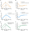
Naa60-(189–242) incorporates in the bilayer through both electrostatic and hydrophobic interactions and adopts helical secondary structure upon membrane interaction. Tryptophan fluorescence spectra (A, C, and E) and circular dichroism (CD) spectra (B, D, and F) of Naa60-(189–242) peptide in the presence (filled line) or absence (dotted line) of various liposomes. A, combining 10 μm Naa60-(189–242) and 180 μm Golgi-like liposomes (52% PC, 20% PE, 15% cholesterol, 8% PI(4)P, and 5% PS) resulted in a blue shift in Trp fluorescence from 355 to 348 nm indicating interaction between Naa60-(189–242) and the Golgi-like liposomes. B, combining 20 μm Naa60-(189–242) and 360 μm Golgi-like liposomes resulted in a characteristic helical spectrum for the peptide. C, combining 10 μm Naa60-(189–242) with 180 μm 100% PC liposomes did not cause a Trp blue shift. D, combining 20 μm Naa60-(189–242) with 360 μm PC liposomes did not affect the CD spectrum of the Naa60 peptide. E, Naa60-(L197A/L201A/L204A) displayed a shift in the λmax from 355 to 348 nm in the presence of Golgi-like liposomes. F, CD spectra of 20 μm Naa60-(L197A/L201A/L204A) with and without 360 μm Golgi-like liposomes. RFI, relative fluorescence intensity.
Figure 3.
Figure 3.
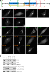
Truncation and deletion constructs of eGFP-tagged Naa60 demonstrated regions in the C-terminal tail important for membrane association in cellulo. HeLa cells were transfected with the indicated eGFP-tagged Naa60 constructs (A) and imaged live (B–D) or subjected to subcellular fractionation followed by high pH/sodium carbonate-mediated protein extraction from membranes (E). A, the sequence of the Naa60 C-terminal tail as shown in Fig. 1, but with the truncated and deleted segments indicated. Red, green, yellow, and orange lines indicate positions of truncations and deletions shown in B–D. B, Naa60-eGFP C-terminal truncated constructs lacking the last 60 (1–182), 26 (1–216), or 17 (1–225) amino acids, compared with the full-length construct (1–242). The truncated segments are indicated with red lines in panel A. Scale bar is 10 μm and is representative for all microscopic images. C, eGFP targeting assay using segments of the Naa60 C-terminal tail attached to the C terminus of eGFP. Segments tested were the last 61 (182–242), 51 (192–242), 41 (202–242), and 26 (217–242) amino acids, each indicated with green lines in A. D, expressed Naa60-eGFP constructs with the indicated segments deleted. Deletions in Pred-α1 are indicated in yellow, whereas deletions in Pred-α2 are indicated in orange, with the corresponding colors used in A to indicate the positions of these segments. E, cells were transfected with the indicated constructs and subjected to high pH/sodium carbonate-mediated protein extraction from membranes following subcellular fractionation and immunoblotting. The peripheral membrane protein GM130 and transmembrane RCAS1 were used as controls for extractable and un-extractable modes of membrane binding, respectively.
Figure 4.
Figure 4.
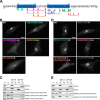
Mutated constructs of Naa60-eGFP revealed amino acids within Pred-α1 and Pred-α2 important for membrane association in cellulo. HeLa cells were transfected with the indicated Naa60-eGFP constructs (A) and imaged live (B and D) or subjected to subcellular fractionation followed by high pH/sodium carbonate-mediated protein extraction from membranes (C and E). A, the sequence of the Naa60 C-terminal tail as described in the legend to Fig. 1. Arrows indicate the positions of mutations shown in B–E, and are color coded accordingly. B, subcellular localization of Naa60-eGFP mutated constructs in the area of Pred-α1 and as indicated with corresponding colors in A. Scale bar is 10 μm and is representative for all microscopic images. C, cells were transfected with the indicated constructs and subjected to high pH/sodium carbonate-mediated protein extraction from membranes following subcellular fractionation and immunoblotting. The peripheral membrane protein GM130 and transmembrane RCAS1 were used as controls for extractable and un-extractable modes of membrane binding. D, as B, but for the area of Pred-α2. E, as C, but for the area of Pred-α2.
Figure 5.
Figure 5.
Effect of the membrane composition on the position of the Naa60 C terminus (185–242) and its interaction with lipids. Three lipid bilayers were considered POPC:PI(4)P, 92:8; POPC:POPS, 75:25; POPC:PSM:CHOL, 70:20:10, in atomistic simulations with the Naa60 C terminus (185–227). A, electron density along the normal to the membrane for the PC lipids (solid lines) and the Naa60 C terminus helices (185–227, dashed line) show that the presence of PI or PS lipids results in a deeper position of the protein. Zero corresponds to the plane between the two lipid layers. B, average number of hydrophobic contacts per trajectory frame between the lipid tails and the two helices (185–227). C and D, snapshot from the atomistic simulation of Naa60-(185–242) on a POPC:PI(4)P bilayer. Helices are shown in the same color scheme as in Fig. 1. C, side view showing that the helices are inserted beyond the phosphate atoms (yellow/tan spheres) of the lipids (PC in gray and PI(4)P in cyan). D, top view showing only the PI(4)P lipids of the upper leaflet. Four of the eight PI(4)P lipids are interacting with the side chains of charged amino acids (blue, stick).
Figure 6.
Figure 6.
Br-PC quenching assay with Naa60 peptides indicate peripheral binding to Golgi-like liposomes. A, WT Naa60-(189–242) peptide (W227) and mutated Naa60-(189–242) peptides (Y214W/W227F, I209W/W227F, or Y193W/W227F) were incubated with Br-Golgi-like liposomes (52% Br-PC, 20% PE, 15% cholesterol, 8% PI(4)P, and 5% PS). Vesicles contained PC molecules carrying a quenching Br-group at either position (11,12), (9,10), (6,7), or (4,5) in the acyl chain. The fluorescence measured for vesicles binding to the brominated vesicles (F) was divided by the fluorescence for vesicles binding to the non-brominated vesicles (F0). n = 2 per peptide. B, based on data in A, the position of Trp residues relative to the center of the bilayer was calculated using the parallax method (34).
Figure 7.
Figure 7.
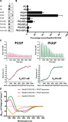
Pulldown ITC and CD experiments demonstrate the Naa60-(189–242) peptide to have a binding preference for PI(4)P. A, pulldown of Naa60-(189–242) using liposomes with 92 mol % PC and 8 mol % of either PI. Peptide and liposomes were incubated at room temperature before pelleting at 200,000 × g into supernatant (S) and pellet (P) fractions. Naa60-(189–242) was detected by means of a dot blot. n = 3–8 per vesicle type. Antibody was validated for detection of the peptide (supplemental Fig. S3). B, dot blot intensities of the pellet fractions in A were quantified (3 replications averaged) using ImageJ and expressed as percentage of bound Naa60 peptide based on P and S intensities. C, ITC experiments measuring dissociation constant for Naa60-(189–242) peptide with either PC-PI(3)P or PC-PI(4)P liposomes. Naa60-(189–242) peptide (50 μm) was injected in 20 successive injections of 2 μl with 180-s intervals into a total lipid concentration of 100 μm and heat rate was measured (upper). Dissociation constants were calculated based on peak area/molar ratio plots (lower). D, far-UV CD spectra of Naa60-(189–242) (20 μm) in the presence of 360 μm liposomes containing 92 mol % PC and 8 mol % of either PI(3)P or PI(4)P. Naa60-(189–242) with no liposomes (yellow), PI(3)P (pink), and PI(4)P (green). Spectra recorded between 180 and 260 nm.
Figure 8.
Figure 8.
Model of Naa60 membrane association and N-terminal acetylation at the membrane. Shown is the structure of Naa60 (PDB code 5ICV) attached to the C-terminal α-helix predictied by PSIPRED. Only the first of the two amphipathic α-helices is shown. The helical anchor dipping horizontally into the membrane positions the enzymatic site of Naa60 available to acetylate N termini of transmembrane proteins. Whether Naa60 makes additional contact points with the membrane besides its two C-terminal helices remains to be revealed.

Similar articles

Cited by

References

    1. Van Damme P., Hole K., Pimenta-Marques A., Helsens K., Vandekerckhove J., Martinho R. G., Gevaert K., and Arnesen T. (2011) NatF contributes to an evolutionary shift in protein N-terminal acetylation and is important for normal chromosome segregation. PLoS Genet. 7, e1002169. - PMC - PubMed
    1. Aksnes H., Drazic A., Marie M., and Arnesen T. (2016) First things first: vital protein marks by N-terminal acetyltransferases. Trends Biochem. Sci. 41, 746–760 - PubMed
    1. Drazic A., Myklebust L. M., Ree R., and Arnesen T. (2016) The world of protein acetylation. Biochim. Biophys. Acta 1864, 1372–1401 - PubMed
    1. Polevoda B., Brown S., Cardillo T. S., Rigby S., and Sherman F. (2008) Yeast N(α)-terminal acetyltransferases are associated with ribosomes. J. Cell Biochem. 103, 492–508 - PubMed
    1. Gautschi M., Just S., Mun A., Ross S., Rücknagel P., Dubaquié Y., Ehrenhofer-Murray A., and Rospert S. (2003) The yeast Nα-acetyltransferase NatA is quantitatively anchored to the ribosome and interacts with nascent polypeptides. Mol. Cell Biol. 23, 7403–7414 - PMC - PubMed

MeSH terms

Associated data

LinkOut - more resources