A process engineering approach to increase organoid yield
- PMID: 28174251
- PMCID: PMC5358111
- DOI: 10.1242/dev.142919
A process engineering approach to increase organoid yield
Abstract
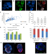
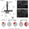
Temporal manipulation of the in vitro environment and growth factors can direct differentiation of human pluripotent stem cells into organoids - aggregates with multiple tissue-specific cell types and three-dimensional structure mimicking native organs. A mechanistic understanding of early organoid formation is essential for improving the robustness of these methods, which is necessary prior to use in drug development and regenerative medicine. We investigated intestinal organoid emergence, focusing on measurable parameters of hindgut spheroids, the intermediate step between definitive endoderm and mature organoids. We found that 13% of spheroids were pre-organoids that matured into intestinal organoids. Spheroids varied by several structural parameters: cell number, diameter and morphology. Hypothesizing that diameter and the morphological feature of an inner mass were key parameters for spheroid maturation, we sorted spheroids using an automated micropipette aspiration and release system and monitored the cultures for organoid formation. We discovered that populations of spheroids with a diameter greater than 75 μm and an inner mass are enriched 1.5- and 3.8-fold for pre-organoids, respectively, thus providing rational guidelines towards establishing a robust protocol for high quality intestinal organoids.
Keywords: Aggregates; Intestinal; Organoids; Process engineering; Sorting; Yield.
© 2017. Published by The Company of Biologists Ltd.
Conflict of interest statement
The authors declare no competing or financial interests.
Figures


Similar articles
-
3D heterogeneous islet organoid generation from human embryonic stem cells using a novel engineered hydrogel platform.Biomaterials. 2018 Sep;177:27-39. doi: 10.1016/j.biomaterials.2018.05.031. Epub 2018 May 25. Biomaterials. 2018. PMID: 29883914
-
Generation of Gastrointestinal Organoids from Human Pluripotent Stem Cells.Methods Mol Biol. 2017;1597:167-177. doi: 10.1007/978-1-4939-6949-4_12. Methods Mol Biol. 2017. PMID: 28361317
-
In vitro generation of human pluripotent stem cell derived lung organoids.Elife. 2015 Mar 24;4:e05098. doi: 10.7554/eLife.05098. Elife. 2015. PMID: 25803487 Free PMC article.
-
The case for applying tissue engineering methodologies to instruct human organoid morphogenesis.Acta Biomater. 2017 May;54:35-44. doi: 10.1016/j.actbio.2017.03.023. Epub 2017 Mar 16. Acta Biomater. 2017. PMID: 28315813 Free PMC article. Review.
-
From Spheroids to Organoids: The Next Generation of Model Systems of Human Cardiac Regeneration in a Dish.Int J Mol Sci. 2021 Dec 7;22(24):13180. doi: 10.3390/ijms222413180. Int J Mol Sci. 2021. PMID: 34947977 Free PMC article. Review.
Cited by
-
Everything You Always Wanted to Know About Organoid-Based Models (and Never Dared to Ask).Cell Mol Gastroenterol Hepatol. 2022;14(2):311-331. doi: 10.1016/j.jcmgh.2022.04.012. Epub 2022 May 25. Cell Mol Gastroenterol Hepatol. 2022. PMID: 35643188 Free PMC article. Review.
-
Gastric Organoids: Progress and Remaining Challenges.Cell Mol Gastroenterol Hepatol. 2022;13(1):19-33. doi: 10.1016/j.jcmgh.2021.09.005. Epub 2021 Sep 20. Cell Mol Gastroenterol Hepatol. 2022. PMID: 34547535 Free PMC article. Review.
-
A comprehensive review on 3D tissue models: Biofabrication technologies and preclinical applications.Biomaterials. 2024 Jan;304:122408. doi: 10.1016/j.biomaterials.2023.122408. Epub 2023 Nov 27. Biomaterials. 2024. PMID: 38041911 Free PMC article. Review.
-
Differential Effects of Extracellular Vesicles of Lineage-Specific Human Pluripotent Stem Cells on the Cellular Behaviors of Isogenic Cortical Spheroids.Cells. 2019 Aug 28;8(9):993. doi: 10.3390/cells8090993. Cells. 2019. PMID: 31466320 Free PMC article.
-
Review: Synthetic scaffolds to control the biochemical, mechanical, and geometrical environment of stem cell-derived brain organoids.APL Bioeng. 2018 Nov 15;2(4):041501. doi: 10.1063/1.5045124. eCollection 2018 Dec. APL Bioeng. 2018. PMID: 31069322 Free PMC article. Review.
References
-
- Anis Y. H., Holl M. R. and Meldrum D. R. (2010). Automated selection and placement of single cells using vision-based feedback control. IEEE Trans. Automat. Sci. Eng. 7, 598-606. 10.1109/TASE.2009.2035709 - DOI
-
- Boehnke K., Iversen P. W., Schumacher D., Lallena M. J., Haro R., Amat J., Haybaeck J., Liebs S., Lange M., Schafer R. et al. (2016). Assay establishment and validation of a high-throughput screening platform for three-dimensional patient-derived colon cancer organoid cultures. J. Biomol. Screen. 21, 931-941. 10.1177/1087057116650965 - DOI - PMC - PubMed
Publication types
MeSH terms
Grants and funding
LinkOut - more resources
Full Text Sources
Other Literature Sources

