. 2017 Mar 6;216(3):709-722.
doi: 10.1083/jcb.201605002. Epub 2017 Feb 7.

The VDAC2-BAK axis regulates peroxisomal membrane permeability

Affiliations

The VDAC2-BAK axis regulates peroxisomal membrane permeability

Ken-Ichiro Hosoi et al. J Cell Biol. .

Abstract

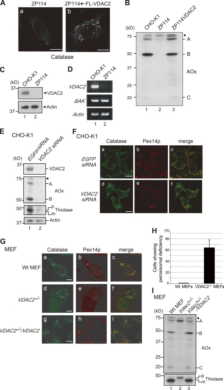
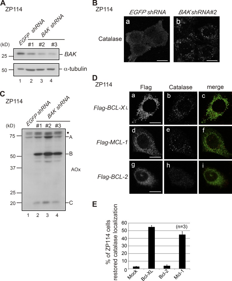
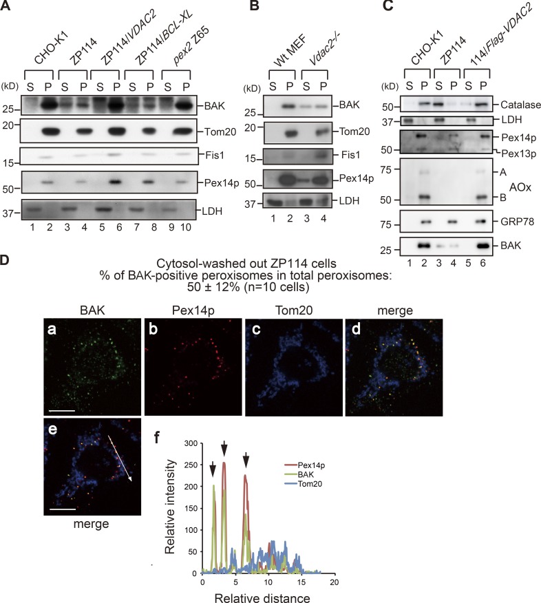
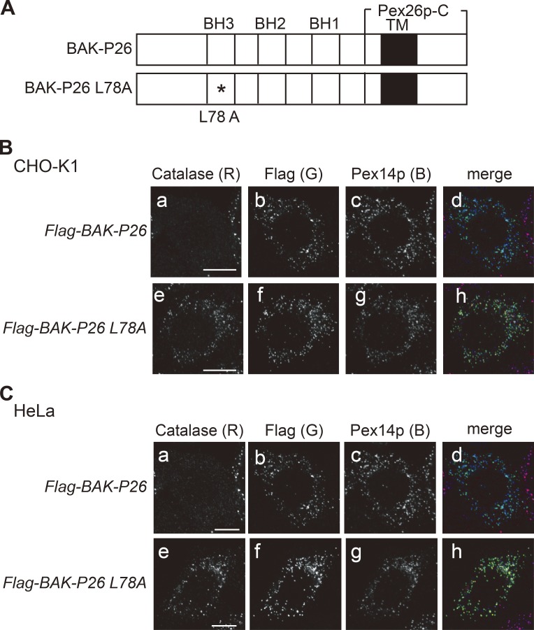
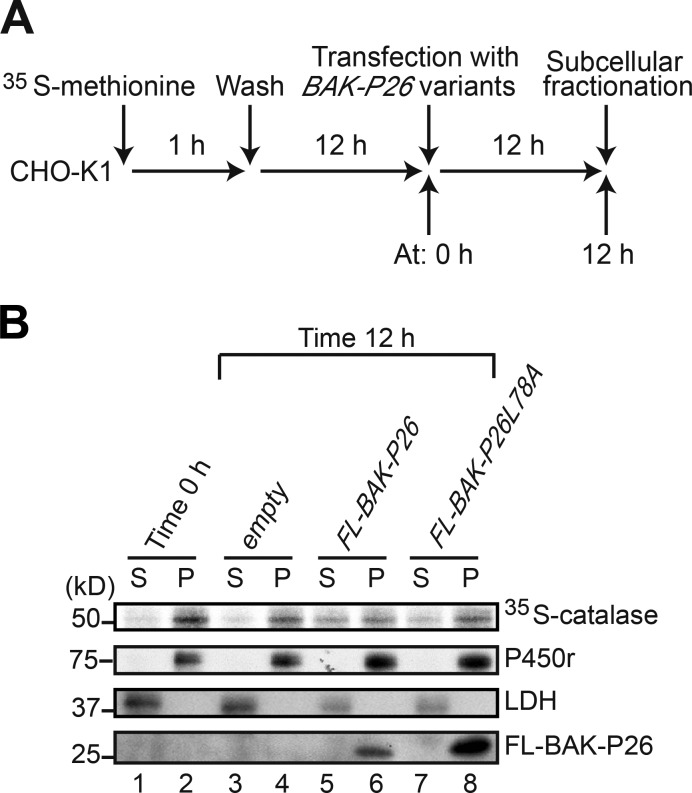
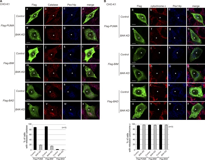
Peroxisomal biogenesis disorders (PBDs) are fatal genetic diseases consisting of 14 complementation groups (CGs). We previously isolated a peroxisome-deficient Chinese hamster ovary cell mutant, ZP114, which belongs to none of these CGs. Using a functional screening strategy, VDAC2 was identified as rescuing the peroxisomal deficiency of ZP114 where VDAC2 expression was not detected. Interestingly, knockdown of BAK or overexpression of the BAK inhibitors BCL-XL and MCL-1 restored peroxisomal biogenesis in ZP114 cells. Although VDAC2 is not localized to the peroxisome, loss of VDAC2 shifts the localization of BAK from mitochondria to peroxisomes, resulting in peroxisomal deficiency. Introduction of peroxisome-targeted BAK harboring the Pex26p transmembrane region into wild-type cells resulted in the release of peroxisomal matrix proteins to cytosol. Moreover, overexpression of BAK activators PUMA and BIM permeabilized peroxisomes in a BAK-dependent manner. Collectively, these findings suggest that BAK plays a role in peroxisomal permeability, similar to mitochondrial outer membrane permeabilization.


Figures

Figure 1.
Figure 1.
VDAC2 deficiency leads to peroxisomal dysfunction. (A) ZP114 cells were mock transfected (a) or transfected with Flag-VDAC2 (b). After 48 h, cells were fixed and immunostained with anticatalase antibody. FL, Flag. (B) Total cell lysates from CHO-K1 cells, ZP114 cells, and ZP114 cells transfected with Flag-VDAC2 were analyzed by SDS-PAGE and Western blotting with an anti-AOx antibody. A full-length 75-kD AOx-A chain and intraperoxisomal processed 52-kD B and 22-kD C chains are indicated. (C) Total cell lysates from CHO-K1 and ZP114 cells were analyzed by Western blotting with antibodies to VDAC2 (top) and actin (bottom). (D) Total RNA was prepared from wild-type CHO-K1 cells and ZP114 cells. RT-PCR was performed with a set of primers for VDAC2, BAK, and Actin. (E) CHO-K1 cells transfected with EGFP siRNA and VDAC2 siRNA were subjected to Western blotting with antibodies to VDAC2, AOx, thiolase, and actin. AOx bands A and B are as in panel B; p and m denote a larger precursor and mature forms of thiolase, respectively. (F) CHO-K1 cells transfected with EGFP siRNA and VDAC2 siRNA were immunostained with antibodies to catalase and Pex14p. Merged views of panels a plus b and panels d plus e are shown in c and f, respectively. (G) Normal control wild-type (Wt) MEFs, Vdac2−/− MEFs, and Vdac2−/− MEFs transfected with VDAC2 (Vdac2−/−/VDAC2) were immunostained with antibodies to catalase and Pex14p. Merged views of panels a plus b, panels d plus e, and panels g plus h are shown in c, f, and i, respectively. Bars: (A and F) 10 µm; (G) 20 µm. (H) Normal control wild-type MEFs and Vdac2−/− MEFs lacking peroxisomal structure as assessed by immunostaining with anti-Pex14p and anticatalase antibodies are counted (200 cells each). The percentages of the cells showing abnormal peroxisome assembly are presented. Data represent means ± SD. n = 3. (I) Total cell lysates from control wild-type MEFs, Vdac2−/− MEFs, and Vdac2−/−/VDAC2 MEFs were analyzed by Western blotting with antibodies to AOx and thiolase. Note that peroxisomal AOx conversion from the A chain to B and C chains and processing of the larger precursor to mature thiolase were restored in Vdac2−/−/VDAC2 MEFs (lane 3). (B, E, and I) Dots indicate nonspecific bands.
Figure 2.
Figure 2.
BAK inactivation restores peroxisome biogenesis in ZP114 cells. (A) The effect of BAK RNA interference was verified by Western blotting. ZP114 cells transfected with EGFP shRNA and each of three different BAK shRNA plasmids were analyzed by Western blotting with antibodies to BAK and α-tubulin. (B) ZP114 cells transfected with EGFP shRNA and BAK shRNA were immunostained with anticatalase antibody. Note that numerous peroxisomes are detectable in b. (C) ZP114 cells transfected with EGFP shRNA and three BAK shRNA plasmids were subjected to Western blotting with anti-AOx antibody as described in Fig. 1 B. A, B, and C denote A, B, and C chains, respectively; the dot indicates nonspecific bands. (D) ZP114 cells were transfected with cDNAs each for Flag–BCL-XL, Flag–MCL-1, and Flag–BCL-2. The cells were immunostained with antibodies to Flag and catalase. Merged views of panels a plus b, panels d plus e, and panels g plus h are shown in c, f, and i, respectively. (B and D) Bars, 10 µm. (E) Percentages of the cells restored in the catalase and the localization of catalase to peroxisomes in total cells expressing Flag-tagged proteins (100 cells each) are presented. Data represent means ± SD.
Figure 3.
Figure 3.
BAK is localized to peroxisomes and the cytosol in VDAC2-deficient cells. (A) Various CHO cells indicated at the top were homogenized and fractionated into cytosol (S) and organelle (P) fractions. Equal aliquots of cytosol and organelle fractions were analyzed by SDS-PAGE and Western blotting with antibodies against BAK, Tom20, Fis1, Pex14p, and lactate dehydrogenase (LDH). (B) Normal control wild-type (Wt) MEFs and Vdac2−/− MEFs were analyzed as in A. (C) CHO-K1, ZP114, and Flag-VDAC2–transfected ZP114 were likewise analyzed as in A using antibodies to respective proteins indicated on the right. A and B denote AOx-A and -B chains, respectively. (D) After permeabilization with digitonin and washing out the cytosol, ZP114 cells were fixed and immunostained with antibodies to BAK, Pex14p, and Tom20. Merged views of a–c are shown in d and e. Signal intensities of BAK, Pex14p, and Tom20 along the arrow in e are plotted in f. Downward arrows in f indicate the superimposed points where BAK is colocalized with Pex14p, but not with Tom20. Bars, 10 µm.
Figure 4.
Figure 4.
Peroxisome-targeted BAK abrogates peroxisome biogenesis. (A) Schematic presentation of peroxisome-targeting BAK-P26. The asterisk designates an L78A mutation in the BH3 domain of BAK. Solid bars indicate a transmembrane segment (TM). (B and C) Flag–BAK-P26 and Flag–BAK-P26–L78A were expressed in CHO-K1 (B) and HeLa (C) cells. The cells were immunostained with antibodies to catalase, Flag, and Pex14p. Merged views of a–c and e–g are shown in d and h, respectively, where three fluorescent colors, red (R), green (G), and blue (B), were merged. Bars, 10 µm.
Figure 5.
Figure 5.
Peroxisome-targeted BAK releases catalase from peroxisomes to cytosol. (A) Time flow of pulse-chase experimentation of catalase translocation. (B) Cells at 0 h (before transfection) and 12 h after transfection with the indicated plasmids (time 12 h) were fractionated into cytosol (S) and organelle (P) fractions. Equal aliquots of cytosol and organelle fractions were solubilized, and catalase was immunoprecipitated with anticatalase antibody. [35S]methionine– and [35S]cysteine–labeled catalase was detected by a Fujix FLA-5000 autoimaging analyzer. As marker proteins for cytosol and the ER, lactate dehydrogenase (LDH) and P450 reductase (P450r) in S and P fractions were detected with the respective antibodies. BAK-P26 variants were detected with anti-Flag antibody.
Figure 6.
Figure 6.
BAK localizes to peroxisomes and regulates catalase latency in wild-type cells. (A, left) The PNS fraction from HeLa cells was separated by ultracentrifugation on an iodixanol density gradient into 12 fractions. Same-volume aliquots of gradient fractions and the PNS fraction (one twentieth of the loaded amount) were analyzed by Western blotting with antibodies against catalase, Pex14p, Tom20, and GRP78. (Right) The PNS fraction, along with fractions (Frc.) 1, 6, and 10 representing the ER, mitochondria, and peroxisomes, respectively, were likewise analyzed by Western blotting with antibodies against catalase, Pex14p, BAK, Tom20, and GRP78. (B) CHO-K1 cells stably expressing empty vector (control) and two independent BAK shRNA vectors (BAK-KD#1 and #2) were analyzed by Western blotting with antibodies to BAK and actin. (C, left) Control and BAK-KD cells were treated with digitonin to selectively permeabilize the plasma membrane. Permeabilized cells were centrifuged to separate into cytosol (S) and organelle (P) fractions. Equal aliquots of S and P fractions were analyzed by Western blotting with antibodies to proteins, including peroxisomal matrix proteins, catalase, AOx, and ADAPS, as indicated. (Right) Catalase in cytosol and organelle fractions from control and BAK-KD cells was quantified and shown as ratios of cytosol/total (cytosol plus organelle). (D) Control, BAK-KD, and pex2 Z65 cells were treated with digitonin at the indicated concentrations, and free catalase activity was determined in the isotonic medium. Free catalase activity is shown as a percentage of the total catalase activity detected in the presence of 1% Triton X-100. (C and D) Data represent means ± SD. n = 3.
Figure 7.
Figure 7.
Overexpression of BH3-only proteins PUMA and BIM, but not of BAD, disrupts peroxisomes in a BAK-dependent manner. (A and B) Control and BAK-KD#2 cells (see Fig. 6, B–D) were transfected with Flag-PUMA, Flag-BIM, and Flag-BAD in the presence of a caspase inhibitor, Q-VD-Oph. At 48 h after transfection, cells were immunostained with antibodies to Flag, Pex14p, and catalase (A) or cytochrome c (B). Asterisks indicate respective transfectants. Merged views of a–c, e–g, i–k, m–o, q–s, and u–w are shown in d, h, l, p, t, and x, respectively. Bars, 10 µm. Bottom graphs show percentages of the cells showing released catalase (A) or cytochrome c (B) in cells expressing Flag-tagged proteins. Data represent means ± SD.
Figure 8.
Figure 8.
Peroxisome-deficient cell mutants show resistance to exogenous H2O2. CHO-K1, pex14 ZP161, and pex2 Z65 cells were treated with 1 mM H2O2 in the presence and absence of 50 mM 3-AT, a catalase inhibitor. Cell viability was determined by MTT assay and represented as percentages relative to those of mock-treated, H2O2-untreated cells. Data represent means ± SD.

Similar articles

Cited by

References

    1. Chen H.C., Kanai M., Inoue-Yamauchi A., Tu H.C., Huang Y., Ren D., Kim H., Takeda S., Reyna D.E., Chan P.M., et al. . 2015. An interconnected hierarchical model of cell death regulation by the BCL-2 family. Nat. Cell Biol. 17:1270–1281. - PMC - PubMed
    1. Cheng E.H.-Y.A., Wei M.C., Weiler S., Flavell R.A., Mak T.W., Lindsten T., and Korsmeyer S.J.. 2001. BCL-2, BCL-XL sequester BH3 domain-only molecules preventing BAX- and BAK-mediated mitochondrial apoptosis. Mol. Cell. 8:705–711. 10.1016/S1097-2765(01)00320-3 - DOI - PubMed
    1. Cheng E.H.-Y., Sheiko T.V., Fisher J.K., Craigen W.J., and Korsmeyer S.J.. 2003. VDAC2 inhibits BAK activation and mitochondrial apoptosis. Science. 301:513–517. 10.1126/science.1083995 - DOI - PubMed
    1. Czabotar P.E., Lessene G., Strasser A., and Adams J.M.. 2014. Control of apoptosis by the BCL-2 protein family: implications for physiology and therapy. Nat. Rev. Mol. Cell Biol. 15:49–63. 10.1038/nrm3722 - DOI - PubMed
    1. Dewson G., Kratina T., Sim H.W., Puthalakath H., Adams J.M., Colman P.M., and Kluck R.M.. 2008. To trigger apoptosis, Bak exposes its BH3 domain and homodimerizes via BH3:groove interactions. Mol. Cell. 30:369–380. 10.1016/j.molcel.2008.04.005 - DOI - PubMed

Publication types

Substances

Supplementary concepts