. 2017 Feb 22;93(4):822-839.e6.
doi: 10.1016/j.neuron.2017.01.008. Epub 2017 Feb 2.

A Brainstem-Spinal Cord Inhibitory Circuit for Mechanical Pain Modulation by GABA and Enkephalins

Affiliations

A Brainstem-Spinal Cord Inhibitory Circuit for Mechanical Pain Modulation by GABA and Enkephalins

Amaury François et al. Neuron. .

Abstract

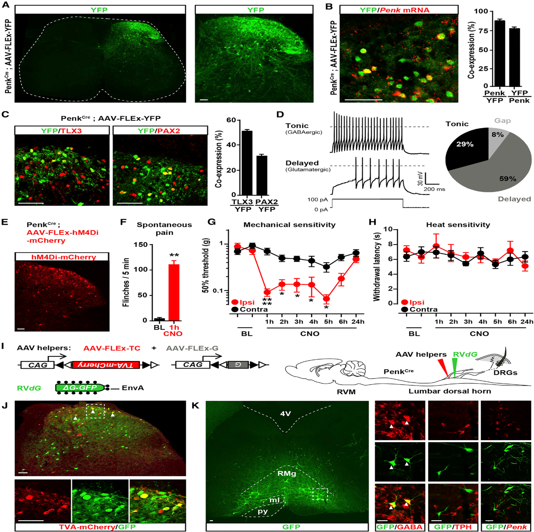
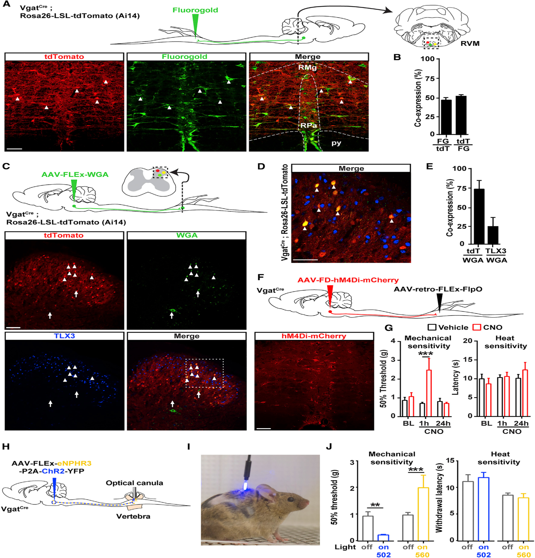
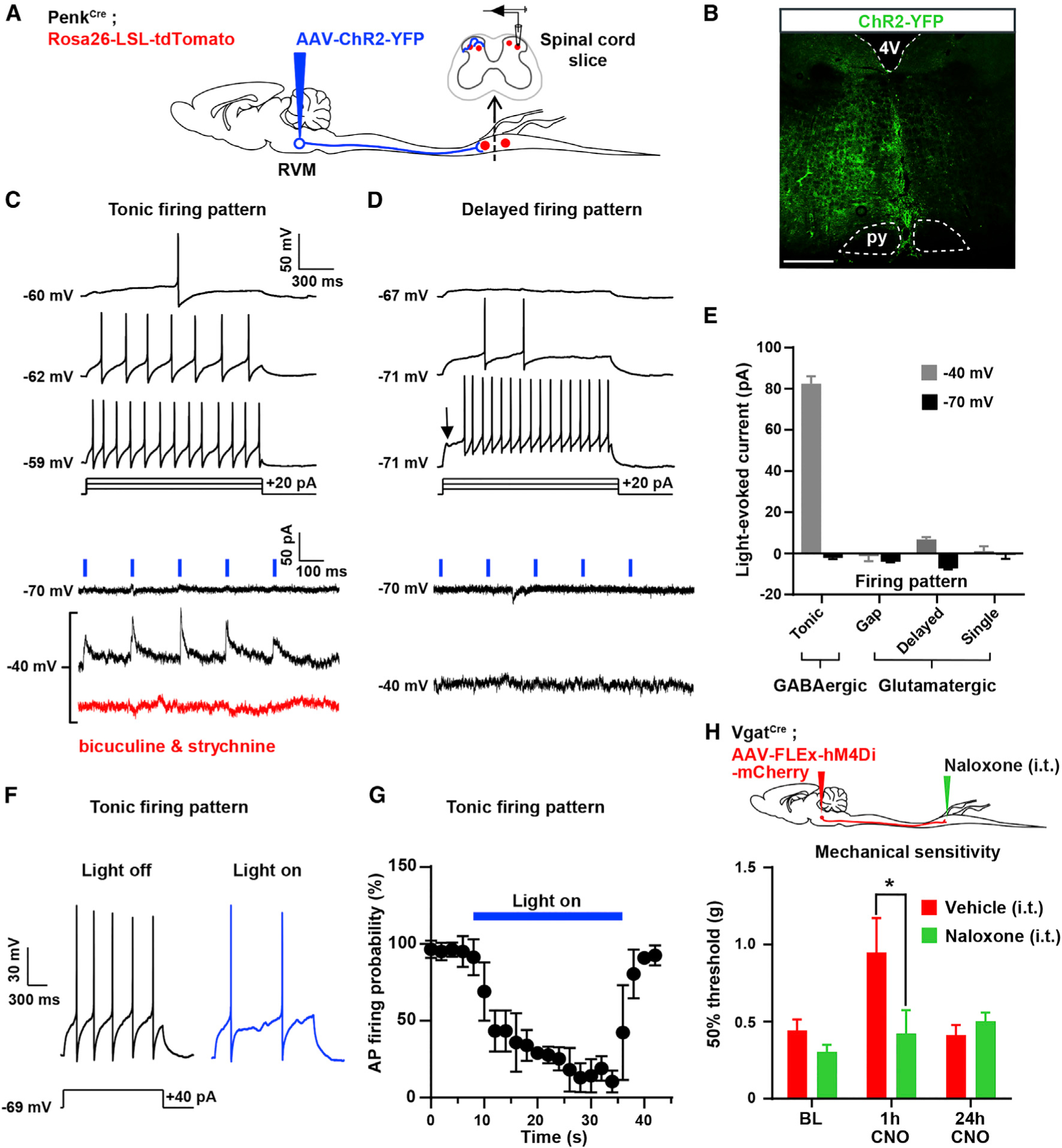
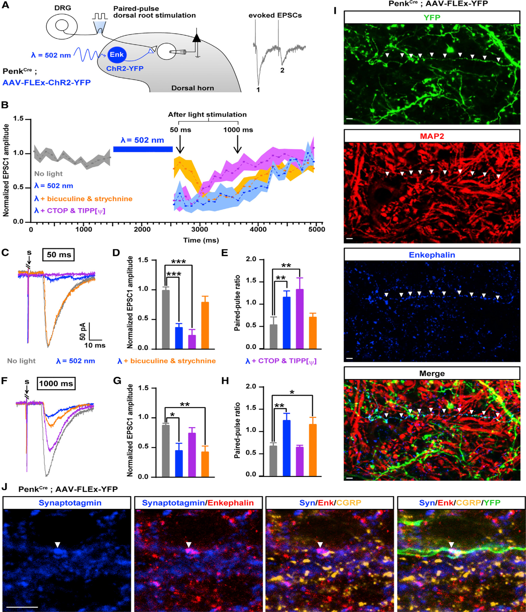
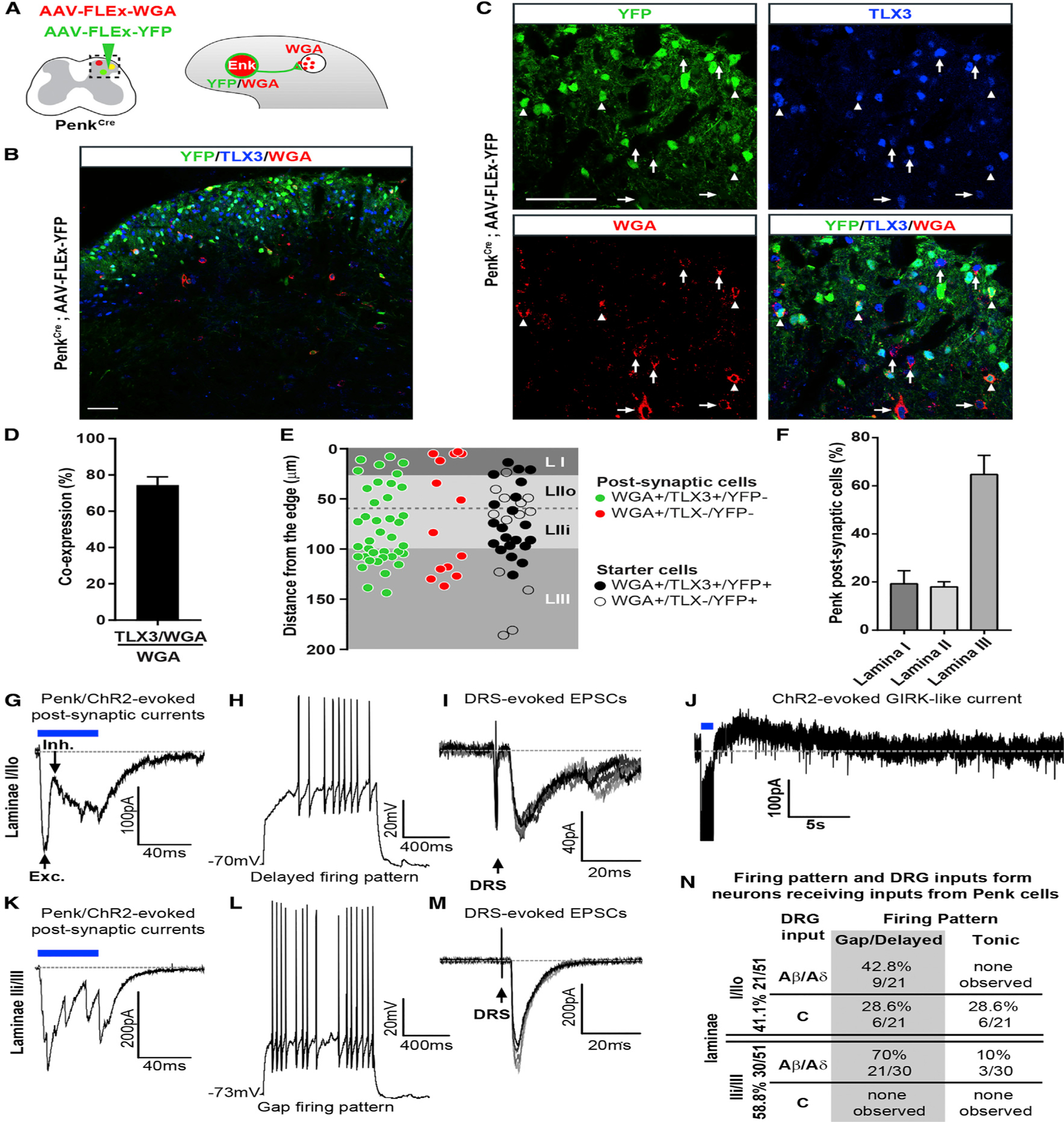
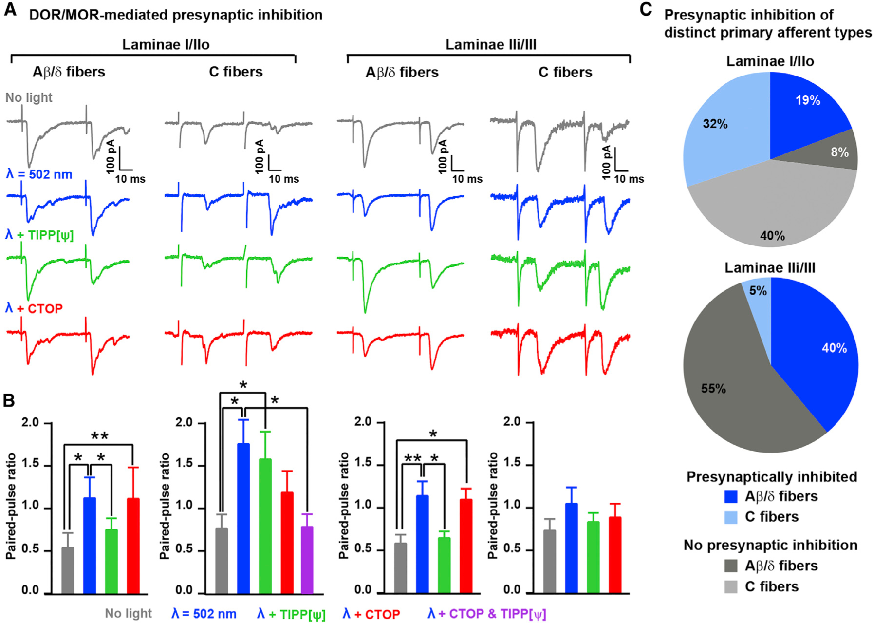
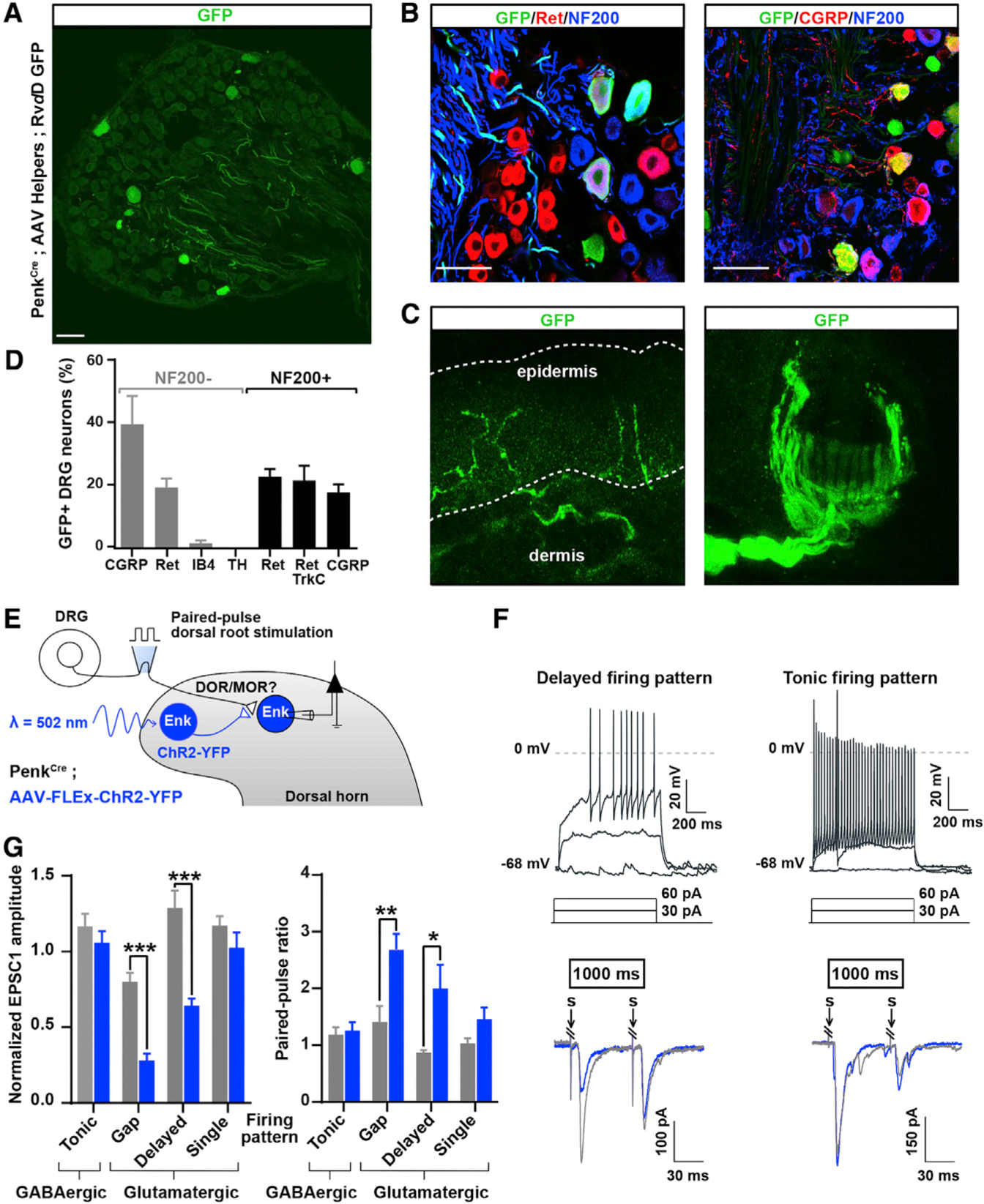
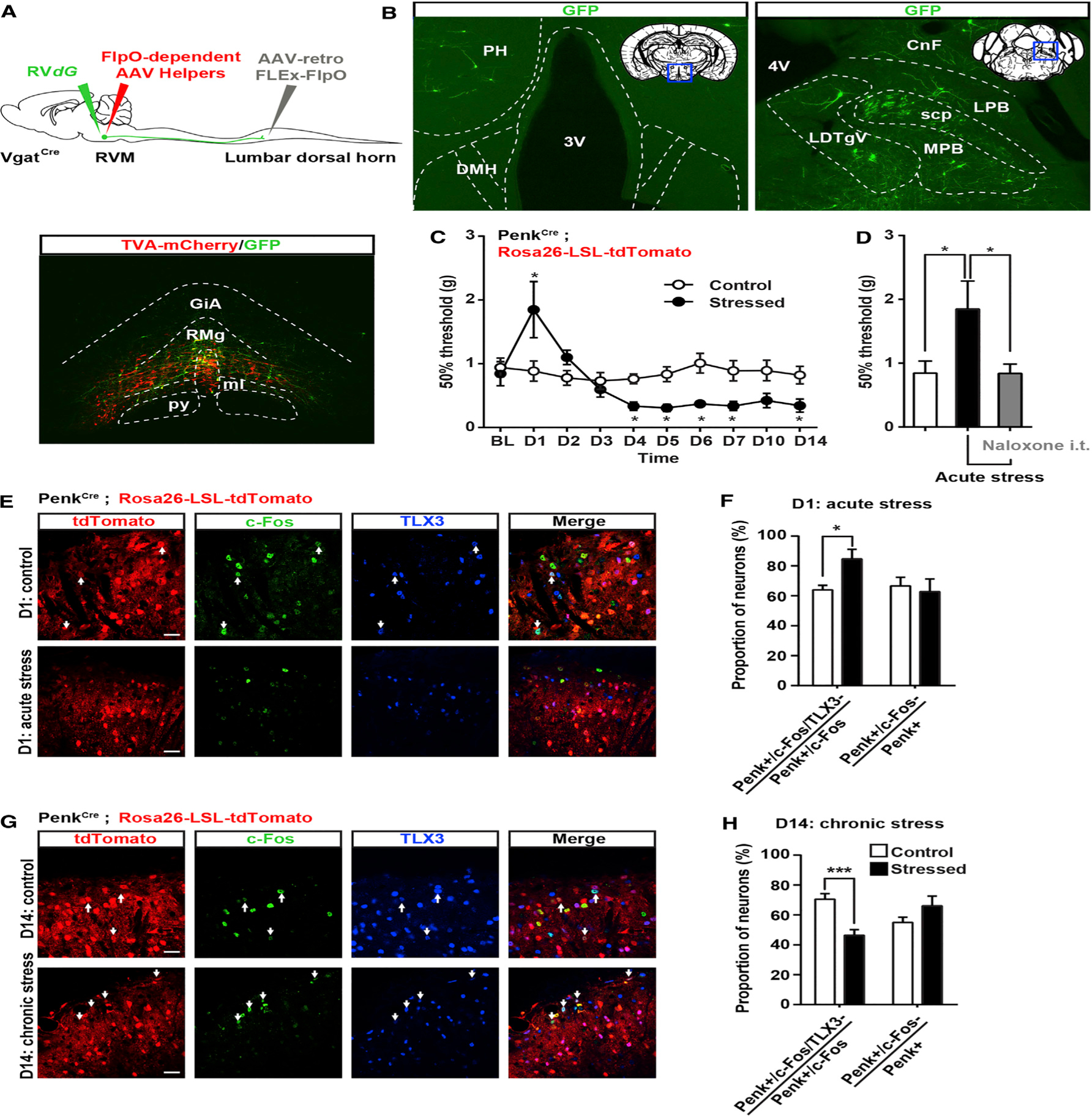
Pain thresholds are, in part, set as a function of emotional and internal states by descending modulation of nociceptive transmission in the spinal cord. Neurons of the rostral ventromedial medulla (RVM) are thought to critically contribute to this process; however, the neural circuits and synaptic mechanisms by which distinct populations of RVM neurons facilitate or diminish pain remain elusive. Here we used in vivo opto/chemogenetic manipulations and trans-synaptic tracing of genetically identified dorsal horn and RVM neurons to uncover an RVM-spinal cord-primary afferent circuit controlling pain thresholds. Unexpectedly, we found that RVM GABAergic neurons facilitate mechanical pain by inhibiting dorsal horn enkephalinergic/GABAergic interneurons. We further demonstrate that these interneurons gate sensory inputs and control pain through temporally coordinated enkephalin- and GABA-mediated presynaptic inhibition of somatosensory neurons. Our results uncover a descending disynaptic inhibitory circuit that facilitates mechanical pain, is engaged during stress, and could be targeted to establish higher pain thresholds. VIDEO ABSTRACT.

Keywords: DREADD; GABA; RVM; endogenous opioid enkephalin; in vivo spinal optogenetics; opioid receptors; pain facilitation; presynaptic inhibition; spinal cord; viral circuit tracing.


Figures

Figure 1.
Figure 1.. Enkephalinergic Neurons in the Dorsal Horn Modulate Mechanical Sensitivity and Receive Inputs from RVM GABAergic Neurons
(A) Coronal section of spinal cord from PenkCre mice injected with AAV-FLEx-YFP (green) showing the distribution of Penk+ neurons in laminae I through V (FLEx Cre on). (B) In situ hybridization shows Penk mRNA (red) in the great majority of YFP+ neurons (green) (88% ± 2.6%; n = 4 mice). (C) Half of Penk+ neurons (green) coexpress the glutamatergic neuron marker TLX3 (54% ± 4.3%; n = 4) (red, left panel) and a third coexpress the GABAergic/glycinergic neuron marker PAX2 (30% ± 2.4%; n = 4) (red, right panel). (D) Electrophysiological characterization of Penk+ neurons in PenkCre;Rosa26-LSL-tdTomato mice. Injection of depolarizing currents in tdTomato+ neurons shows that 58% (20/34 neurons) of PenkCre neurons presented a tonic (top panel, pie chart), 29% (10/34) a delayed (bottom panel, pie chart), and 8% (3/34) a gap firing pattern (pie chart). (E) Injection of AVV-FLEx-hM4Di-mCherry into the right side of the spinal cord dorsal horn of PenkCre mice causes Cre-dependent expression of hM4Di-mCherry 4 weeks after injection. (F) CNO generated spontaneous nociceptive behaviors 1 hr after administration (Mann-Whitney test, **p < 0.01; n = 5). (G) CNO induced profound mechanical hypersensitivity in the von Frey test (two-way ANOVA, Bonferroni post hoc test, *p < 0.05, ****p < 0.0001; n = 9). (H) CNO did not alter heat sensitivity (Hargreaves test). (I) Strategy for identifying neurons presynaptic to enkephalinergic spinal neurons with rabies virus-mediated trans-synaptic retrograde tracing. (J) Coronal section of spinal cord dorsal horn from PenkCre mice injected with AAV helpers (red) and RVdG (green) (top panel). Arrows indicate examples of co-infected starter cells (yellow). Bottom panels show a close-up view of the dashed box shown in the top panel. (K) GFP expression in RVM neurons revealing that enkephalinergic spinal neurons receive input from brainstem descending neurons (left panel). Right panels show a close-up of the dashed box in the left panel. Arrows indicate RVM RVdG GFP+ neurons coexpressing GABA (left column). RVM RVdG GFP+ neurons are TPH-negative (middle column) and rarely Penk positive (right column). RMg, Raphe Magnus nucleus; py, pyramidal tract; ml, medial lemniscus; 4V, fourth ventricle. All scale bars represent 50 μm. All bar graphs represent mean ± SEM. See also Figures S1 and S2.
Figure 2.
Figure 2.. Inhibition of RVM GABAergic Neurons Projecting onto GABAergic Dorsal Horn Neurons Causes Mechanical Hyposensitivity
(A) Top: strategy to identify GABAergic RVM neurons projecting to the spinal cord using the retrograde tracer Fluorogold and VgatCre; Rosa26-LSL-tdTomato reporter mice. Bottom: representative image of Fluorogold in the RVM of VgatCre;Rosa26-LSL-tdTomato mice. (B) Approximately half of the RVM neurons projecting to the dorsal horn (Fluorogold+, green) are GABAergic (VgatCre+, red) (n = 3 mice). (C) Top: experimental approach to identify the output of GABAergic RVM neurons projecting to the spinal cord using an AAV to express the anterograde tracer WGA in a Cre-dependent manner. Bottom: representative image of WGA in the dorsal horn of VgatCre;Rosa26-LSL-tdTomato mice. (D) Close-up view of the dashed box shown in (C). The majority of WGA-containing dorsal horn neurons are tdTomato+ (arrowheads) and TLX3-negative, indicating that GABAergic RVM neurons predominantly project onto GABAergic spinal neurons. Arrows indicate neurons containing WGA that express neither tdTomato nor TLX3. (E) Quantification of (D) (n = 3 mice). (F) Spinal injection of the retrograde AAV-retro-FLEx-FlpO in VgatCre mice allows expression of hM4Di-mCherry in a Cre and FlpO-dependent manner to target only RVM GABAergic spinal projections (see also Figure S4). (G) CNO caused strong mechanical hyposensitivity in the von Frey test, but not in the Hargreaves test (two-way ANOVA, Bonferroni post hoc test, ***p < 0.001; n = 5). (H) Experimental approach to stimulate spinal cord terminals of RVM Vgat neurons expressing ChR2 and eNPHR3 in freely moving animals. (I) Photograph of a VgatCre mouse injected with AAV-FLEx-ChR2-P2A-eNPHAR3-YFP in the RVM and with an optical fiber implanted in the lumbar vertebra. (J) Stimulation of RVM Vgat spinal terminals caused a strong mechanical hypersensitivity while inhibition causes mechanical hyposensitivity (von Frey test; Mann-Whitney test, **p < 0.01, ***p < 0.001; n = 5), but no change in heat thresholds. Scale bars represent 100 μm. All bar graphs represent mean ± SEM. See also Figures S3, S4, and S5.
Figure 3.
Figure 3.. GABAergic RVM Neurons Control the Excitability of Spinal Enkephalinergic Neurons
(A) Experimental approach used to test the functional connectivity between RVM neurons and Penk+ spinal neurons. An AAV was injected into the RVM of PenkCre;Rosa26-LSL-tdTomato mice to express ChR2-YFP in RVM neurons. Recordings from tdTomato+ neurons in spinal cord slices were performed during optogenetic stimulation of ChR2-YFP+ axons of RVM descending neurons. (B) Expression of ChR2-YFP in the RVM. (C) 5 Hz blue light pulses induced robust positive inward (inhibitory) currents at −40 mV without failure. These currents were blocked by bath application of 10 μM bicuculline and 2 μM strychnine. All neurons presenting blue light-evoked IPSCs showed a tonic action potential firing pattern, a hallmark of GABAergic spinal neurons. (D) No EPSCs or IPSCs were evoked by light stimulation at either −70 mV or −40 mV in Penk+ neurons presenting a delayed firing pattern (glutamatergic spinal neurons). (E) Summary graph showing the amplitude of light-evoked currents recorded in spinal Penk+ neurons presenting a tonic, gap, delayed, or single firing pattern. IPSCs were only observed in neurons with a tonic firing pattern. (F) Light stimulation reduced action potential firing triggered by current injection in Penk+ neurons showing a tonic firing pattern. (G) Light stimulation reduced the probability of action potential firing in Penk+ GABAergic neurons (n = 16 neurons). (H) I.t. naloxone reversed the mechanical hyposensitivity induced by CNO in VgatCre mice injected with AAV-FLEx-hM4Di-mCherry in the RVM (two-way ANOVA, Bonferroni post hoc test, *p < 0.05, n = 12). 16 neurons were recorded for these electrophysiological experiments. Scale bar represents 500 μm. All graphs represent mean ± SEM.
Figure 4.
Figure 4.. Temporally Coordinated Presynaptic Inhibition of Primary Afferents by GABA/Glycine and Enkephalins from Penk+ Neurons
(A) Experimental design used to assess the effect of ChR2-mediated activation of Penk+ neurons on synaptic transmission between primary afferent and spinal neurons based on the amplitude of EPSCs evoked by dorsal root stimulation (Penk-negative neurons were recorded). (B) Activation of Penk+ neurons reduced synaptic transmission between primary afferent and spinal neurons for up to 2 s after light stimulation. Results are expressed as mean ± SEM. (C) Example traces of EPSCs evoked by dorsal root stimulation and modulated by light during the early phase of inhibition of synaptic transmission (50 ms after light stimulation). Bicuculline and strychnine, but not the DOR and MOR antagonists Tipp-psi and CTOP, blocked the reduction in EPSC amplitude during the early phase of synaptic transmission inhibition. “S” indicates dorsal root stimulation artifacts. (D) Quantification of (C). (E) Light-evoked increase in the paired-pulse ratio (PPR), which indicates presynaptic inhibition, was also blocked by bicuculline and strychnine during the early phase of synaptic transmission inhibition. (F) Example traces of EPSCs evoked by dorsal root stimulation and regulated by light during the late phase of presynaptic inhibition (1,000 ms after light). DOR and MOR antagonists Tipp-psi and CTOP, but not bicuculline and strychnine, prevented the reduction in EPSC amplitude during the early phase of presynaptic inhibition. “S” indicates dorsal root stimulation artifacts. (G) Quantification of (H). (H) The increase in the PPR during the late phase of light-induced presynaptic inhibition was also blocked by Tipp-psi and CTOP. (I) Immunostaining in spinal cord sections from PenkCre mice injected with AAV-FLEx-YFP (green) showed that enkephalins detected in the processes of Penk+ neurons do not co-localize with the somato-dendritic marker MAP2 (red), suggesting enkephalin presence in axons. (J) Enkephalins (red) co-localized with the presynaptic marker synaptotagmin (blue) and were present in close proximity to primary afferent axon terminals containing CGRP (gold) and synaptotagmin. Arrow heads indicate a process from YFP+ Penk+ neuron (green) forming an enkephalinergic en passant synapse with a CGRP+ primary afferent axon terminal. Kruskal-Wallis test, *p < 0.05, **p < 0.01, ***p < 0.001. Scale bars represent 10 μm. All bar graphs represent mean ± SEM. See also Figure S6.
Figure 5.
Figure 5.. Dorsal Horn Postsynaptic Targets of Penk+ Neurons
(A) Spinal injection of AAV-FLEx-WGA and AAV-FLEx-YFP in PenkCre mice allows expression of WGA and YFP in Penk+ neurons and transport of WGA to postsynaptic cells. (B and C) Co-staining for WGA and TLX3 in PenkCre mice 3 weeks after injections of AAV-FLEx-WGA and AAV-FLEx-YFP (B). (C) is a close-up of (B). Arrowheads indicate cell initially infected (starter cells; YFP+ and WGA+), arrows indicate WGA+ and TLX3+ postsynaptic neurons. (D) More than 75% of spinal neurons receiving input from Penk+ neurons are TLX3+. (E and F) The majority of postsynaptic cells receiving inputs from Penk+ neurons are located in lamina III. Distribution of WGA cells among spinal laminae (E). Summary of the laminar distribution of WGA+ neurons (F) (n = 3). (G–M) Electrophysiological recordings from Penk-negative neurons in PenkCre mice injected with AAV-FLEx-ChR2-YFP; laminae I/IIouter neurons (G–J) and laminae IIinner/IIIi neurons (K–M). Blue light stimulation evoked polysynaptic excitatory and inhibitory postsynaptic currents in laminae I/IIouter neurons (G) presenting a delayed/gap firing pattern (H) and receiving Aδ/β inputs (I). Blue light stimulation also evoked outward potassium current in some laminae II neurons (3/14) (J). ChR2 stimulation in laminae IIi/III (K) triggered polysynaptic excitatory postsynaptic currents in neurons also presenting a gap/delayed firing pattern (L) and receiving large Aβ/δ inputs (M). (N) The majority of interneurons receiving inputs from Penk interneurons are observed in laminae Iii/III, present a gap/delayed firing pattern, and receive Aβ/δ inputs. Scale bars represent 50 μm. All bar graphs represent mean ± SEM.
Figure 6.
Figure 6.. Laminar Organization and DOR/MOR Contribution to Enkephalinergic Presynaptic Inhibition of C- and A-fibers
(A) Example traces showing that the MOR antagonist CTOP (1 μM) reversed the light-induced enkephalinergic presynaptic inhibition of C fibers in lamina I/IIo, whereas the DOR antagonist Tipp-psi (1 μM) reversed presynaptic inhibition of A-fibers in lamina IIi/III. (B) Quantification of (A) indicating the effect of CTOP and Tipp-psi on the light-induced increase in PPR 1,000 ms after light stimulation (Kruskal-Wallis test, *p < 0.05; **p < 0.01). (C) Pie charts indicating the proportions of neurons in which light induced a significant increase in PPR (i.e., presynaptically inhibited) in laminae I/IIo and IIi/III and for C- and A-fibers (n = 26 neurons for lamina I/IIo and 18 for lamina IIi/III). All bar graphs represent mean ± SEM.
Figure 7.
Figure 7.. Differential Primary Sensory and Descending Neuron Inputs onto Glutamatergic and GABAergic Penk+ Neurons
(A) DRG sections from PenkCre mice in which AAV helpers and GFP-expressing RVdG were injected in the dorsal horn, as in Figure 1J, showing that DRG neurons with both small- and large-diameter cell bodies express GFP and thus project onto Penk+ spinal neurons. (B) Immunostaining indicating that GFP+ DRG neurons shown in (A) include Ret+ myelinated (NF200+) mechanoreceptors and CGRP+ unmyelinated (NF200−) nociceptors. (C) Skin analysis confirmed that GFP+ DRG neurons were cutaneous afferents, including myelinated mechanoreceptors forming circumferential and longitudinal lanceolate endings around hair follicles, and nociceptors forming epidermal free nerve endings. (D) Molecular identity of GFP+ DRG neurons. (E) Experimental approach used to determine whether Penk+ neurons receive inputs from primary afferent neurons expressing DOR or MOR. (F) Penk+ neurons showing a delayed firing pattern (top left) present a decrease in EPSC amplitude and increase in PPR (bottom left) following light stimulation. In contrast, Penk+ neurons presenting a tonic firing pattern (top right) do not show any change in EPSC amplitude or PPR (bottom right). Gray and blue traces represent paired EPSCs before and 1,000 ms after light stimulation, respectively. (G) Quantification of (F) (two-way ANOVA, Bonferroni post hoc test, *p < 0.05, **p < 0.01, ***p < 0.001). “S” indicates dorsal root stimulation artifacts. Scale bars represent 50 μm. All bar graphs represent mean ± SEM. See also Figure S7.
Figure 8.
Figure 8.. RVM GABAergic Neurons Receive Inputs from Brain Structures Critical for Stress Responses and Differentially Engage Penk+ GABAergic Neurons in the Spinal Cord
(A) Similar strategy as in Figures 2 and S4 to infect VgatCre neurons projecting to the spinal cord with AAV-retro-FLEx-FlpO in the spinal cord, flpO-dependent AAV helpers (red), and RVdG (green) in the RVM. (B) Expression of GFP in the lateral hypothalamus (left) or lateral parabrachial nucleus (right) reveals that RVM GABAergic neurons receive inputs from brain structures critical for stress. (C) Mechanical sensitivity in mice restrained 2 hr daily for 2 weeks and in unstressed littermate controls. Acute stress induced analgesia, whereas chronic stress increased mechanical sensitivity (two-way ANOVA (F(9, 144) = 4.525; * = p < 0.05). (D) Stress-induced analgesia can be reversed by intrathecal injection of 5 μg naloxone (one-way ANOVA *p < 0.05). (E and G) Coimmunostaining of c-Fos (green) and TLX3 (blue) PenkCre;Rosa26-LSL-tdTomato mice (red) before and after acute (E) or chronic (G) stress. (F) Acute stress-induced analgesia was accompanied by an increase in the number of c-Fos+ Penk+ neurons that do not express TLX3 (presumably GABAergic). The total number of c-Fos+ Penk+ neurons is similar in both conditions. (H) Chronic stress-induced hyperalgesia was accompanied by a decrease in the number of c-Fos+ Penk+ neurons not expressing TLX3 (blue) without affecting the overall population of c-Fos+ Penk+ neurons (Mann-Whitney test * = p < 0.05; *** = p < 0.0001). PH, posterior hypothalamus; DMH, dorsomedial hypothalamus; LPB, lateral parabrachial nu; MPB, medial parabrachial nu; scp, superior cerebellar peduncle; LDTg, laterodorsal tegmental nu ventral part; CnF, cuneiform nu. Scale bars represent 20 μm. All bar graphs represent mean ± SEM. See also Figure S8.

Comment in

  • Pain: A gatekeeper circuit.
    Whalley K. Whalley K. Nat Rev Neurosci. 2017 Apr;18(4):195. doi: 10.1038/nrn.2017.28. Epub 2017 Feb 23. Nat Rev Neurosci. 2017. PMID: 28228638 No abstract available.

Similar articles

Cited by

References

    1. Abraira VE, and Ginty DD (2013). The sensory neurons of touch. Neuron 79, 618–639. - PMC - PubMed
    1. Antal M, Petkó M, Polgár E, Heizmann CW, and Storm-Mathisen J (1996). Direct evidence of an extensive GABAergic innervation of the spinal dorsal horn by fibres descending from the rostral ventromedial medulla. Neuroscience 73, 509–518. - PubMed
    1. Baker KB, Schuster D, Cooperrider J, and Machado AG (2010). Deep brain stimulation of the lateral cerebellar nucleus produces frequency-specific alterations in motor evoked potentials in the rat in vivo. Exp. Neurol 226, 259–264. - PMC - PubMed
    1. Barbaro NM, Heinricher MM, and Fields HL (1986). Putative pain modulating neurons in the rostral ventral medulla: reflex-related activity predicts effects of morphine. Brain Res 366, 203–210. - PubMed
    1. Bardoni R, Takazawa T, Tong CK, Choudhury P, Scherrer G, and Macdermott AB (2013). Pre- and postsynaptic inhibitory control in the spinal cord dorsal horn. Ann. N Y Acad. Sci 1279, 90–96. - PMC - PubMed

MeSH terms

LinkOut - more resources