. 2017 Apr 15;26(8):1419-1431.
doi: 10.1093/hmg/ddx039.

Interaction of the polyglutamine protein ataxin-3 with Rad23 regulates toxicity in Drosophila models of Spinocerebellar Ataxia Type 3

Affiliations

Interaction of the polyglutamine protein ataxin-3 with Rad23 regulates toxicity in Drosophila models of Spinocerebellar Ataxia Type 3

Joanna R Sutton et al. Hum Mol Genet. .

Abstract

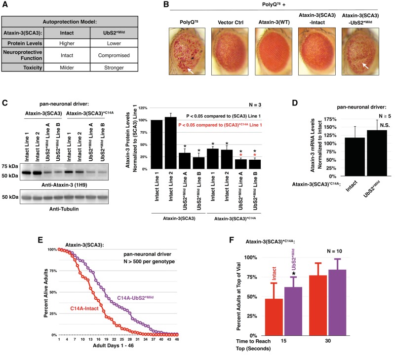
Polyglutamine (polyQ) repeat expansion in the deubiquitinase ataxin-3 causes neurodegeneration in Spinocerebellar Ataxia Type 3 (SCA3), one of nine inherited, incurable diseases caused by similar mutations. Ataxin-3's degradation is inhibited by its binding to the proteasome shuttle Rad23 through ubiquitin-binding site 2 (UbS2). Disrupting this interaction decreases levels of ataxin-3. Since reducing levels of polyQ proteins can decrease their toxicity, we tested whether genetically modulating the ataxin-3-Rad23 interaction regulates its toxicity in Drosophila. We found that exogenous Rad23 increases the toxicity of pathogenic ataxin-3, coincident with increased levels of the disease protein. Conversely, reducing Rad23 levels alleviates toxicity in this SCA3 model. Unexpectedly, pathogenic ataxin-3 with a mutated Rad23-binding site at UbS2, despite being present at markedly lower levels, proved to be more pathogenic than a disease-causing counterpart with intact UbS2. Additional studies established that the increased toxicity upon mutating UbS2 stems from disrupting the autoprotective role that pathogenic ataxin-3 has against itself, which depends on the co-chaperone, DnaJ-1. Our data reveal a previously unrecognized balance between pathogenic and potentially therapeutic properties of the ataxin-3-Rad23 interaction; they highlight this interaction as critical for the toxicity of the SCA3 protein, and emphasize the importance of considering protein context when pursuing suppressive avenues.


Figures

Figure 1
Figure 1
Rad23 influences ataxin-3(SCA3)-dependent toxicity. (A) Model depicting how Rad23 influences ataxin-3’s degradation, based on studies from Blount et al, 2014 (20). The ubiquitin-interacting motifs (UIMs) of ataxin-3 bind ubiquitin chains on proteasome substrates, facilitating the interaction of ataxin-3 with this machinery. Once at the proteasome, in the absence of interaction with Rad23, ataxin-3 is degraded. If ataxin-3 interacts with Rad23 through UbS2, it is rescued. (B) Summary of the transgenic Drosophila lines used. (C) Summary of observations with ubiquitous expression of pathogenic ataxin-3 with or without Rad23 overexpression. In this panel and rest of the figures, unless otherwise noted, flies were heterozygous for all transgenes. (D) Longevity of flies ubiquitously expressing pathogenic ataxin-3 or the empty host vector inserted into attP2. (E) Longevity of flies ubiquitously expressing pathogenic ataxin-3 without or with UAS-RNAi targeting Rad23. A control line was included that expresses Rad23 RNAi in the absence of ataxin-3. Flies were siblings from the same crosses and on a different background (y-w-) than flies in the rest of this figure (w1118). (F) Western blots from independent lysates of whole flies ubiquitously expressing the specified transgenes. Means ± SD. Asterisks: P< 0.05 from Student’s t-tests compared to UAS-ataxin-3(SCA3) without exogenous Rad23. When quantifying ataxin-3 protein levels for (-)Rad23 flies, lane 4 was set to 100%. Lane 5 was not quantified, due to the bubble disrupting the ataxin-3 band. (G) Longevity of flies ubiquitously expressing pathogenic ataxin-3 with or without Rad23 overexpression.
Figure 2
Figure 2
Mutating UbS2 of pathogenic ataxin-3 increases its toxicity. (A) Lethality observed when pathogenic ataxin-3 with or without a mild UbS2 mutation is ubiquitously expressed. (B) Western blots from larvae ubiquitously expressing pathogenic ataxin-3 with or without a UbS2 mutation. Expression of constructs in (A) and (B) was driven by the sqh-Gal4 driver. Means ± SD. Asterisks: P <  0.05 from Student’s t-tests compared to line 1. Lines 1, 2, and A, B are from different founders, which had similar phenotypic outcomes and were combined in later studies. (C) qRT-PCR data from one day old flies pan-neuronally expressing pathogenic ataxin-3 with or without a mild UbS2 mutation. Endogenous control: rp49. Means ± SD. N.S: non statistically significant. (D) Western blots from adult flies pan-neuronally expressing pathogenic ataxin-3 with or without a UbS2 mutation. Means ± SD. Asterisks: P <  0.05 from Student’s t-tests compared to line 1. Lines 1, 2, and A, B are from different founders, which had similar phenotypic outcomes and were combined in later studies. (E) Longevity of flies pan-neuronally expressing pathogenic ataxin-3 with or without a mild UbS2 mutation. (F) Results from negative geotaxis motility assays, which were repeated at least three times, totaling at least 100 flies per genotype. Means ± SD. Asterisks: P < 0.05 from Student’s t-tests compared to Ctrl (black) or Intact (red) for each time point. Ctrl: empty vector inserted into site attP2. Expression of constructs in (C)–(F) was driven by the elav-Gal4 driver.
Figure 3
Figure 3
Mutating UbS2 increases pathogenicity of ataxin-3 in fly eyes. (A) Western blots from fly heads expressing pathogenic ataxin-3 without or with a UbS2 mutation. Means ± SD. Asterisks: P <  0.05 from Student’s t-tests compared to line 1. Lines 1, 2, and A, B are from different founders, which had similar phenotypic outcomes and were combined in studies in the other panels of this figure. (B) External photos and (C) histological sections of fly eyes expressing pathogenic ataxin-3 without or with a mild UbS2 mutation. Ctrl: empty vector inserted into attP2. White circle in (B): example of the pseudo-pupil. Red boxes in (C): examples of densely staining aggregates. The precise subcellular localization of the aggregates is not clear from this assay. Red bracketed lines in (C): examples of disruption of the ommatidial boundaries. Expression of constructs was driven by the GMR-Gal4 driver.
Figure 4
Figure 4
Mutating UbS2 of catalytically inactive, pathogenic ataxin-3 reduces its protein levels and toxicity. (A) Table summarizing the working model for the role of ataxin-3’s UbS2 in toxicity. (B) Photos of external eyes expressing a truncated polyQ fragment of ataxin-3 with 78 repeats and co-expressing the indicated lines. Flies were heterozygous for all transgenes, except for flies homozygous for polyQ78 (far left). Constructs were expressed using the GMR-gal4 driver. White arrows: examples of indentation. (C) Western blots from adult flies pan-neuronally expressing pathogenic ataxin-3 that is catalytically active or inactive. Means ± SD. Black asterisks: P <  0.05 from Student’s t-tests compared to catalytically active ataxin-3 line 1. Red asterisks: P <  0.05 from Student’s t-tests compared to catalytically inactive ataxin-3 line 1. Lines 1, 2, and A, B are from different founders, which had similar phenotypic outcomes and were combined in later studies in this figure. (D) qRT-PCR data from one day old flies expressing pathogenic, catalytically inactive ataxin-3 with or without a mild UbS2 mutation. Endogenous control: rp49. Means ± SD. N.S: non statistically significant. (E) Longevity data from flies expressing pathogenic, catalytically inactive ataxin-3 without or with a mild UbS2 mutation. (F) Motility of flies expressing pathogenic, catalytically inactive ataxin-3 without or with a mild UbS2 mutation. Flies were 7 days old. N = 10, totaling at least 100 flies per genotype. Asterisk: P <  0.05 from Student’s t-tests compared to C14A-Intact.
Figure 5
Figure 5
A stronger mutation of UbS2 reduces pathogenic ataxin-3 protein levels and toxicity compared to UbS2*Mild. (A) Western blot from in vitro binding assays of ataxin-3(WT) and full-length, GST-tagged Rad23A. GST or GST-Rad23 were pulled down on glutathione agarose beads, washed thrice with NETN and incubated with ataxin-3(WT)-Intact or ataxin-3(WT)-UbS2*Strong for 1 h on ice. Colored arrows denote the different amounts of ataxin-3. Percentages compare UbS2-mutated ataxin-3 pulled down by GST-Rad23A to its non-mutated counterpart of the same amount. (B) Western blots from whole cell lysates. HeLa cells were transfected with the indicated constructs and harvested 24 h later. Means ± SD. Asterisks: P <  0.05 from Student’s t-tests when compared to Intact (black) or UbS2*Mild (purple). (C) Western blots from HeLa cells transfected with the indicated constructs and treated 24 h later with cycloheximide (CHX) for the indicated amounts of time. Transfection and loading were done such that the protein level of the two different versions of ataxin-3 were comparable at 0h. Example shown is from experiment repeated independently at least 4 times with similar results. (D) Lethality observed when ubiquitously expressing pathogenic ataxin-3 without or with a UbS2 mutation. (E) Western blots from larvae ubiquitously expressing pathogenic ataxin-3 without or with a strong UbS2 mutation. Means ± SD. Asterisks: P <  0.05 from Student’s t-test compared to Intact. Lines 1 and 2 are from different founders, which had similar phenotypic outcomes and were combined in phenotypic studies. (F) qRT-PCR data from larvae ubiquitously expressing pathogenic ataxin-3 without or with a strong UbS2 mutation. Endogenous control: rp49. N.S: non statistically significant. (G) Longevity of flies pan-neuronally expressing pathogenic ataxin-3 without or with a UbS2 mutation. (H) Results from motility assays of flies expressing pathogenic ataxin-3 without or with a UbS2 mutation. Means ± SD. Asterisks: P <  0.05 from Student’s t-tests compared to Intact (red) or UbS2*Mild (purple). N = 10, totaling at least 100 flies per genotype.
Figure 6
Figure 6
Exogenous DnaJ-1 suppresses toxicity from pathogenic ataxin-3 with mutated UbS2. (A) External eye photos and histological sections of fly eyes expressing pathogenic ataxin-3 with a mild UbS2 mutation in the absence or presence of exogenous DnaJ-1. Constructs were expressed using the GMR-Gal4 driver. Vector Ctrl: empty vector inserted into attP2. Red boxes: examples of densely staining structures. Red bracketed line: an example of disruption of the ommatidial boundaries. (B) Motility of flies expressing pathogenic ataxin-3 with a mild UbS2 mutation in the presence or absence of exogenous DnaJ-1. Here, elav-Gal4 was on the third chromosome, expressing at lower levels than in previous figures, where elav-Gal4 was on the X chromosome (46). Means ± SD. Asterisks: P <  0.05 from Student’s t-test, compared to UbS2*Mild without exogenous DnaJ-1. N = 10, totaling at least 100 flies per genotype. (C) Longevity of flies expressing pathogenic ataxin-3 with a mild UbS2 mutation in the presence or absence of exogenous DnaJ-1. (D) Lethality observed when ubiquitously expressing pathogenic ataxin-3 with a mild UbS2 mutation in the presence or absence of exogenous DnaJ-1, or wild type ataxin-3, or knockdown of DnaJ-1 through UAS-RNAi.
Figure 7
Figure 7
Model of Rad23 and UbS2 regulating ataxin-3’s levels and toxicity. (A) Knocking down Rad23 reduces its availability to bind ataxin-3 at UbS2, increasing ataxin-3’s degradation. This reduces ataxin-3 protein levels while leaving its autoprotection intact, resulting in lowered ataxin-3 toxicity. (B) Ataxin-3 with intact UbS2 is present at baseline levels; its pathogenicity is regulated by its autoprotection, which requires Rad23 binding to UbS2. (C) Exogenous Rad23 increases its availability to bind ataxin-3, inhibiting ataxin-3’s proteasomal degradation and increasing its toxicity. (D) Mutating UbS2 reduces Rad23 binding, resulting in lower ataxin-3 levels. However, mutating this site compromises ataxin-3’s protection against itself, thus increasing its toxicity.

Similar articles

Cited by

References

    1. Todi S.V., Williams A., Paulson H. (2007) Waxman S.G. (ed.), In Molecular Neurology. Academic Press, London, in press., pp. 257–276.
    1. Costa Mdo C., Paulson H.L. (2012) Toward understanding Machado-Joseph disease. Prog. Neurobiol., 97, 239–257. - PMC - PubMed
    1. Matos C.A., de Macedo-Ribeiro S., Carvalho A.L. (2011) Polyglutamine diseases: The special case of ataxin-3 and Machado-Joseph disease. Prog. Neurobiol., 95, 26–48. - PubMed
    1. Winborn B.J., Travis S.M., Todi S.V., Scaglione K.M., Xu P., Williams A.J., Cohen R.E., Peng J., Paulson H.L. (2008) The deubiquitinating enzyme ataxin-3, a polyglutamine disease protein, edits Lys63 linkages in mixed linkage ubiquitin chains. J. Biol. Chem., 283, 26436–26443. - PMC - PubMed
    1. Scaglione K.M., Zavodszky E., Todi S.V., Patury S., Xu P., Rodriguez-Lebron E., Fischer S., Konen J., Djarmati A., Peng J., et al. (2011) Ube2w and Ataxin-3 Coordinately Regulate the Ubiquitin Ligase CHIP. Mol. Cell, 43, 599–612. - PMC - PubMed

Publication types

MeSH terms

LinkOut - more resources