. 2017 Feb;216(2):343-353.
doi: 10.1083/jcb.201610060. Epub 2017 Jan 31.

Localization of mTORC2 activity inside cells

Affiliations

Localization of mTORC2 activity inside cells

Michael Ebner et al. J Cell Biol. 2017 Feb.

Abstract

Activation of protein kinase Akt via its direct phosphorylation by mammalian target of rapamycin (mTOR) complex 2 (mTORC2) couples extracellular growth and survival cues with pathways controlling cell growth and proliferation, yet how growth factors target the activity of mTORC2 toward Akt is unknown. In this study, we examine the localization of the obligate mTORC2 component, mSin1, inside cells and report the development of a reporter to examine intracellular localization and regulation by growth factors of the endogenous mTORC2 activity. Using a combination of imaging and biochemical approaches, we demonstrate that inside cells, mTORC2 activity localizes to the plasma membrane, mitochondria, and a subpopulation of endosomal vesicles. We show that unlike the endosomal pool, the activity and localization of mTORC2 via the Sin1 pleckstrin homology domain at the plasma membrane is PI3K and growth factor independent. Furthermore, we show that membrane recruitment is sufficient for Akt phosphorylation in response to growth factors. Our results indicate the existence of spatially separated mTORC2 populations with distinct sensitivity to PI3K inside cells and suggest that intracellular localization could contribute to regulation of mTORC2 activity toward Akt.


Figures

Figure 1.
Figure 1.
LocaTOR2 reports local mTORC2 activity. (A) Scheme of the compartment-specific reporter. Cytosolic FRB:Akt2 can be recruited to the plasma membrane using AP21967 rapalog, and FRB:Akt2 phosphorylation can be monitored using specific antibodies. (B and C) AP21967 induces translocation of FRB:Akt2-citrine to subcellular membrane compartments. Recruitment half time was determined by calculating Pearson’s coefficient of the recruiter channel (mCherry, red in second and third row; B) and the citrine channel (green; B). Bars, 10 µm. Data are presented as means ± SD; the number of cells analyzed is shown in parentheses (C). (D) FRB:Akt2 phosphorylation is sensitive to Torin1, but not to rapamycin. HEK293T cells were transfected with LocaTOR2 for 18 h, treated for 40 min with 250 nM Torin1, 275 nM rapamycin, or the vehicle, and lysed, and the lysates were analyzed using PAGE/WB. endo, endogenously expressed Akt.
Figure 2.
Figure 2.
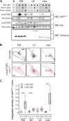
Probing endogenous mTORC2 activity using LocaTOR. (A) LocaTOR2 phosphorylation in subcellular membrane compartments is mTOR dependent. HEK293T cells were cotransfected with LocaTOR2 and the corresponding recruiter constructs, serum starved overnight, and incubated with 250 nM Torin1 before (pre) or after (post) a 40-min treatment with 250 nM AP21967. endo, endogenously expressed Akt. (B) LocaTOR2 phosphorylation is restricted to the recruiting compartments. HEK293T cells were cotransfected with FRB:Akt2-citrine (bottom, gray) and the corresponding recruiter constructs (top) for 18 h and serum starved overnight. After 40 min of incubation with 250 nM AP21967, the cells were fixed, permeabilized, and immunostained with AktpS473 phosphospecific antibody (bottom, red). Bars, 5 µm. (C) Quantification of the results shown in B. The images of mCherry-tagged recruiter constructs were segmented to create binary masks, in which mean fluorescence intensity in the phosphospecific antibody channel was determined and normalized on a cell-by-cell basis by the mean intensity of FRB:Akt2-citrine fluorescence. Data are presented as box plots (red, median; whiskers, 5th and 95th percentiles). Numbers in parentheses indicate the number of cells analyzed from two to three independent experiments. Mann-Whitney U test: ***, P < 0.001. EE, early endosome; LE, late endosome; PM, plasma membrane; mito, outer mitochondrial membrane; N, nontransfected control.
Figure 3.
Figure 3.
Regulation of intracellular mTORC2 activity by PI3K. (A and B) Intracellular membrane compartments display distinct mTORC2 sensitivity to PI3K inhibition. HEK293T cells were cotransfected with LocaTOR2 and the corresponding recruiter constructs for 18 h, serum starved overnight, and then preincubated for 30 min with 500 nM GDC-0941 or vehicle and treated with 250 nM AP21967 for 40 min. The cells were lysed, and the level of LocaTOR2 pSer474 phosphorylation was analyzed by quantitative WBs and normalized against total Akt. The data are presented as fold increase over untreated vehicle control averaged over several independent experiments (shown in parentheses); shown are means ± SEM. *, P < 0.05 (one-sample t test with Benjamin-Hochberg correction). EE, early endosome; LE, late endosome; PM, plasma membrane; mito, outer mitochondrial membrane; N, nontransfected control; RE, recycling endosome. (C) mTORC2 activity at the plasma membrane is insensitive to PI3K activity. HEK293T cells were cotransfected with LocaTOR2 and the plasma membrane recruiter construct for 18 h, grown with or without 10% serum (FBS) overnight, and then preincubated for 30 min with 500 nM GDC-0941 or the vehicle and treated with 250 nM AP21967 for 40 min. The cells were lysed, and the level of LocaTOR2pSer474 was determined by quantitative WBs. The results are plotted as the fold increase over vehicle control. The numbers in parentheses indicate independent experiments; error bars are SEM. n.s., not significant. One-way analysis of variance: P > 0.5. (D) mTORC2 activity at the plasma membrane is insensitive to growth factors. HEK293T cells were cotransfected with LocaTOR2 and the plasma membrane recruiter construct for 18 h, serum starved overnight, pretreated for 40 min with 250 nM AP21967, and then stimulated by 100 ng/ml insulin. The cells were lysed, and pSer473 phosphorylation of the endogenous Akt and LocaTOR2 was determined using PAGE/WBs. ctrl, control; endo, endogenously expressed Akt.
Figure 4.
Figure 4.
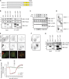
PH domain targets mSin1/mTORC2 to cellular membranes. (A) Schematic representation of mSin1 isoforms 1, 2, and 5. Yellow boxes represent the PH domain. (B) Isoforms 1, 2, and 5 of mSin1-GFP are incorporated into mTORC2. HEK293T cells were transiently transfected with GFP or the corresponding mSin1-GFP isoform, and lysed, and GFP-tagged proteins were pulled down using GFP-Trap beads. Shown is a representative pull-down result. n = 2 for mSin1.1 and mSin1.5; n = 3 for mSin1.2. (C) In vitro kinase assay with immunoprecipitated mTORC2. HEK293T cells transiently transfected with GFP or mSin1.2WT-GFP were stimulated with 100 ng/ml insulin for 10 min and lysed, and GFP-tagged proteins were pulled down using GFP-Trap beads. The beads were preincubated with or without 100 nM Torin1 and incubated for 1 h at 37°C with purified dephosphorylated recombinant full-length human Akt1 with or without 0.5 mM ATP, and pSer473 phosphorylation was determined using PAGE/WBs. Shown is a representative result of two independent experiments. (D) Intracellular localization of isoforms 1, 2, and 5 of mSin1-GFP in HeLa cells. (E) mSin1 localizes to the plasma membrane and endosomal vesicles. HeLa cells were transiently cotransfected with mSin1.2-GFP and markers of the plasma membrane (PM; mCherry-KRas4BC30) along with early (mCherry-Rab5) and late endosomes (mCherry-Rab7). (F) The PH domain targets mSin1 to cellular membranes. HeLa cells were transiently transfected with mSin1.2WT-GFP or mSin1ΔPH-GFP. (D–F) Bars, 10 µm. (G) The PH domain is dispensable for mSin1 incorporation into mTORC2. HeLa cells were transiently transfected with mSin1.2WT-GFP or mSin1.2ΔPH-GFP and lysed, and GFP-tagged proteins were pulled down using GFP-Trap beads. IP, immunoprecipitate. (H) Full-length mSin1 does not change localization upon growth factor treatment or PI3K inhibition. MCF7 cells cotransfected with mSin1.2WT-GFP and PHAkt-mCherry for 18 h were starved overnight and then treated first with 100 ng/ml insulin and then with 500 nM GDC-0941. Shown are the time traces of mean fluorescence intensity in a cytosolic region of interest. Data are presented as means ± SEM. n = 2; 18 cells. Normalized to t = –150 s.
Figure 5.
Figure 5.
Akt membrane recruitment is sufficient for Ser473 phosphorylation by mTORC2. (A and B) Model showing reversible recruitment strategy (A). HeLa cells were cotransfected with FKBP:mCherry-Akt2K14A and TagBFP-2×DHFR-CAAX constructs for 18 h and then treated with 1 µM SLF'-TMP for 12 min, after which 10 µM TMP was added (mean fluorescence intensity in the cytosolic region of interest). Data are presented as means ± SD; seven cells. PM, plasma membrane. (C) Akt membrane recruitment is necessary and sufficient for pSer474 phosphorylation. HeLa cells were cotransfected with FKBP:mCherry-Akt2K14A and TagBFP-2×DHFR-CAAX constructs for 18 h and then treated with 1 µM SLF'-TMP for 30 min, after which 10 µM TMP was added for the time indicated. The cells were lysed, and pSer473 phosphorylation was determined using PAGE/WBs. Shown is one representative out of three experiments. endo, endogenously expressed Akt.

Similar articles

Cited by

References

    1. Ahmed N.N., Franke T.F., Bellacosa A., Datta K., Gonzalez-Portal M.E., Taguchi T., Testa J.R., and Tsichlis P.N.. 1993. The proteins encoded by c-akt and v-akt differ in post-translational modification, subcellular localization and oncogenic potential. Oncogene. 8:1957–1963. - PubMed
    1. Andjelković M., Alessi D.R., Meier R., Fernandez A., Lamb N.J., Frech M., Cron P., Cohen P., Lucocq J.M., and Hemmings B.A.. 1997. Role of translocation in the activation and function of protein kinase B. J. Biol. Chem. 272:31515–31524. 10.1074/jbc.272.50.31515 - DOI - PubMed
    1. Arias E., Koga H., Diaz A., Mocholi E., Patel B., and Cuervo A.M.. 2015. Lysosomal mTORC2/PHLPP1/Akt regulate Chaperone-mediated autophagy. Mol. Cell. 59:270–284. 10.1016/j.molcel.2015.05.030 - DOI - PMC - PubMed
    1. Bar-Peled L., Chantranupong L., Cherniack A.D., Chen W.W., Ottina K.A., Grabiner B.C., Spear E.D., Carter S.L., Meyerson M., and Sabatini D.M.. 2013. A tumor suppressor complex with GAP activity for the Rag GTPases that signal amino acid sufficiency to mTORC1. Science. 340:1100–1106. 10.1126/science.1232044 - DOI - PMC - PubMed
    1. Bellacosa A., Chan T.O., Ahmed N.N., Datta K., Malstrom S., Stokoe D., McCormick F., Feng J., and Tsichlis P.. 1998. Akt activation by growth factors is a multiple-step process: the role of the PH domain. Oncogene. 17:313–325. 10.1038/sj.onc.1201947 - DOI - PubMed

Publication types

MeSH terms

Substances