

Sensing Membrane Curvature in Macroautophagy
- PMID: 28088480
- PMCID: PMC5276735
- DOI: 10.1016/j.jmb.2017.01.006
Sensing Membrane Curvature in Macroautophagy
Abstract
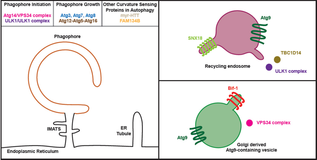
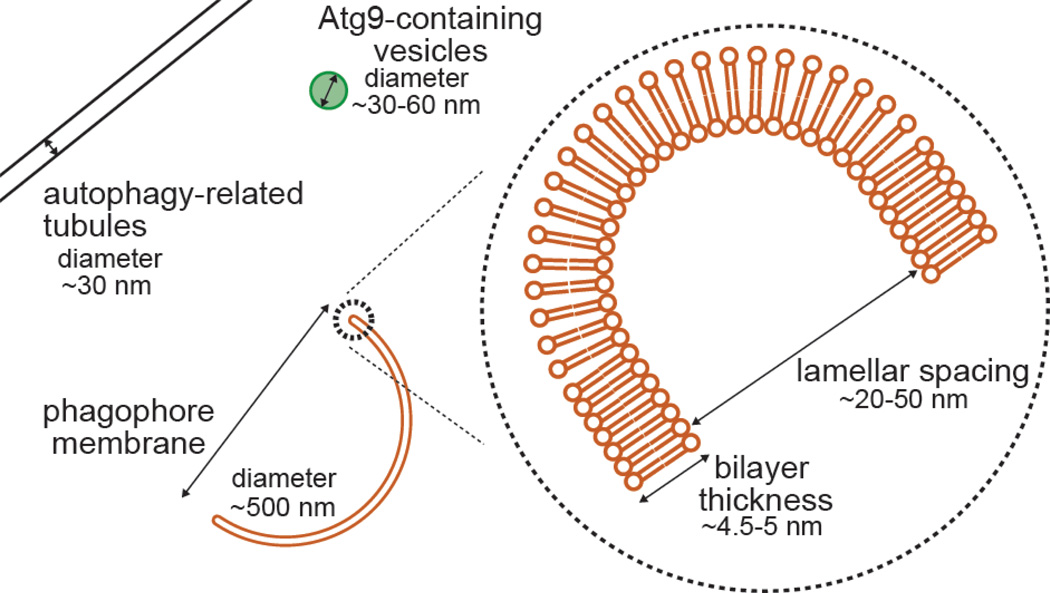
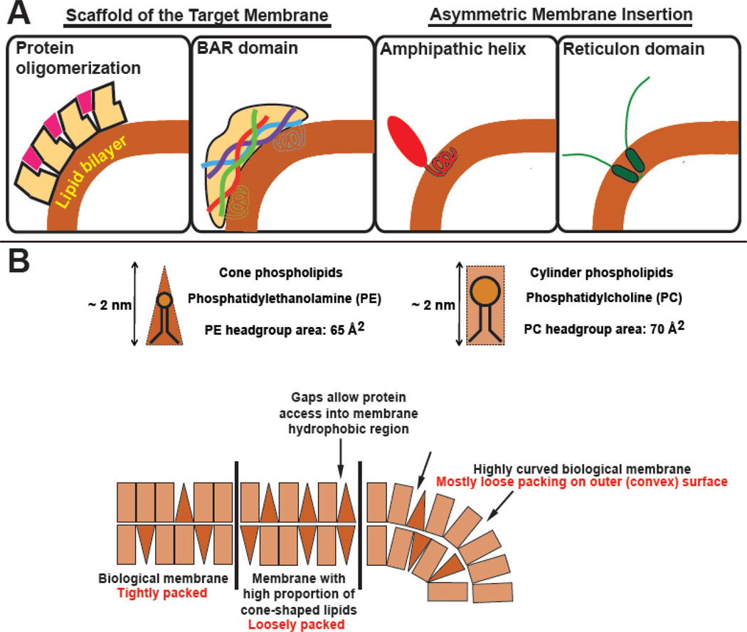
In response to intracellular stress events ranging from starvation to pathogen invasion, the cell activates one or more forms of macroautophagy. The key event in these related pathways is the de novo formation of a new organelle called the autophagosome, which either surrounds and sequesters random portions of the cytoplasm or selectively targets individual intracellular challenges. Thus, the autophagosome is a flexible membrane platform with dimensions that ultimately depend upon the target cargo. The intermediate membrane, termed the phagophore or isolation membrane, is a cup-like structure with a clear concave face and a highly curved rim. The phagophore is largely devoid of integral membrane proteins; thus, its shape and size are governed by peripherally associated membrane proteins and possibly by the lipid composition of the membrane itself. Growth along the phagophore rim marks the progress of both organelle expansion and ultimately organelle closure around a particular cargo. These two properties, a reliance on peripheral membrane proteins and a structurally distinct membrane architecture, suggest that the ability to target or manipulate membrane curvature might be an essential activity of proteins functioning in this pathway. In this review, we discuss the extent to which membranes are naturally curved at each of the cellular sites believed to engage in autophagosome formation, review basic mechanisms used to sense this curvature, and then summarize the existing literature concerning which autophagy proteins are capable of curvature recognition.
Keywords: BAR domain; amphipathic helix; membrane curvature; reticulon domain.
Copyright © 2017 Elsevier Ltd. All rights reserved.
Figures

Similar articles
-
In situ structural analysis reveals membrane shape transitions during autophagosome formation.Proc Natl Acad Sci U S A. 2022 Sep 27;119(39):e2209823119. doi: 10.1073/pnas.2209823119. Epub 2022 Sep 19. Proc Natl Acad Sci U S A. 2022. PMID: 36122245 Free PMC article.
-
A conserved membrane curvature-generating protein is crucial for autophagosome formation in fission yeast.Nat Commun. 2023 Aug 8;14(1):4765. doi: 10.1038/s41467-023-40530-4. Nat Commun. 2023. PMID: 37553386 Free PMC article.
-
Autophagosome Biogenesis.Cells. 2023 Feb 20;12(4):668. doi: 10.3390/cells12040668. Cells. 2023. PMID: 36831335 Free PMC article. Review.
-
In situ snapshots along a mammalian selective autophagy pathway.Proc Natl Acad Sci U S A. 2023 Mar 21;120(12):e2221712120. doi: 10.1073/pnas.2221712120. Epub 2023 Mar 14. Proc Natl Acad Sci U S A. 2023. PMID: 36917659 Free PMC article.
-
Mechanistic Insights into the Role of Atg11 in Selective Autophagy.J Mol Biol. 2020 Jan 3;432(1):104-122. doi: 10.1016/j.jmb.2019.06.017. Epub 2019 Jun 22. J Mol Biol. 2020. PMID: 31238043 Free PMC article. Review.
Cited by
-
The autophagic membrane tether ATG2A transfers lipids between membranes.Elife. 2019 Jul 4;8:e45777. doi: 10.7554/eLife.45777. Elife. 2019. PMID: 31271352 Free PMC article.
-
The Atg8 Family of Proteins-Modulating Shape and Functionality of Autophagic Membranes.Front Genet. 2017 Aug 28;8:109. doi: 10.3389/fgene.2017.00109. eCollection 2017. Front Genet. 2017. PMID: 28894458 Free PMC article. Review.
-
In human astrocytes neurotropic flaviviruses increase autophagy, yet their replication is autophagy-independent.Cell Mol Life Sci. 2022 Oct 25;79(11):566. doi: 10.1007/s00018-022-04578-7. Cell Mol Life Sci. 2022. PMID: 36283999 Free PMC article.
-
In situ structural analysis reveals membrane shape transitions during autophagosome formation.Proc Natl Acad Sci U S A. 2022 Sep 27;119(39):e2209823119. doi: 10.1073/pnas.2209823119. Epub 2022 Sep 19. Proc Natl Acad Sci U S A. 2022. PMID: 36122245 Free PMC article.
-
Targeting of the Mon1-Ccz1 Rab guanine nucleotide exchange factor to distinct organelles by a synergistic protein and lipid code.J Biol Chem. 2023 Mar;299(3):102915. doi: 10.1016/j.jbc.2023.102915. Epub 2023 Jan 14. J Biol Chem. 2023. PMID: 36649906 Free PMC article.
References
-
- Hayashi-Nishino M, Fujita N, Noda T, Yamaguchi A, Yoshimori T, Yamamoto A. A subdomain of the endoplasmic reticulum forms a cradle for autophagosome formation. Nat Cell Biol. 2009;11:1433–1437. - PubMed
-
- Israelachvili JN, Mitchell DJ. A model for the packing of lipids in bilayer membranes. Biochim Biophys Acta. 1975;389:13–19. - PubMed
-
- Yla-Anttila P, Vihinen H, Jokitalo E, Eskelinen EL. 3D tomography reveals connections between the phagophore and endoplasmic reticulum. Autophagy. 2009;5:1180–1185. - PubMed
Publication types
MeSH terms
Substances
Grants and funding
LinkOut - more resources
Full Text Sources
Other Literature Sources
