

Structural insight into an evolutionarily ancient programmed cell death regulator - the crystal structure of marine sponge BHP2 bound to LB-Bak-2
- PMID: 28079890
- PMCID: PMC5386376
- DOI: 10.1038/cddis.2016.469
Structural insight into an evolutionarily ancient programmed cell death regulator - the crystal structure of marine sponge BHP2 bound to LB-Bak-2
Abstract
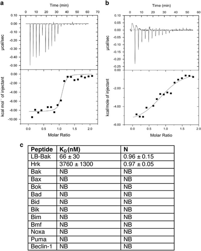
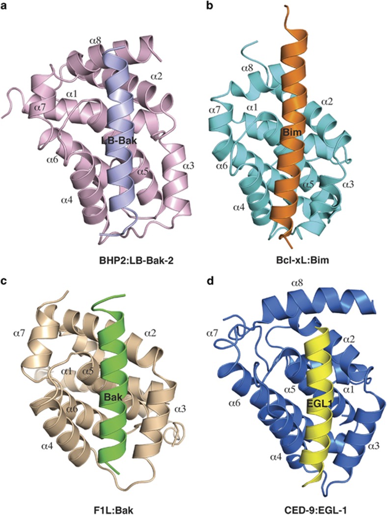
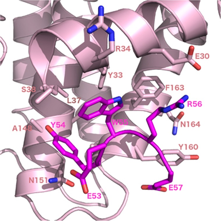
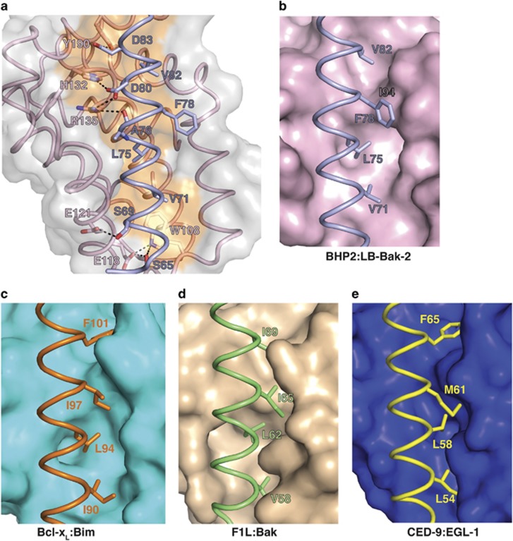
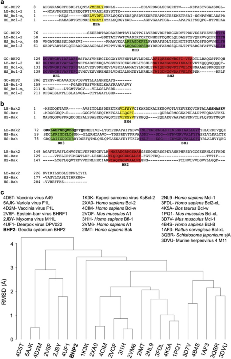
Sponges of the porifera family harbor some of the evolutionary most ancient orthologs of the B-cell lymphoma-2 (Bcl-2) family, a protein family critical to regulation of apoptosis. The genome of the sponge Geodia cydonium contains the putative pro-survival Bcl-2 homolog BHP2, which protects sponge tissue as well as mammalian Hek-293 and NIH-3T3 cells against diverse apoptotic stimuli. The Lake Baikal demosponge Lubomirskia baicalensis has been shown to encode both putative pro-survival Bcl-2 (LB-Bcl-2) and pro-apoptotic Bcl-2 members (LB-Bak-2), which have been implied in axis formation (branches) in L. baicalensis. However, the molecular mechanism of action of sponge-encoded orthologs of Bcl-2 remains to be clarified. Here, we report that the pro-survival Bcl-2 ortholog BHP2 from G. cydonium is able to bind the BH3 motif of a pro-apoptotic Bcl-2 protein, LB-Bak-2 of the sponge L. baicalensis. Furthermore, we determined the crystal structure of BHP2 bound to LB-Bak-2, which revealed that using a binding groove conserved across all pro-survival Bcl-2 proteins, BHP2 binds multi-motif Bax-like proteins through their BH3-binding regions. However, BHP2 discriminates against BH3-only bearing proteins by blocking access to a hydrophobic pocket that is critical for BH3 motif binding in pro-survival Bcl-2 proteins from higher organisms. This differential binding mode is reflected in a structure-based phylogenetic comparison of BHP2 with other Bcl-2 family members, which revealed that BHP2 does not cluster with either Bcl-2 members of higher organisms or pathogen-encoded homologs, and assumes a discrete position. Our findings suggest that the molecular machinery and mechanisms for executing Bcl-2-mediated apoptosis as observed in mammals are evolutionary ancient, with early regulation of apoptotic machineries closely resembling their modern counterparts in mammals rather than Caenorhabditis elegans or drosophila.
Conflict of interest statement
The authors declare no conflict of interest.
Figures
Similar articles
-
Structural and Functional Insight into Canarypox Virus CNP058 Mediated Regulation of Apoptosis.Viruses. 2017 Oct 20;9(10):305. doi: 10.3390/v9100305. Viruses. 2017. PMID: 29053589 Free PMC article.
-
The structural basis of Bcl-2 mediated cell death regulation in hydra.Biochem J. 2020 Sep 18;477(17):3287-3297. doi: 10.1042/BCJ20200556. Biochem J. 2020. PMID: 32776134 Free PMC article.
-
Structural biology of the Bcl-2 family of proteins.Biochim Biophys Acta. 2004 Mar 1;1644(2-3):83-94. doi: 10.1016/j.bbamcr.2003.08.012. Biochim Biophys Acta. 2004. PMID: 14996493 Review.
-
Molecular evolution of apoptotic pathways: cloning of key domains from sponges (Bcl-2 homology domains and death domains) and their phylogenetic relationships.J Mol Evol. 2000 Jun;50(6):520-31. doi: 10.1007/s002390010055. J Mol Evol. 2000. PMID: 10835482
-
Structural biology of the Bcl-2 family and its mimicry by viral proteins.Cell Death Dis. 2013 Nov 7;4(11):e909. doi: 10.1038/cddis.2013.436. Cell Death Dis. 2013. PMID: 24201808 Free PMC article. Review.
Cited by
-
Diversity in the intrinsic apoptosis pathway of nematodes.Commun Biol. 2020 Aug 28;3(1):478. doi: 10.1038/s42003-020-01208-5. Commun Biol. 2020. PMID: 32859965 Free PMC article.
-
Ancient and conserved functional interplay between Bcl-2 family proteins in the mitochondrial pathway of apoptosis.Sci Adv. 2020 Sep 30;6(40):eabc4149. doi: 10.1126/sciadv.abc4149. Print 2020 Sep. Sci Adv. 2020. PMID: 32998881 Free PMC article.
-
Structural Investigation of Orf Virus Bcl-2 Homolog ORFV125 Interactions with BH3-Motifs from BH3-Only Proteins Puma and Hrk.Viruses. 2021 Jul 15;13(7):1374. doi: 10.3390/v13071374. Viruses. 2021. PMID: 34372579 Free PMC article.
-
Structural and Functional Insight into Canarypox Virus CNP058 Mediated Regulation of Apoptosis.Viruses. 2017 Oct 20;9(10):305. doi: 10.3390/v9100305. Viruses. 2017. PMID: 29053589 Free PMC article.
-
Metazoans and Intrinsic Apoptosis: An Evolutionary Analysis of the Bcl-2 Family.Int J Mol Sci. 2022 Mar 28;23(7):3691. doi: 10.3390/ijms23073691. Int J Mol Sci. 2022. PMID: 35409052 Free PMC article.
References
-
- Lasi M, David CN, Bottger A. Apoptosis in pre-Bilaterians: hydra as a model. Apoptosis 2010; 15: 269–278. - PubMed
-
- Koziol C, Borojevic R, Steffen R, Muller WE. Sponges (Porifera) model systems to study the shift from immortal to senescent somatic cells: the telomerase activity in somatic cells. Mech Ageing Dev 1998; 100: 107–120. - PubMed
-
- Vaux DL, Haecker G, Strasser A. An evolutionary perspective on apoptosis. Cell 1994; 76: 777–779. - PubMed
-
- Adams JM, Cory S. The Bcl-2 protein family: arbiters of cell survival. Science 1998; 281: 1322–1326. - PubMed
Publication types
MeSH terms
Substances
LinkOut - more resources
Full Text Sources
Other Literature Sources
Research Materials
Miscellaneous
