. 2016 Dec 28;11(12):e0167636.
doi: 10.1371/journal.pone.0167636. eCollection 2016.

Cytochrome c Negatively Regulates NLRP3 Inflammasomes

Affiliations

Cytochrome c Negatively Regulates NLRP3 Inflammasomes

Chong-Shan Shi et al. PLoS One. .

Abstract

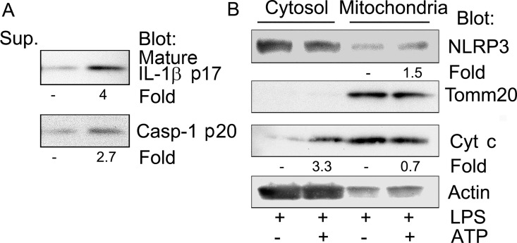
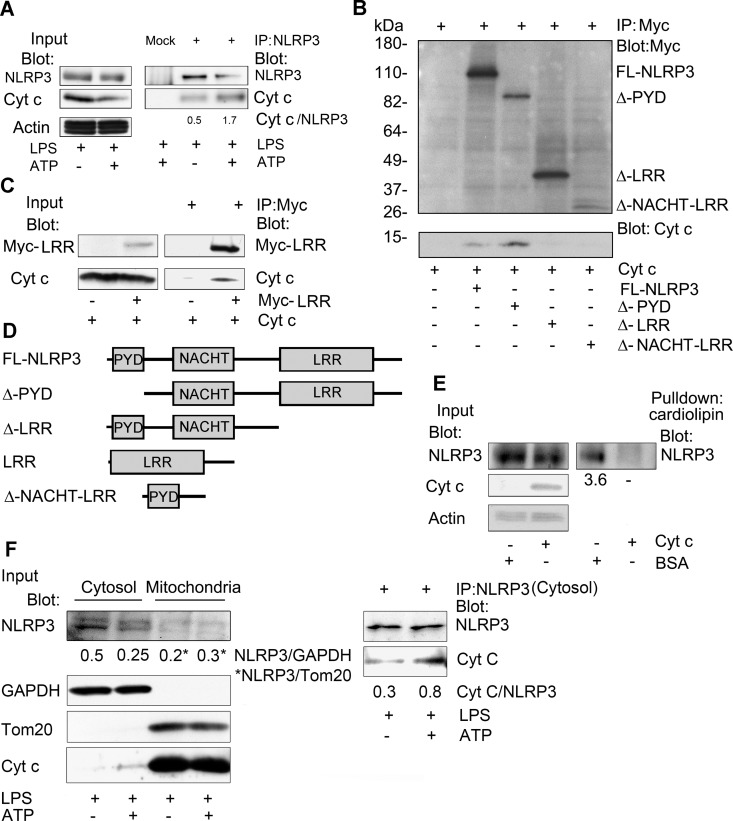
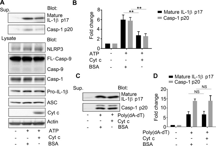
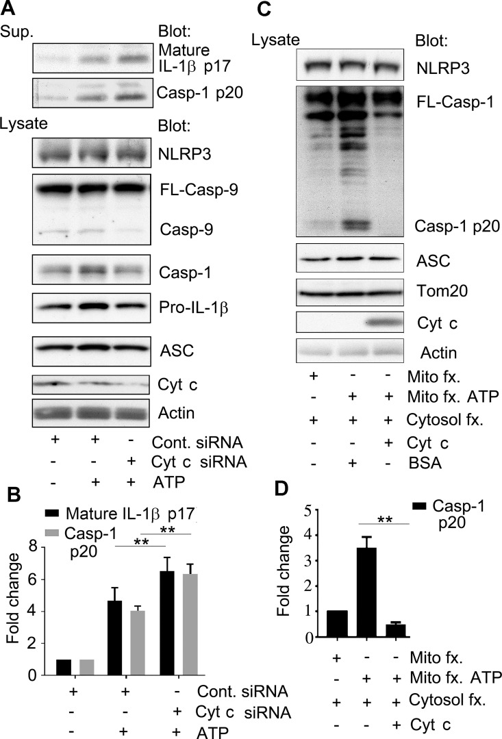
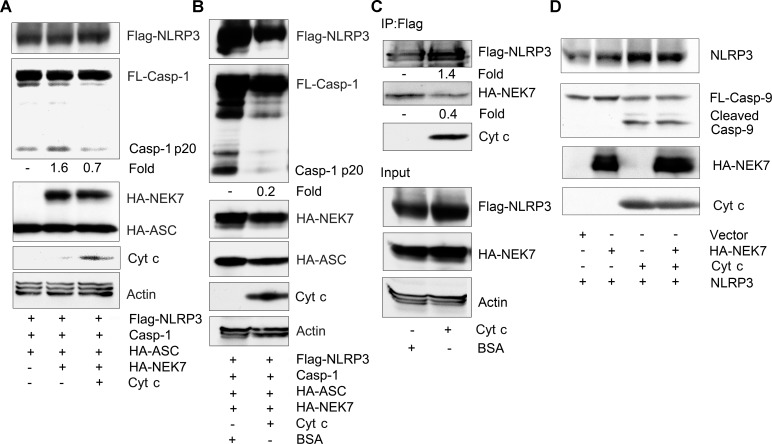
The release of cytochrome c from the inner mitochondrial membrane, where it is anchored by caridolipin, triggers the formation of the Apaf-1 apoptosome. Cardiolipin also interacts with NLRP3 recruiting NLRP3 to mitochondria and facilitating inflammasome assembly. In this study we investigated whether cytosolic cytochrome c impacts NLRP3 inflammasome activation in macrophages. We report that cytochrome c binds to the LRR domain of NLRP3 and that cytochrome c reduces the interactions between NLRP3 and cardiolipin and between NLRP3 and NEK7, a recently recognized component of the NLRP3 inflammasome needed for NLRP3 oligomerization. Protein transduction of cytochrome c impairs NLRP3 inflammasome activation, while partially silencing cytochrome c expression enhances it. The addition of cytochrome c to an in vitro inflammasome assay severely limited caspase-1 activation. We propose that there is a crosstalk between the NLRP3 inflammasome and apoptosome pathways mediated by cytochrome c, whose release during apoptosis acts to limit NLRP3 inflammasome activation.


Conflict of interest statement

The authors have declared that no competing interests exist.

Figures

Fig 1
Fig 1. ATP treatment induces cytochrome c release.
PMA differentiated THP-1 cells were primed with LPS 50 ng/ml overnight. One hour before harvesting the cells 5 mM ATP was added. Cell lysates were fractionated into a cytosolic and mitochondria enriched fractions. (A) Immunoblots of IL-1β and caspase-1 p20 present in the collected cell supernatants to verify NLRP3 inflammasome activation. (B) Immunoblots of cytosol and mitochondria enriched fractions from the above cells to assess cytochrome c release following NLRP3 inflammasome activation.
Fig 2
Fig 2. Cytochrome c binds to the LRR repeats of NLRP3 and interferes with cardiolipin binding.
(A) Immunoblots of cell lysates and either NLRP3 or sham immunoprecipitates prepared from LPS-primed THP-1 macrophage cells, stimulated with ATP or not, to detect an interaction between endogenous NLRP3 and cytochrome c. The ratios between the NLRP3 and cytochrome c levels in the immunoprecipitates were quantitated using Image J. (B) Immunoblots of cell lysates and either control or Myc immunoprecipitates to map the portion of NLRP3 important for the interaction with cytochrome c. Myc-tagged NLRP3 constructs were transfected into HEK 293T cells. The immunoprecipitates were washed, incubated with cytochrome c (50 ng), washed again, fractionated by SDS-PAGE, and immunoblotted. (C) Immunoblots of cell lysates and Myc-LRR domain immunoprecipitates incubated with cytochrome c, or not. A construct expressing a myc tagged NLRP3 LRR domain was transfected into HEK 293T cells. The Myc and control immunoprecipitates were incubated with cytochrome c (50 ng), washed, and immunoblotted. (D) Schematic of the constructs used in the above experiments (B & C). (E) Immunoblots of cell lysates and cardiolipin bead pull-downs to assess whether cytochrome c interferes with the interaction between cardiolipin and NLRP3. BSA (50 ng) or purified cytochrome c (50 ng) was added to lysates prepared from HEK 293T cells expressing NLRP3-Flag. Following a 30 minute incubation cardiolipin conjugated beads were added to the lysates for an additional 30 minutes. The caridolipin beads were washed; and the bound NLRP3 eluted in SDS-sample buffer, size fractionated by SDS PAGE, and quantitated by immunoblotting. The amount of NLRP3 in the cardiolipin pulldowns was normalized to the BSA control. The above experiments were respectively performed twice. (F) Cell lysates from cell treated with ATP, or not, were fractionated into cytosolic and mitochondrial fractions and the indicated proteins were immunoblotted. NLRP3 immunoprecipitates were prepared using the cytosolic fraction. An interaction between endogenous NLRP3 and cytochrome c was assess by immunoblotting. The ratios between the NLRP3 and cytochrome c levels in the immunoprecipitates were quantitated using Image J.
Fig 3
Fig 3. Increased cytosolic cytochrome c inhibits NLRP3, but not AIM2 inflammasome activation.
(A) Immunoblots of cell lysates and supernatants for the expression of IL-1β, caspase-1, caspase-9, ASC, cytochrome c, and actin as indicated to assess the impact of cytochrome c on NLRP3 inflammasome activity. Cytochrome c or BSA was transduced into LPS-primed THP-1 cells for 3 hours, and during the last hour ATP was present in the cell culture. Cell lysates and supernatants were collected. Endogenous cytochrome c was visualized on longer exposure (not shown). (B) Quantification of the results from (A) and 2 other similar experiments. Results are shown as mean +/- SEM. The difference between the BSA and cytochrome c transduced cells were compared by Student t test using Prism software. ** indicates that p < 0.01. (C) Immunoblots of cell lysates for processed IL-1β and caspase-1 p20 to examine the impact of cytochrome c on AIM2 inflammasome activation. LPS primed (2 h) THP-1 cells were transduced with cytochrome c or BSA, and transfected with poly(dA-dT) for six hours. Supernatants were collected and analyzed. (D) Quantification of the results from (C) and 2 other similar experiments. Results are shown as mean +/- SEM. The difference between the BSA and cytochrome c transduced cells were compared by Student t test using Prism software. N.S. indicates non-significant. The above experiments were respectively performed 3 times.
Fig 4
Fig 4. Reducing cytochrome c enhances NLRP3 inflammasome activation while exogenous cytochrome c inhibits the caspase-1 activation in a reconstitution assay.
(A) Immunoblots of cell supernatants and cell lysates from LPS primed THP-1 previously transfected with cytochrome c or control siRNAs. The indicated proteins were immunoblotted. (B) Quantification of the results from (A) and 2 other similar experiments. Results are shown as mean +/- SEM of the fold increase of the indicated protein as assessed by Image J. The difference between the control and cytochrome c siRNA treated cells were compared by Student t test using Prism software. ** indicates that p < 0.01. (C) Immunoblot to assess caspase-1 activation in vitro using a cell fraction enriched for mitochondria and another for cytosolic proteins. The mitochondrial fraction was prepared from HEK 293T cells that had been treated with or without ATP. The cytosolic fraction was prepared from LPS primed THP-1 cells. The two fractions were mixed and incubated with either BSA or cytochrome c. The levels of the indicate proteins in the various mixtures are shown. (D) Quantification of the results from (C) and 2 other similar experiments. Results are shown as mean +/- SEM. The intensity of the bands determined using Image J. The differences in processed caspase-1 (p20) levels in the reactions containing BSA or cytochrome c were compared by Student t test using Prism software. ** indicates that p < 0.01. The above experiments were respectively performed 3 times.
Fig 5
Fig 5. Cytochrome c reduces NEK7 mediated NLRP3 inflammasome activation and the interaction between NEK7 and NLRP3.
(A) Immunoblots of cell lysates from HEK 293T cells transfected with constructs expressing FLAG-NLRP3, caspase-1, and HA-ASC in the presence or absence of HA-NEK7, and the presence or absences of construct expressing cytochrome c to assess caspase-1 cleavage. (B) Immunoblots of cell lysate from Flag-NLRP3, caspase-1, and HA-ASC transfected HEK 293T cells mixed with cell lysates from HA-NEK7 transfected HEK 293T cells and incubated with either BSA or cytochrome c to assess caspase-1 cleavage. The mixed cells lysates were incubated for 30 minutes at 30°C prior to immunoblotting for the indicated proteins. (C) Immunoblots of cell lysates and Flag-NLRP3 immunoprecipitates mixed with HA-NEK7 expressing cell lysates to assess whether cytochrome c affects the interaction between NLRP3 and NEK7. Cell lysates from HEK 293T cells expressing HA-NEK7 were added to Flag-NLRP3 immunoprecipitates and incubated with BSA or cytochrome c for 1 hour at room temperature. After which the anti-Flag beads were washed, the bound proteins eluted, and used for immunoblotting. Input levels in the cell lysates are also shown. (D) Immunoblots of a mixture of cell lysates prepared from LPS stimulated differentiated THP-1 cells and HEK 293T cell expressing HA-NEK7 to assess caspase-9 cleavage. The mixed cell lysates were incubated with cytochrome c (0.25 mg/ml) for 30 minutes at 30°C, or not. The above experiments were respectively performed twice.

Similar articles

Cited by

References

    1. Wang X. The expanding role of mitochondria in apoptosis. Genes Dev. 2001;15(22):2922–33. Epub 2001/11/17. - PubMed
    1. Fink SL, Cookson BT. Apoptosis, pyroptosis, and necrosis: mechanistic description of dead and dying eukaryotic cells. Infect Immun. 2005;73(4):1907–16. Epub 2005/03/24. 73/4/1907 [pii]. 10.1128/IAI.73.4.1907-1916.2005 - DOI - PMC - PubMed
    1. Lamkanfi M, Dixit VM. Mechanisms and Functions of Inflammasomes. Cell. 2014;157(5):1013–22. Epub 2014/05/27. S0092-8674(14)00475-9 [pii]. 10.1016/j.cell.2014.04.007 - DOI - PubMed
    1. Lamkanfi M, Dixit VM. Inflammasomes and their roles in health and disease. Annu Rev Cell Dev Biol. 2012;28:137–61. Epub 2012/09/15. 10.1146/annurev-cellbio-101011-155745 - DOI - PubMed
    1. Py BF, Kim MS, Vakifahmetoglu-Norberg H, Yuan J. Deubiquitination of NLRP3 by BRCC3 critically regulates inflammasome activity. Mol Cell. 2013;49(2):331–8. 10.1016/j.molcel.2012.11.009 - DOI - PubMed

Grants and funding

The authors received no specific funding for this work.

LinkOut - more resources