. 2016 Dec 22:6:39796.
doi: 10.1038/srep39796.

A20 Curtails Primary but Augments Secondary CD8+ T Cell Responses in Intracellular Bacterial Infection

Affiliations

A20 Curtails Primary but Augments Secondary CD8+ T Cell Responses in Intracellular Bacterial Infection

Sissy Just et al. Sci Rep. .

Abstract

The ubiquitin-modifying enzyme A20, an important negative feedback regulator of NF-κB, impairs the expansion of tumor-specific CD8+ T cells but augments the proliferation of autoimmune CD4+ T cells. To study the T cell-specific function of A20 in bacterial infection, we infected T cell-specific A20 knockout (CD4-Cre A20fl/fl) and control mice with Listeria monocytogenes. A20-deficient pathogen-specific CD8+ T cells expanded stronger resulting in improved pathogen control at day 7 p.i. Imaging flow cytometry revealed that A20-deficient Listeria-specific CD8+ T cells underwent increased apoptosis and necroptosis resulting in reduced numbers of memory CD8+ T cells. In contrast, the primary CD4+ T cell response was A20-independent. Upon secondary infection, the increase and function of pathogen-specific CD8+ T cells, as well as pathogen control were significantly impaired in CD4-Cre A20fl/fl mice. In vitro, apoptosis and necroptosis of Listeria-specific A20-deficient CD8+ T cells were strongly induced as demonstrated by increased caspase-3/7 activity, RIPK1/RIPK3 complex formation and more morphologically apoptotic and necroptotic CD8+ T cells. In vitro, A20 limited CD95L and TNF-induced caspase3/7 activation. In conclusion, T cell-specific A20 limited the expansion but reduced apoptosis and necroptosis of Listeria-specific CD8+ T cells, resulting in an impaired pathogen control in primary but improved clearance in secondary infection.


Figures

Figure 1
Figure 1. Improved control of Listeria monocytogenes during primary infection but impaired clearance upon rechallenge in CD4-Cre A20fl/fl mice.
(a) Experimental design: CD4-Cre A20fl/fl and A20fl/fl control mice were infected with Lm and spleens were analyzed at the indicated time points. Reinfection was performed 50 days after the primary infection. (b) CFU in spleen was determined at day 3, 7 and 14 after infection with Lm WT. (c) CFU in spleen after Lm OVA infection was determined at day 7 and 14 p.i. (d) CFU in spleen of Lm WT infected mice at day 50 and 3 days after reinfection at day 53. (e) CFU in spleen of Lm OVA infected mice at day 50 and 3 days after reinfection at day 53. Data are compiled of 3 independent experiments with 3-5 animals per group and experiment. Error bars indicate + SEM. Non-parametric Mann Whitney test, with *p < 0.05, **p < 0.01.
Figure 2
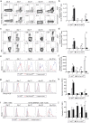
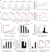
Figure 2. Improved primary but impaired secondary CD8+ T cell response in CD4-Cre A20fl/fl mice.
CD4-Cre A20fl/fl and A20fl/fl control mice were infected with a non-lethal dose of Lm OVA and CD8+ T cell response in spleen was analyzed at the indicated time points. (a) Representative dot plots and (b) absolute number of H2-Kb SIINFEKL pentamer+ CD8+ T cells after primary infection (day 0, 7 and 21 p.i.) and after reinfection (day 50 and 53 p.i.) with Lm OVA. (c) Representative dot plots and (d) absolute number of IFN-γ producing CD8+ T cells p.i. with Lm OVA and ex vivo restimulation with SIINFEKL peptide for 4 h in the presence of Brefeldin A. (e) IFN-γ-producing CD8+ T cells were gated and representative histograms of IFN-γ is displayed for the indicated time points after Lm OVA infection. (f) IFN-γ MFI from IFN-γ-producing CD8+ T cells. (g) Representative histograms and (h) granzyme B MFI of CD8+ T cells after Lm OVA infection and ex vivo restimulation with SIINFEKL peptide. (i) Representative histograms and (j) MFI of PD-1 expression on bulk CD8+ T cells (0 d.p.i.) or Lm OVA-specific CD8+ T cells (7, 21, 50 and 53 d.p.i.). A representative of 3 independent experiments is shown with 3 mice per group. Error bars indicate + SEM. Student’s t-test, *p < 0.05.
Figure 3
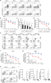
Figure 3. Reduced numbers of memory and memory-precursor CD8+ T cells in CD4-Cre A20fl/fl mice.
CD4-Cre A20fl/fl and A20fl/fl control mice were infected with a non-lethal dose of Lm OVA and spleens were analyzed for Tmem formation at the indicated time points. (a) Kinetic of Lm OVA-specific CD8+ T cells, stained with H2-Kb SIINFEKL pentamer and gated on singlets and CD3+ CD8+ cells. (b) Absolute numbers of total (living, apoptotic and necroptotic) Lm OVA-specific CD8+ T cells. (c) Relative numbers of living (annexin/7-AAD) Lm OVA-specific CD8+ T cells. (d) Absolute numbers of living (annexin/7-AAD) Lm OVA-specific CD8+ T cells. (e) Representative dot plots with frequencies of Lm OVA-specific CD8+ SLEC (KLRG-1+ CD127) and MPEC (KLRG-1 CD127+) were determined. (f) Absolute numbers of Lm OVA-specific CD8+ SLEC. (g) Absolute numbers of Lm OVA-specific CD8+ MPEC. (h) Representative dot plots with frequencies of Teff (CD62Llow and CD127low), TEM (CD62Llow and CD127high) and TCM (CD62Lhigh and CD127high) were determined at day 50 and 53 p.i. (i) Absolute numbers of Lm OVA-specific CD8+ Teff, TEM and TCM cells. Data are compiled of 2 independent experiments with 3–5 animals per group and experiment. Error bars indicate + SEM. Student’s t-test, *p < 0.05; **p < 0.01.
Figure 4
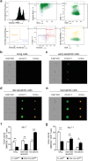
Figure 4. Enhanced necroptosis and apoptosis of A20-deficient CD8+ T cells.
CD4-Cre A20fl/fl and A20fl/fl control mice were infected with a sub-lethal dose of Lm OVA. On day 7 and 11 p.i., Lm OVA-specific CD8+ T cells were analyzed with IFC for apoptotic and necroptotic morphology. (a) IFC gating strategy for Lm OVA-specific CD8+ T cells and determination of double negative, annexin V+ (early apoptotic), and annexin V/7-AAD double positive cells. Double positive cells were further distinguished into late apoptotic and necroptotic cells. (b–e) Examples of microscopical analysis of cell morphology with IFC. (b) Living cells: negative for annexin V and 7-AAD. (c) Early apoptotic cells: annexin V+/7-AAD with beginning of nucleus condensation and membrane blebbing. (d) Late apoptotic cells: annexin V+/7-AAD+ with condensed nuclei and membrane blebbing. (e) Necroptotic cells: annexin V+/7-AAD+ with swollen cell and nuclei. (f–g) Frequency of living, apoptotic (early and late stage) and necroptotic Lm OVA-specific CD8+ T cells in spleen at day 7 p.i. (f) and day 11 p.i. (g). A representative of 2 independent experiments is shown, with 3–4 mice per group. Error bars indicate + SEM. Student’s t-test, *p < 0.05; **p < 0.01.
Figure 5
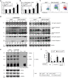
Figure 5. A20-deficient T cells are highly susceptible to apoptosis and necroptosis in vitro.
CD8+ T cells from CD4-Cre A20fl/fl and A20fl/fl control mice were isolated and stimulated with anti-CD3/CD28 and/or TNF, respectively, for the indicated time points. (a,b) Active caspase-3/7 was determined by flow cytometry using DEVD substrate after stimulation with (a) anti-CD3/CD28 only, or (b) anti-CD3/CD28 with TNF. (c) Dot Plots showing proliferation and caspase-3/7 activity after 72 h of stimulation. (d) Western blot analysis of full length caspase-8, cleaved caspase-8, full-length caspase-3 and cleaved caspase-3 after TCR or TNF stimulation, respectively. (e) Lysates from unstimulated and 48 h anti-CD3/CD28-stimulated CD8+ T cells were immunoprecipitated with anti-RIPK1. Protein concentrations were equalized by staining lysates for GAPDH. Beads plus lysate without antibody (B+L) and beads plus anti-RIPK1 without lysates (B+A) were used as controls. (f) CD8+ T cells were left untreated or stimulated with anti-CD3/CD28 for 72 h and IFC was performed. A representative of 2 independent experiments is shown, with 3 mice per group. Error bars indicate + SEM. Student’s t-test, *p < 0.05; **p < 0.01.
Figure 6
Figure 6. A20 reduces CD95 expression by inhibition of NF-κB activation.
(a) Histograms display MFI of CD95 ex vivo on Lm OVA-specific CD8+ T cells at the indicated time points p.i. (b) Ex vivo kinetic of active caspase-3/7 in Lm OVA-specific CD8+ T cells at the indicated time points p.i. (c) CD8+ T cells were in vitro stimulated with anti-CD3/CD28 and flow cytometric analysis for CD95 was performed at the indicated time points. (d) Frequency of living cells was determined after in vitro stimulation with anti-CD3/CD28. Cell death was determined by flow cytometry, staining for annexin V and 7-AAD. (e) T cells were stimulated with anti-CD3/CD28, treated with IKK-inhibitor or were left untreated. mRNA was isolated and levels of CD95 were determined. (f) CD8+ T cells were stimulated in vitro with anti-CD3/CD28 and recombinant CD95L for the indicated time points. Active caspase 3/7 was measured using DEVD substrate. (g) Frequency of living cells was determined after CD8+ T cells were stimulated with CD95L for 2 h. To inhibit apoptosis, Z-VAD-FMK was added prior to the stimulation. (h) Active caspase-3/7 was determined before and after AICD. (a,b) Cells were gated on singlets and H2-Kb SIINFEKL+ CD8+ T cells. A representative of 2 independent experiments is shown, with 3 mice per group. Error bars indicate + SEM. (a–d,f,h) Student’s t-test. (e,g) Tukey’s multiple comparison test; *p < 0.05; **p < 0.01.

Similar articles

Cited by

References

    1. Zhang N. & Bevan M. J. CD8(+) T cells: foot soldiers of the immune system. Immunity 35, 161–8 (2011). - PMC - PubMed
    1. Porter B. B. & Harty J. T. The onset of CD8+ -T-cell contraction is influenced by the peak of Listeria monocytogenes infection and antigen display. Infect. Immun. 74, 1528–1536 (2006). - PMC - PubMed
    1. Kaech S. M., Hemby S., Kersh E. & Ahmed R. Molecular and functional profiling of memory CD8 T cell differentiation. Cell 111, 837–851 (2002). - PubMed
    1. Hettmann T., Opferman J. T., Leiden J. M. & Ashton-Rickardt P. G. A critical role for NF-κB transcription factors in the development of CD8+ memory-phenotype T cells. Immunol. Lett. 85, 297–300 (2003). - PubMed
    1. Krishna S. et al.. Chronic activation of the kinase IKKβ impairs T cell function and survival. J. Immunol. 189, 1209–1219 (2012). - PMC - PubMed

Publication types

MeSH terms

Substances