. 2017 Apr 20;45(7):4142-4157.
doi: 10.1093/nar/gkw1287.

MicroRNA-202 maintains spermatogonial stem cells by inhibiting cell cycle regulators and RNA binding proteins

Affiliations

MicroRNA-202 maintains spermatogonial stem cells by inhibiting cell cycle regulators and RNA binding proteins

Jian Chen et al. Nucleic Acids Res. .

Abstract

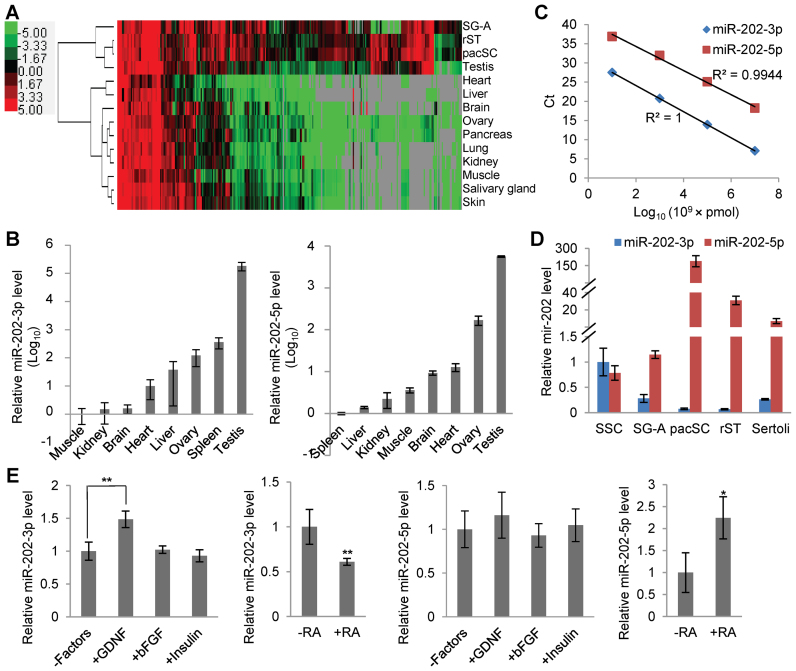
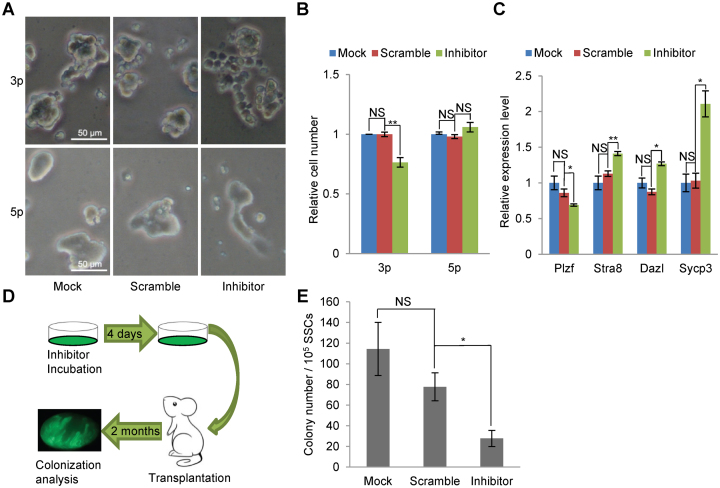
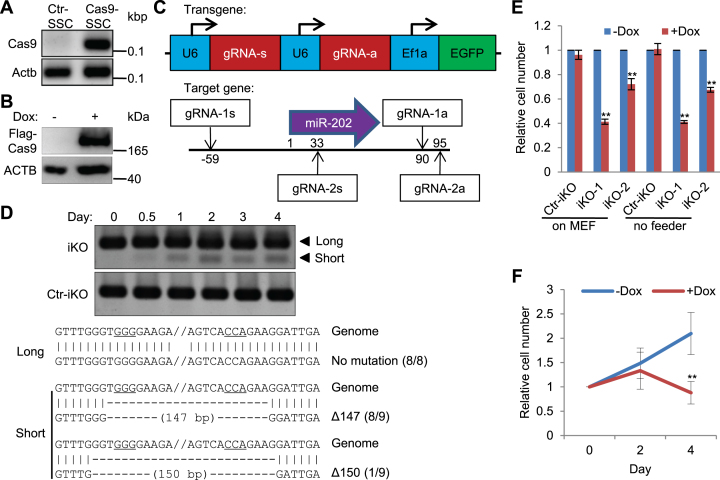
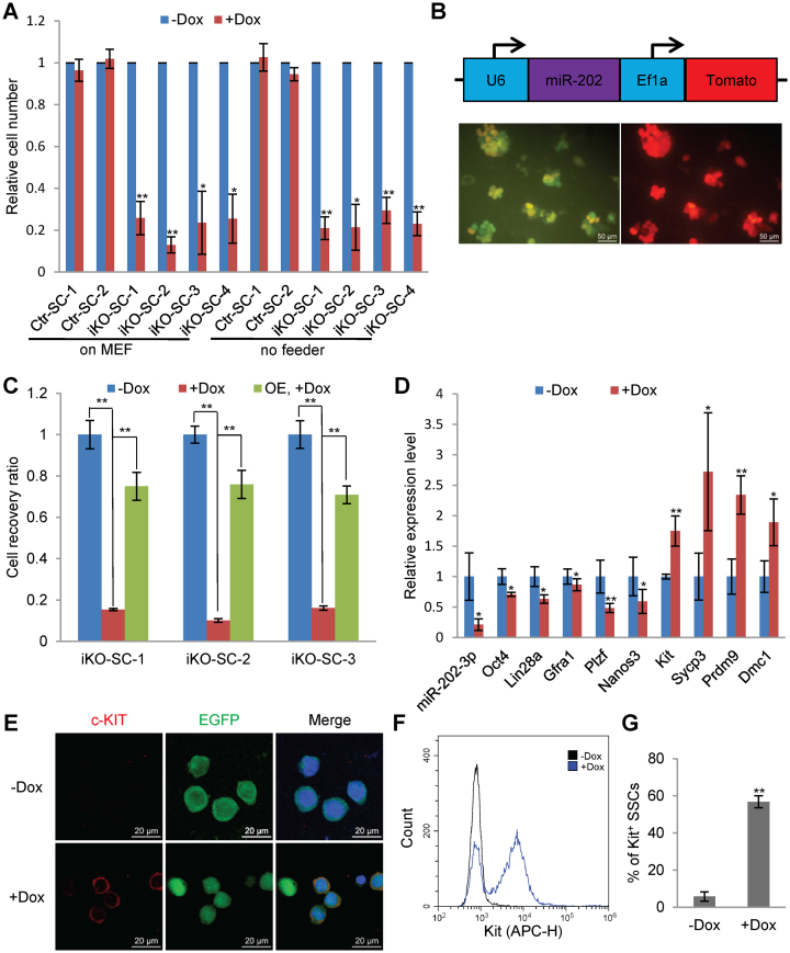
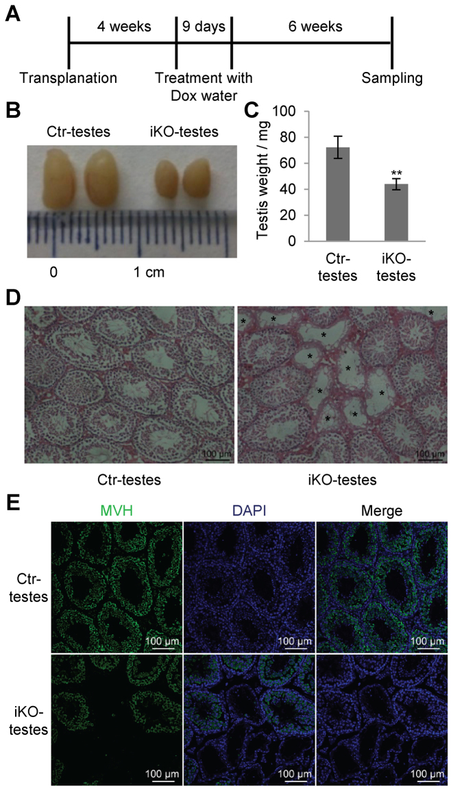
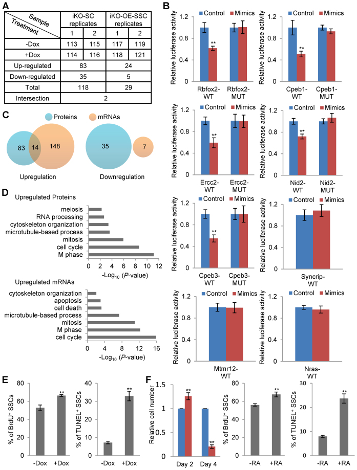
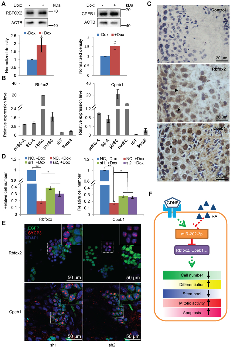
miRNAs play important roles during mammalian spermatogenesis. However, the function of most miRNAs in spermatogenesis and the underlying mechanisms remain unknown. Here, we report that miR-202 is highly expressed in mouse spermatogonial stem cells (SSCs), and is oppositely regulated by Glial cell-Derived Neurotrophic Factor (GDNF) and retinoic acid (RA), two key factors for SSC self-renewal and differentiation. We used inducible CRISPR-Cas9 to knockout miR-202 in cultured SSCs, and found that the knockout SSCs initiated premature differentiation accompanied by reduced stem cell activity and increased mitosis and apoptosis. Target genes were identified with iTRAQ-based proteomic analysis and RNA sequencing, and are enriched with cell cycle regulators and RNA-binding proteins. Rbfox2 and Cpeb1 were found to be direct targets of miR-202 and Rbfox2 but not Cpeb1, is essential for the differentiation of SSCs into meiotic cells. Accordingly, an SSC fate-regulatory network composed of signaling molecules of GDNF and RA, miR-202 and diverse downstream effectors has been identified.


Figures

Figure 1.
Figure 1.
Expression and regulation of miR-202-3p and miR-202-5p. (A) Clustering analysis of miRNAs highly expressed in at least one of the three spermatogenic cell types of SG-A, pacSC and rST. The values of the heat map are Log2 (CFs ×10 000). (B) qPCR analyses of miR-202-3p and miR-202-5p in multiple organs. (C) Standard expression curves of miR-202-3p and miR-202-5p plotted as Ct values verse the concentrations of miRNAs. (D) qPCR analyses of miR-202-3p and miR-202-5p in spermatogenic cells. (E) Regulation of miR-202-3p and miR-202-5p by growth factors and RA determined by qPCR. n = 4 for miR-202-3p and n = 3 for miR-202-5p.
Figure 2.
Figure 2.
Inhibition of miR-202-3p induces differentiation of SSCs. (A) Bright field images of SSCs treated with inhibitors for miR-202-3p and miR-202-5p on day 4 of treatment. (B) Quantification of cell numbers of feeder-free SSCs on day 4 of treatment with inhibitors. n = 5 for miR-202-3p and n = 3 for miR-202-5p. Data are normalized to mock SSCs. (C) qPCR analysis of the expression of marker genes in SSCs on day 4 of treatment with miR-202-3p inhibitor (n = 3). (D) The scheme of transplantation assays for assessing the stem cell contents of cultured SSCs. (E) Quantitative results of stem cell content assessment of SSCs treated with miR-202-3p inhibitor and controls by following the scheme in (D) (n = 3).
Figure 3.
Figure 3.
Deletion of miR-202 using inducible CRISPR-Cas9 system reduces the proliferation of SSCs. (A) PCR detection of Cas9 sequence integrated in the genome of SSCs. (B) Western blotting detection of Flag-Cas9 induced by Dox treatment. (C) Schematic illustration of gRNA expression from the lentiviral vector and their target sites in the genome. (D) Detection of miR-202-deleted genomic sequence in Doxycycline-treated iKO-SSCs and Ctr-iKO-SSCs by PCRs and sequencing. PAM sites are underlined. (E) Quantification of cell numbers of Ctr-iKO-SSCs, iKO-SSCs and iKO-SSCs-2 on day 4 of treatment. n = 5 for Ctr-iKO-SSCs (Ctr-iKO) and iKO-SSCs (iKO-1); n = 3 for iKO-SSCs-2 (iKO-2). (F) Time course of cell number changes of iKO-SSCs with and without Dox treatment. Data are normalized to the value on day 0 of treatment (n = 3).
Figure 4.
Figure 4.
Reduced proliferation of and the generation of c-KIT+ spermatogonia from miR-202-KO SSCs expanded from single cells. (A) Quantitative analyses of the proliferation of SSC clones expanded from single cells of iKO-SSCs (iKO-SC-1-4) and Ctr-iKO-SSCs (Ctr-iKO-SC-1-2). Data are normalized to SSCs without induction (n = 4). (B) Ectopic expression of miR-202 in SSCs of clone iKO-SC-1-3. Upper panel, the structure of the expression cassette; lower panel, fluorescent images of cells, which express EGFP from the double gRNA construct (see also Figure 3C) and Tomato from the ectopic expression construct. (C) Rescue of proliferation changes of SSCs in response to Dox treatment by the ectopic expression of miR-202 in clones iKO-SC-1-3. Data are normalized to SSCs without Dox treatment (n = 3). (D) qPCR analysis of marker gene expression on day 2 of Dox treatment (n = 3). (E) Immunofluorescence of c-KIT in cells on day 2 of Dox treatment. Blue color is from DAPI staining. (F) A representative plot of flow cytometry analysis of c-KIT+ cells on day 2 of Dox treatment. (G) Quantitative results of flow cytometry analyses of c-KIT+ SSCs represented by (E) (n = 3).
Figure 5.
Figure 5.
Stem cell activity assessment of SSCs by transplantation assays. (A) The scheme of transplantation assay for Ctr-iKO-SSCs and iKO-SSCs. (B) Images of Ctr-testes and iKO-testes. (C) Testis weight of Ctr-testes and iKO-testes after Dox treatment (n = 5). (D) Hematoxylin and Eosin (HE) images of testicular sections of Ctr-testes and iKO-testes after Dox induction. Germ cell-depleted tubules of iKO-testes are marked by asterisks. (E) MVH immunostaining of sections from Ctr-testes and iKO-testes to show the germ cell changes in response to miR-202 KO induced by Dox treatment. Paraffin sections of 5 μm were analyzed by immunofluorescence.
Figure 6.
Figure 6.
Proteomic and transcriptomic analyses of targets of miR-202. (A) The scheme of sample preparation for the differential proteomic analyses and the numbers of up- and downregulated proteins in response to miR-202 KO. The number 113–119, and 121 represent the isotopes used to label protein samples in the iTRAQ experiments. (B) Validation of proteomic approach-identified targets of miR-202 by dual luciferase assays (n = 6). (C) Venn diagrams showing the overlaps between up-/downregulated genes in response to miR-202 KO identified using proteomic and transcriptomic analyses. (D) GO terms enriched in upregulated gene sets. (E) BrdU incorporation and TUNEL assays of Dox-treated iKO-SC-1 by flow cytometry analyses on day 2 of treatment (n = 5 for BrdU and n = 4 for TUNEL). (F) Assessment of cell proliferation (n = 4), mitotic division (n = 5) and apoptosis (n = 4) of RA-treated SSCs on day 2 of treatment.
Figure 7.
Figure 7.
The miR-202 target Rbfox2 is essential for meiosis initiation. (A) RBFOX2 and CPEB1 increased their protein levels in response to miR-202 KO induced by Dox treatment (n = 3 for RBFOX2 and n = 4 for CPEB1). (B) qPCR analyses of Rbfox2 and Cpeb1 expression in different germ cell types. Data are normalized by the value of priSG-A (primitive SG-A) (n = 3). (C) Immunostaining of RBFOX2 and CPEB1 in mouse testis. Frozen sections of 10 μm were analyzed by immunohistochemistry. Arrowheads indicate pacSC with strong signals. (D) Proliferation assessment of iKO-SC-1 cells with and without the knockdown of Rbfox2 and Cpeb1 on day 4 of Dox treatment (n = 3 for Rbfox2 and n = 4 for Cpeb1). (E) Evaluation of meiosis initiation of SSCs by EGFP immunofluorescence and SYCP3 immunostaining. Insects are enlarged images of SYCP3+ SSCs. (F) A schematic model of a regulatory network that regulates the fate of SSCs in response to GDNF and RA. The signals were transduced to many effectors such Rbfox2 and Cpeb1 by miR-202 to change the proliferation, differentiation, stem cell activity, mitotic division and apoptosis.

Similar articles

Cited by

References

    1. Griswold M.D. Spermatogenesis: the commitment to meiosis. Physiol. Rev. 2016; 96:1–17. - PMC - PubMed
    1. de Rooij D.G., Griswold M.D.. Questions about spermatogonia posed and answered since 2000. J. Androl. 2012; 33:1085–1095. - PubMed
    1. Manku G., Culty M.. Mammalian gonocyte and spermatogonia differentiation: recent advances and remaining challenges. Reproduction. 2015; 149:R139–R157. - PubMed
    1. Wang S., Wang X., Ma L., Lin X., Zhang D., Li Z., Wu Y., Zheng C., Feng X., Liao S. et al. . Retinoic acid is sufficient for the in vitro induction of mouse spermatocytes. Stem Cell Rep. 2016; 7:80–94. - PMC - PubMed
    1. Busada J.T., Geyer C.B.. The role of retinoic acid (RA) in spermatogonial differentiation. Biol. Reprod. 2016; 94:10. - PMC - PubMed

Publication types