. 2016 Nov 15;7(6):e01553-16.
doi: 10.1128/mBio.01553-16.

Viral DNA Sensors IFI16 and Cyclic GMP-AMP Synthase Possess Distinct Functions in Regulating Viral Gene Expression, Immune Defenses, and Apoptotic Responses during Herpesvirus Infection

Affiliations

Viral DNA Sensors IFI16 and Cyclic GMP-AMP Synthase Possess Distinct Functions in Regulating Viral Gene Expression, Immune Defenses, and Apoptotic Responses during Herpesvirus Infection

Benjamin A Diner et al. mBio. .

Abstract

The human interferon-inducible protein IFI16 is an important antiviral factor that binds nuclear viral DNA and promotes antiviral responses. Here, we define IFI16 dynamics in space and time and its distinct functions from the DNA sensor cyclic dinucleotide GMP-AMP synthase (cGAS). Live-cell imaging reveals a multiphasic IFI16 redistribution, first to viral entry sites at the nuclear periphery and then to nucleoplasmic puncta upon herpes simplex virus 1 (HSV-1) and human cytomegalovirus (HCMV) infections. Optogenetics and live-cell microscopy establish the IFI16 pyrin domain as required for nuclear periphery localization and oligomerization. Furthermore, using proteomics, we define the signature protein interactions of the IFI16 pyrin and HIN200 domains and demonstrate the necessity of pyrin for IFI16 interactions with antiviral proteins PML and cGAS. We probe signaling pathways engaged by IFI16, cGAS, and PML using clustered regularly interspaced short palindromic repeat (CRISPR)/Cas9-mediated knockouts in primary fibroblasts. While IFI16 induces cytokines, only cGAS activates STING/TBK-1/IRF3 and apoptotic responses upon HSV-1 and HCMV infections. cGAS-dependent apoptosis upon DNA stimulation requires both the enzymatic production of cyclic dinucleotides and STING. We show that IFI16, not cGAS or PML, represses HSV-1 gene expression, reducing virus titers. This indicates that regulation of viral gene expression may function as a greater barrier to viral replication than the induction of antiviral cytokines. Altogether, our findings establish coordinated and distinct antiviral functions for IFI16 and cGAS against herpesviruses.

Importance: How mammalian cells detect and respond to DNA viruses that replicate in the nucleus is poorly understood. Here, we decipher the distinct functions of two viral DNA sensors, IFI16 and cGAS, during active immune signaling upon infection with two herpesviruses, herpes simplex virus 1 (HSV-1) and human cytomegalovirus (HCMV). We show that IFI16 rapidly oligomerizes at incoming herpesvirus genomes at the nuclear periphery to transcriptionally repress viral gene expression and limit viral replicative capacity. We further demonstrate that IFI16 does not initiate upstream activation of the canonical STING/TBK-1/IRF3 signaling pathway but is required for downstream antiviral cytokine expression. In contrast, we find that, upon DNA sensing during herpesvirus infection, cGAS triggers apoptosis in a STING-dependent manner. Our live-cell imaging, mass spectrometry-based proteomics, CRISPR-based cellular assays, and optogenetics underscore the value of integrative approaches to uncover complex cellular responses against pathogens.


Figures

FIG 1
FIG 1
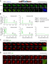
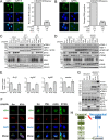
Multiphasic redistribution of IFI16 during both HSV-1 and HCMV infections. (A) Schematic of IFI16-eGFP fusion and HSV-1::bfp-nls recombinant virus containing a pCMV-bfp-nls expression cassette (top). HFFs expressing IFI16-eGFP were infected with HSV-1::bfp-nls (MOI of 10), and IFI16 was monitored by live-cell confocal fluorescence microscopy. Dynamic puncta are indicated (white arrowheads). Bars, 5 µm. (B) Maximum signal/noise pixel intensity ratios calculated in ImageJ are plotted as a function of time (in minutes). The characteristic phases of IFI16 are labeled and defined. (C and D) HFFs expressing IFI16-FusionRed were infected with either WT HCMV::gfp (MOI of 3) (C) or ΔpUL83 HCMV::gfp (MOI of 3) (D), and IFI16 was monitored and annotated as described above for panel A. See also Movie S1 and Fig. S1 in the supplemental material.
FIG 2
FIG 2
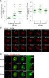
IFI16 localizes to sites of HSV-1 DNA deposition in an MOI-dependent manner. (A) Number of IFI16-eGFP peripheral (phase 1, left panel) and nucleoplasmic puncta (phase 2, right panel) observed in HSV-1::bfp-nls-infected HFFs at various viral loads (plaque-forming units/cell). Each green dot symbol represents the number of puncta observed per cell. Values are means (black line) ± standard errors of the means (SEMs) (n = 30) (error bars). Values that are significantly different by one-way ANOVA are indicated by asterisks as follows: ***, P < 0.001; ****, P < 0.0001. (B) HFFs expressing IFI16-FusionRed were infected with HSV-1 d109 infection (MOI of 10) and monitored by live-cell fluorescence confocal microscopy. Dynamic puncta are indicated (white arrowheads). Images are at the same magnification as shown in panel C. (C) As in panel B, IFI16-eGFP during HSV-1::mRFP-vp26 infection (MOI of 10). IFI16 was monitored and annotated as described above for panel B. Red fluorescent HSV-1 capsids are indicated (white arrowheads). Bar, 5 µm. See also Movie S2 in the supplemental material.
FIG 3
FIG 3
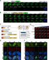
IFI16 PY and HIN domains display distinct behaviors during HSV-1 infection. (A and B) HFFs expressing either IFI16-PY-eGFP (A) or IFI16-HINAB-eGFP (b) infected with HSV-1::bfp-nls (MOI of 10) and imaged live by confocal fluorescence microscopy. Schematic representations of eGFP-tagged IFI16 derivatives are displayed above the images; pyrin (PY), HINAB (two HIN200 domains), and nuclear localization signal (NLS) are shown. Dynamic puncta are indicated (white arrowheads). Bars, 5 µm. (C, left) Schematic of FusionRed-CRY2olig-IFI16 fusions. FRed, FusionRed. (Right) Concept of blue light-induced CRY2olig-mediated aggregation and experimental scheme. LED, light-emitting diode. (D) FusionRed-CRY2olig-IFI16 fusions expressed in HFFs were induced to assemble (min 0 to 10) and disassemble (min 10 to 20). “Activation” represents application of blue light. Bars, 5 µm. (E) HFFs expressing IFI16 domain-eGFP fusions or eGFP alone infected with WT HSV-1 (MOI of 10) are stained for centromeres (anticentromere antibodies) at 3 hpi. Overlap between eGFP constructs (green) and centromeres (red) are shown (white arrowheads). DAPI, 4′,6′-diamidino-2-phenylindole. Bars, 5 µm. (F) As in panel E, WT, RF, d106, and d109 infection (MOI of 10). See also Movie S3 in the supplemental material.
FIG 4
FIG 4
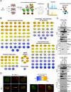
IFI16 interacts with PML and cGAS through the PY domain. (A) Scheme for immunoaffinity purification-mass spectrometry-based identification of IFI16 domain protein interactions in RF HSV-1-infected HFFs (MOI of 10) at 6 hpi. (B) Specificity (SAINT)-filtered IFI16-PY and IFI16-HIN interaction networks during RF HSV-1 were generated with the STRING database and rendered with Cytoscape. Colors represent relative spectral abundances of interactions enriched in either PY (orange) or HIN (blue) domain isolations. (C) Western blots of reciprocal immunoaffinity isolations in HEK293T cells cotransfected with the indicated IFI16-eGFP fusions (green arrowheads) and either FLAG-PML or FLAG-cGAS. The positions of molecular mass markers (in kDa) are shown to the left of the blots. (D) Immunofluorescence microscopy of indicated IFI16-eGFP fusions and PML in HFFs. A 3D image was rendered from Z-stacks. Bars, 5 µm. See also Fig. S2 in the supplemental material.
FIG 5
FIG 5
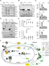
IFI16 is required for antiviral cytokine expression, but not activation of STING/TBK-1/IRF3 signaling. (A and B) CRISPR-mediated knockout of IFI16 (sgIFI16) (A) or PML (sgPML) (B) compared to scrambled sgRNA control cells (sgScr). (Left) CRISPR-HFFs were imaged by immunofluorescence microscopy. Bar, 5 µm. (Right) The average percentage of IFI16- or PML-positive cells per field (plus SEM) is plotted. Values that are significantly different (P ≤ 0.0001) from the value for the scrambled control by Student’s t test are indicated (****). The numbers of scored cells and fields are shown below the bars. (C) Western blots of CRISPR-HFFs infected with WT or RF HSV-1 (MOI of 10) at 6 hpi. (D) As in panel C, WT or ΔpUL83 HCMV infection (MOI of 3) at 6 hpi. (E) Cytokine mRNA levels in CRISPR-HFFs infected with RF HSV-1 (MOI of 10) at 6 hpi. Data were normalized to β-actin. Values are means ± SEMs (n = 2). Values that are significantly different from the value for the scrambled control by Student’s t test are indicated by asterisks as follows: *, P ≤ 0.05; **, P ≤ 0.01. (F) Immunofluorescence microscopy of ICP4 and PML (left panel) and of ICP4 and IFI16 (right panel) in CRISPR-HFFs (sgIFI16 versus sgScr and sgPML, sgcGAS, and sgSTING versus sgScr, respectively) upon RF HSV-1 infection (MOI of 0.1) at 24 hpi. A representative cell shown is at the edge of plaque. Bars, 10 µm. (G) Western blots of HEK293T or HEK293T-STING cells transfected with the indicated constructs. STING and IRF3 dimerization was tested by nonreducing SDS-PAGE and native PAGE, respectively. EV, empty vector. (H) Two proposed signaling cascades initiated in response to pathogenic DNA (red) by either cGAS (green) or IFI16 (blue). Question marks denote unknown signaling components. See also Fig. S2 in the supplemental material.
FIG 6
FIG 6
cGAS, not IFI16, is required for HSV-1 and DNA-dependent apoptosis. (A) Western blots of WT, RF, or d106 HSV-1-infected HFFs (MOI of 10) at the indicated times. (B) Western blots of cycloheximide (CHX) (10 µg/ml) or mock-treated HFFs infected with HSV-1 as described above for panel A for 24 h. (C) Western blots of d106 HSV-1-infected CRISPR-HFFs. (D) Western blot of HEK293T or HEK293T-STING cells transfected with indicated constructs for 16 h. (E) Western blots of HEK293T cells transfected as described above for panel D. AA, hcGAS GS212/213AA mutant; gt, human STING I200N mutation. For all panels, PARP cleavage is indicated by black arrowheads.
FIG 7
FIG 7
IFI16 suppresses HSV-1 gene expression and viral replication. (A) Western blots of CRISPR-HFFs infected with WT or RF HSV-1 (MOI of 1) for the indicated times. (B) As in panel A, induced Flp-In 293s expressing the indicated constructs (green arrowheads). (C) mRNA levels of immediate early (IE) and delayed early (DE) viral genes in RF HSV-1-infected CRISPR-HFFs (MOI of 1) at 8 hpi. Data normalized to β-actin. Values are means ± standard deviations (SD) (n = 3). (D) Western blots of RF HSV-1-infected Flp-In 293s expressing indicated IFI16-eGFP fusions (green arrowheads). (E) Progeny WT or RF HSV-1 titers from infected CRISPR-HFFs (MOI of 0.5). Cell-associated and cell-free virus were pooled at 24 hpi, and the titers of the virus on U2OS cells were determined by plaque assay. Values are means ± SEMs (n = 3). Values that are significantly different (P ≤ 0.001) from the value for the scrambled control by Student’s t test are indicated (***). (F) Model for IFI16 and cGAS antiviral functions. Nucleus-replicating DNA viruses, such as HSV-1 and HCMV, deposit their double-stranded DNA (dsDNA) genome into the nucleus (step 1). IFI16 PY domain oligomerizes at the nuclear periphery (step 2a), and HINAB domains bind the viral genome (step 2b). The PY domain interacts with ND10 bodies and their associated protein components (step 3), which may play auxiliary functions in coordinating with full-length (FL) IFI16 to transcriptionally repress viral gene expression (step 4a). IFI16 binding to viral DNA may trigger a noncanonical cytokine signaling pathway that is independent of STING (step 4b). Viral DNA is also sensed by cGAS. The resulting cGAMP production stimulates the STING/TBK-1/IRF3 signaling axis to induce antiviral cytokines and apoptosis. ER, endoplasmic reticulum.

Similar articles

Cited by

References

    1. Kerur N, Veettil MV, Sharma-Walia N, Bottero V, Sadagopan S, Otageri P, Chandran B. 2011. IFI16 acts as a nuclear pathogen sensor to induce the inflammasome in response to Kaposi Sarcoma-associated herpesvirus infection. Cell Host Microbe 9:363–375. doi:10.1016/j.chom.2011.04.008. - DOI - PMC - PubMed
    1. Li T, Diner BA, Chen J, Cristea IM. 2012. Acetylation modulates cellular distribution and DNA sensing ability of interferon-inducible protein IFI16. Proc Natl Acad Sci U S A 109:10558–10563. doi:10.1073/pnas.1203447109. - DOI - PMC - PubMed
    1. Orzalli MH, DeLuca NA, Knipe DM. 2012. Nuclear IFI16 induction of IRF-3 signaling during herpesviral infection and degradation of IFI16 by the viral ICP0 protein. Proc Natl Acad Sci U S A 109:E3008–E3017. doi:10.1073/pnas.1211302109. - DOI - PMC - PubMed
    1. Li T, Chen J, Cristea IM. 2013. Human cytomegalovirus tegument protein pUL83 inhibits IFI16-mediated DNA sensing for immune evasion. Cell Host Microbe 14:591–599. doi:10.1016/j.chom.2013.10.007. - DOI - PMC - PubMed
    1. Orzalli MH, Conwell SE, Berrios C, DeCaprio JA, Knipe DM. 2013. Nuclear interferon-inducible protein 16 promotes silencing of herpesviral and transfected DNA. Proc Natl Acad Sci U S A 110:E4492–E4501. doi:10.1073/pnas.1316194110. - DOI - PMC - PubMed

MeSH terms