. 2016 Dec 13;113(50):14402-14407.
doi: 10.1073/pnas.1611106113. Epub 2016 Nov 23.

Activation of mutant TERT promoter by RAS-ERK signaling is a key step in malignant progression of BRAF-mutant human melanomas

Affiliations

Activation of mutant TERT promoter by RAS-ERK signaling is a key step in malignant progression of BRAF-mutant human melanomas

Yinghui Li et al. Proc Natl Acad Sci U S A. .

Abstract

Although activating BRAF/NRAS mutations are frequently seen in melanomas, they are not sufficient to drive malignant transformation and require additional events. Frequent co-occurrence of mutations in the promoter for telomerase reverse transcriptase (TERT), along with BRAF alterations, has recently been noted and correlated with poorer prognosis, implicating a functional link between BRAF signaling and telomerase reactivation in melanomas. Here, we report that RAS-ERK signaling in BRAF mutant melanomas is critical for regulating active chromatin state and recruitment of RNA polymerase II at mutant TERT promoters. Our study provides evidence that the mutant TERT promoter is a key substrate downstream of the RAS-ERK pathway. Reactivating TERT and hence reconstituting telomerase is an important step in melanoma progression from nonmalignant nevi with BRAF mutations. Hence, combined targeting of RAS-ERK and TERT promoter remodeling is a promising avenue to limit long-term survival of a majority of melanomas that harbor these two mutations.

Keywords: BRAF mutations; ERK-MAPK pathway; TERT promoter mutations; cancer; telomerase reactivation.


Conflict of interest statement

The authors declare no conflict of interest.

Figures
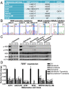
Fig. 1.
Fig. 1.
Disruption of the RAS-ERK pathway inhibits the transcriptional activation of TERT at mutant TERT promoters in BRAF/NRAS mutant melanoma cells. (A) Table shows the profiles of BRAF, NRAS, and TERT promoter mutations in human melanoma cell lines. (B) Representative DNA chromatogram sequences of BRAF, NRAS, and TERT promoter mutations of melanoma cell lines analyzed. (C) A375 cells were treated with 0.4 µM verumafenib, 0.1 µM dabrafenib, and 20 nM trametinib alone or in combination with trametinib for 1, 3, and 6 h. Cell lysates of control and treated samples were analyzed for changes in MEK1/2 and ERK1/2 phosphorylation by Western blotting. (D) BRAF/NRAS-mutant, TERT promoter mutant melanoma cells were treated with 0.1 µM dabrafenib, 0.4 µM verumafenib, and 20 nM trametinib alone or in combination with trametinib for 1 d and analyzed for TERT expression by quantitative real-time PCR (qPCR). Data from three experiments are represented as the relative fold change in mRNA expression (mean ± SEM). Student’s t test (two-tailed) was used for all statistical analyses: **P < 0.01; ***P < 0.001.
Fig. S1.
Fig. S1.
Downregulation of RAS-ERK signaling reduces the proliferation of BRAF/NRAS-mutant melanoma cells carrying the TERT promoter mutation. (A) Cell viability assay of A375, UACC257, G361, and BLM cells following 3 d treatment of 0.1 µM dabrafenib, 0.4 µM verumafenib, and 20 nM trametinib alone or in combination with trametinib. Data from two experiments are represented as average fluorescence intensity (mean ± SEM). (B) si-Ctrl or si-BRAF–treated UACC257 and BLM cells were analyzed for levels of phosphorylated or total MEK1/2 and ERK1/2 by Western blotting.
Fig. 2.
Fig. 2.
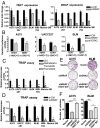
Inhibition of RAS-ERK signaling reduces the telomerase activity and proliferation of BRAF/NRAS-mutant melanoma cells carrying TERT promoter mutations. (A) A375, UACC257, G361, BLM, WM793, and Malme-3M cells were treated with si-Control (Ctrl) or si-BRAF and analyzed for TERT and BRAF expression after 3 d. Plots represent relative fold changes in mRNA expression (mean ± SEM) from two independent experiments. (B) Luciferase reporter assays were performed in A375, UACC257, and BLM cells that were transfected with pGL3 reporter vector containing the TERT promoter region (−340 to −55) with −146C > T or −124C > T mutation. Transfected cells were treated with siRNA-targeting BRAF or ERK2. Data shown are representative of two independent experiments. (C) A375, UACC257, G361, WM793, and Malme-3M cells were treated with BRAF and MEK inhibitors (0.1 µM dabrafenib and 20 nM trametinib), and BLM cells were treated with MEK inhibitor (20 nM trametinib). DMSO control (Ctrl) or BRAF/MEK inhibitor-treated cells were analyzed for telomerase activity after 2 d by a Telomere Repeat Amplification Protocol (TRAP) assay. Data shown are from two experiments and represent relative telomerase activity (mean ± SEM). (D) A375, UACC257, G361, BLM, WM793, and Malme-3M cells were transfected with si-Ctrl or si-BRAF and analyzed for telomerase activity after 3 d. Data from two experiments are shown (mean ± SEM). (E) Colony formation assay was performed in vector or shBRAF-expressing A375 and vector or shERK2 BLM cells. TERT was re-expressed in shBRAF and shERK2 cells via lentiviral transduction of Flag-TERT WT construct, and colony formation assay was performed in parallel. Representative images from two independent experiments are shown. (F) Plot depicts average number of colonies counted per field (mean ± SEM) from all experiments in the earlier assay. Student’s t test (two-tailed) was used for all statistical analyses: *P < 0.05; ***P < 0.001.
Fig. S2.
Fig. S2.
Inhibition of RAS-ERK signaling via BRAF downregulation reduces TERT expression of BRAF/NRAS-mutant melanoma cells with TERT promoter mutation. (A) A375, BLM, and UACC257 cells were transduced with shRNA targeting BRAF and analyzed for TERT, BRAF, and uPA expression by qPCR. Plots show relative fold changes in mRNA expression (mean ± SEM) from two experiments. *P < 0.05; **P < 0.01; Student’s t test (two-tailed). (B) Vector and shBRAF-expressing melanoma cells were analyzed for protein levels of BRAF and phosphorylated or total MEK1/2 and ERK1/2. (C) A375 and BLM were transduced with shBRAF, shERK2 alone, or in combination with the TERT WT overexpression construct and analyzed for protein levels of TERT, BRAF, and phosphorylated or total ERK1/2.
Fig. 3.
Fig. 3.
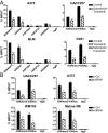
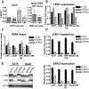
RAS-ERK signaling regulates the active chromatin state of mutant TERT promoters. (A) ChIP was performed in control or BRAF/MEK inhibitor-treated A375, UACC257, BLM, and G361 cells using antibodies specific for different histone marks and IgG as negative control. Enrichment of TERT promoter DNA fragments in ChIP DNA was normalized to DNA input. (B) ChIP was performed in si-Ctrl or si-BRAF–transfected UACC257, A375, WM793, and Malme-3M cells using antibodies specific for H3K4me3 and H3K9ac and IgG as negative control. Data shown represent at least two independent ChIP assays for all cell lines. Student’s t test (two-tailed) was used for all statistical analyses of control versus treated samples for each ChIP: *P < 0.05; **P < 0.01; ***P < 0.001.
Fig. 4.
Fig. 4.
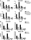
The RAS-RAF-MEK-ERK pathway regulates the recruitment of RNA polymerase II at mutant TERT promoters in human melanoma cells. (A) ChIP was performed in control or BRAF/MEK inhibitor-treated A375, UACC257, BLM, and G361 cells using antibodies specific for RNA polymerase II (Pol II), Pol II phosphorylated on Serine 5 (Pol II S5P) and Serine 2 (Pol II S2P), and IgG as negative control. Enrichment of TERT promoter DNA fragments in ChIP DNA was normalized to DNA input. (B) ChIP was performed in si-Ctrl or si-BRAF–transfected UACC257, A375, WM793, and Malme-3M cells using antibodies specific for Pol II, Pol II S5P, and IgG as negative control. Plots shown represent at least two independent ChIP assays for all cell lines. *P < 0.05; **P < 0.01; ***P < 0.001; Student’s t test (two-tailed).
Fig. 5.
Fig. 5.
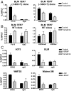
Conversion of TERT promoter mutation to WT promoter abolishes the regulation of the RAS-ERK pathway on TERT reactivation. ChIP was performed in control or trametinib-treated BLM mutant TERT (−146C > T) promoter CRISPR cells (A) or BLM WT TERT promoter CRISPR cells (B) using antibodies specific for RNA polymerase II (Pol II), H3K4me3, and H3K9ac and IgG as negative control. Enrichment of TERT promoter DNA fragments in ChIP DNA was normalized to DNA input. (C) ERK2 is bound to mutant TERT promoters of BRAF/NRAS-mutant melanoma cells. ChIP was performed in si-Ctrl or si-BRAF–treated A375, WM793, and Malme-3M cells and control or trametinib-treated BLM cells using ERK-2–specific antibody and IgG as negative control. Data shown represent two independent experiments for all cell lines. *P < 0.05, Student’s t test (two-tailed).
Fig. S3.
Fig. S3.
Knockdown of ERK2 reduces telomerase function of TERT promoter mutant melanoma cells. (A) ChIP was performed in BRAF-mutant, WT TERT promoter C8161 cells using antibodies specific for ERK2, GABPA, and RNA polymerase II (Pol II) and IgG as negative control. ERK2 ChIP was performed in BLM TERT WT CRISPR cells. Data shown represent two independent experiments. (BE) TERT promoter mutant melanoma cells were transfected with si-Ctrl, si-ERK1, or si-ERK2 and analyzed for TERT expression (B), telomerase activity (C), ERK1 and ERK2 expression (D), and protein levels of phosphorylated and total ERK1/2 (E). Expression and TRAP assay data shown are from three experiments. *P < 0.05; **P < 0.01; ***P < 0.001; Student’s t test.
Fig. 6.
Fig. 6.
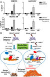
Inhibition of the RAS-ERK pathway promotes the recruitment of the HDAC1 repressor complex to mutant TERT promoters of melanoma cells. (A) HDAC1 and Sp1 ChIP was performed in the BRAF-mutant, TERT promoter mutant melanoma cells WM793, UACC257, Malme-3M, and A375, which were treated either with si-BRAF or BRAF and MEK inhibitors to disrupt ERK activation. Enrichment of TERT promoter DNA fragments in ChIP DNA was normalized to DNA input. Data shown are representative of two independent experiments. *P < 0.05; Student’s t test (two-tailed). (B) A model for TERT reactivation at mutant TERT promoter during constitutive RAS-ERK signaling in BRAF-mutant melanoma tumors is shown.

Similar articles

Cited by

References

    1. Paluncic J, et al. Roads to melanoma: Key pathways and emerging players in melanoma progression and oncogenic signaling. Biochim Biophys Acta. 2016;1863(4):770–784. - PubMed
    1. Johannessen CM, et al. A melanocyte lineage program confers resistance to MAP kinase pathway inhibition. Nature. 2013;504(7478):138–142. - PMC - PubMed
    1. Poulikakos PI, Rosen N. Mutant BRAF melanomas: Dependence and resistance. Cancer Cell. 2011;19(1):11–15. - PubMed
    1. Wellbrock C, Arozarena I. The complexity of the ERK/MAP kinase pathway and the treatment of melanoma skin cancer. Front Cell Dev Biol. 2016;4:33. - PMC - PubMed
    1. Davies H, et al. Mutations of the BRAF gene in human cancer. Nature. 2002;417(6892):949–954. - PubMed

Publication types

MeSH terms