. 2017 Jan 10;25(1):166-181.
doi: 10.1016/j.cmet.2016.10.023. Epub 2016 Nov 23.

Cellular Aging Contributes to Failure of Cold-Induced Beige Adipocyte Formation in Old Mice and Humans

Affiliations

Cellular Aging Contributes to Failure of Cold-Induced Beige Adipocyte Formation in Old Mice and Humans

Daniel C Berry et al. Cell Metab. .

Erratum in

Abstract

Cold temperatures induce progenitor cells within white adipose tissue to form beige adipocytes that burn energy and generate heat; this is a potential anti-diabesity therapy. However, the potential to form cold-induced beige adipocytes declines with age. This creates a clinical roadblock to potential therapeutic use in older individuals, who constitute a large percentage of the obesity epidemic. Here we show that aging murine and human beige progenitor cells display a cellular aging, senescence-like phenotype that accounts for their age-dependent failure. Activating the senescence pathway, either genetically or pharmacologically, in young beige progenitors induces premature cellular senescence and blocks their potential to form cold-induced beige adipocytes. Conversely, genetically or pharmacologically reversing cellular aging by targeting the p38/MAPK-p16Ink4a pathway in aged mouse or human beige progenitor cells rejuvenates cold-induced beiging. This in turn increases glucose sensitivity. Collectively, these data indicate that anti-aging or senescence modalities could be a strategy to induce beiging, thereby improving metabolic health in aging humans.

Keywords: Ink4a/Arf; adipose; beige adipocytes; cellular aging; cold exposure; mural cells; senescence; thermogenesis.


Figures

Figure 1
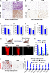
Figure 1. Age dependent failure of cold-inducible beige fat formation
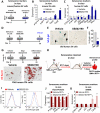
Two (Young) or six (Old) month old C57/Bl6 male mice were maintained at room temperature (23°C) or cold exposed (6°C) for seven days. (A-B) H&E staining. (A-B) UCP1 IHC. (C) Rectal temperature. (D) Sera glucose. (E) Free fatty acids. (F) Fat content. Data are means ± SEM; n=6 mice/group. Scale bar = 200 μm. *P<0.05 cold compared to room temperature controls. #P<0.02 Old compared to young cold exposed cohorts. (G) Un-induced UCP1-CreERT2; RFP male mice were aged to postnatal day (P), 60, 120 and 180 and subsequently cold exposed (6°C). Immediately after cold exposure mice were TM induced. IGW were examined for direct RFP fluorescence. L = Lymph node. Scale bar = 4 mm. (H) Beige and thermogenic gene expression from mice describe in (G). §P<0.01 Young compared to Old cold exposed cohorts. (I-J) Beige adipocyte differentiation of SV cells from two or six month old C57/Bl6 male mice: Oil Red O (I) and beige adipocyte markers (J). ##P <0.001 young beige adipocytes compared to undifferentiated SV cells.
Figure 2
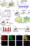
Figure 2. Old beige progenitors express a cellular aging/senescence phenotype
(A) Senescence activated (SA) βgal staining of inguinal (IGW) adipose depots from two (Young) and six (Old) month old male mice. Arrow points to SA-βgal positive areas. Scale bar = 4 mm. (B) Expression of senescence genes from two and six month old mice. Data are means ± SEM; n=5 mice/group. *P-value <0.05 Old compared to young cells. (C) Expression of senescence genes from young and old BMI matched human SV cells. **P <0.01 Old compared to young cells. (D-E) Two month and six month old SMA-CreERT2; R26RRFP male mice were administered TM; RFP+ cells were FACS isolated at pulse. Cells were stained for Senescence Activated (SA)-βgal (D) or mRNA expression of senescence genes (E) was analyzed. #P<0.05 old SMA/RFP+ compared to young SMA/RFP+ cells. (F) Two or six month old SMA-CreERT2; R26RRFP TM pulsed male mice were randomized to the cold for seven days. (G) Sections from mice described in (F) were analyzed for RFP and UCP1. DAPI was used to visualize nuclei. Scale bar = 200 μm.
Figure 3
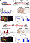
Figure 3. Senescence is sufficient to block cold-inducible beige formation
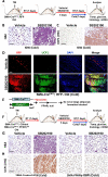
(A-B) Generation of SMA-rtTA; TRE-Cre; TRE-p21; R26RRFP (SMA-p21-OE) (A). Two-month-old SMA-p21-OE male mice were administered Doxycycline for 14 days then mice were analyzed for senescence markers or were randomized to RT or cold for seven days (B). (C) Mice described in (B) were analyzed for senescence markers. (D-H) UCP1 IHC (D), fate mapping of RFP fluorescence and UCP1 IHC (E), rectal temperature (F), sera glucose (G), and IGW weight (H) from mice described in (B). Data are means ±S.E.M; n=8 mice/group. #P <0.01 young TRE-p21 mutant mice compared to control mice. §P<0.02 cold compared to room temperature. (I-J) Schema- Two-month-old SMA-CreERT2; R26RRFP male mice were TM induced, one week later mice were administered vehicle (5% DMSO /water) or PD (250 μg/mouse/day) for five consecutive days. Mice were subsequently analyzed for senescence markers. *P ≤0.05 SMA/RFP+ cells from PD0332991 treated mice compared to SMA/RFP+ cells from vehicle treated mice. (K-O) UCP1 IHC (K), fate mapping of RFP fluorescence and UCP1 IHC (L), temperature (M), glucose (N), and IGW weight (O). Data are means ± S.E.M; n=9 mice/group. *P ≤0.05 PD0332991 treated compared to vehicle treated mice. §P <0.02 cold compared to room temperature. Scale bar = 200 μm.
Figure 4
Figure 4. Deleting Ink4a/Arf in beige progenitors reverses cellular senescence
(A) Ink4a and Bmi1 expression from C57/Bl6 male mice at denoted ages. *P <0.001 Old compared to young cells. (B) Illustration of genetic alleles used to generate SMA-CreERT2; Ink4a/Arffl/fl; RR26RRFP (SMA-Ink/Arf) mice. (C-F) Schema- Six-month-old SMA-CreERT2; Ink4a/Arffl/fl male mice were TM induced. Fourteen days later, SMA/RFP+ cells were FACS isolated and mRNA (D and E) and protein (F) expression of senescence markers were examined. #P<0.05 mutant SMA/RFP+ cells compared to control SMA/RFP+ cells. (G) 12-month-old SMA-CreERT2; Ink4a/Arffl/fl male mice were TM induced. Fourteen days later, SMA/RFP+ cells were FACS isolated and expression of senescence genes were examined. #P<0.05 mutant SMA/RFP+ cells compared to control SMA/RFP+ cells. (H-J) Un-induced SMA-CreERT2; Ink4a/Arffl/fl cells were cultured in vehicle or TM for 48 hrs. Beige adipocyte differentiation was assessed by Oil Red O (H), triglyceride levels (I) and beige and thermogenic mRNA expression (J). *P <0.001 Old compared to young cells. **P<0.0001 mutant compared to control mice.
Figure 5
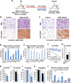
Figure 5. Reversing cellular aging/senescence is sufficient to restore cold-inducible beige formation
(A-L) Six and twelve month old SMA-CreERT2; Ink4a/Arffl/fl male mice were TM induced and then cold exposed for seven days (A). (B-C) H&E staining and UCP1 IHC. (D-E) mRNA of beige and thermogenic genes. F-G) Rectal temperatures. H-I) Sera glucose. J-K) Fat content. L) Glucose uptake. Data are means ±S.E.M; n=4-9 mice/group. *P<0.001 SMA-CreERT2; Ink4a/Arffl/fl mutant mice compared to control mice. #P<0.02 cold exposed mice compared to room temperature mice. §P<0.01 twelve month old SMA-CreERT2; Ink4a/Arffl/fl mutant mice compared to aged matched control mice. **P≤0.05 SMA-CreERT2; Ink4a/Arffl/fl mutant mice compared to control mice. Scale bar = 200 μm.
Figure 6
Figure 6. Pharmacologically targeting p38/MAPK in old SMA+ mural cells rejuvenates beige formation
(A-C) Total SV cells were isolated from six month old C57/Bl6 male mice (A, B) or SV cells generated from aged human samples. Cells were treated with vehicle or SB202190 (5 μM) for 15 consecutive days. mRNA of senescence markers was examined. *P <0.01 Old compared to young murine cells. *P <0.05 Old compared to young murine cells. (D-F) Human SV cells were treated with vehicle or SB for 15 consecutive days. Subsequently, SA-βgal staining was performed (E) and positive cells were quantified (F). (G) Cells described in (D) were assessed for beige adipogenesis by Oil Red O staining. (H-K) Six month old TM induced SMA-CreERT2; R26RRFP male mice were administered vehicle or SB (75 μg/mouse/day) for five consecutive days (H). RFP+ cells were counted (I) and FACS isolated and examined for phosphor-p38/MAPK and denoted proteins (I, J) and expression of senescence genes (K). Data are means ±S.E.M; n=4 mice/group. #P<0.03 SB202190 treated RFP+ cells compared to vehicle treated RFP+ cells.
Figure 7
Figure 7. Inhibiting p38/MAPK in SMA cells stimulates cold-induced beiging
(A-D) Schema- Six month old TM induced SMA-CreERT2; R26RRFP male mice were administered vehicle or SB202190 (75 μg/mouse/day) for five consecutive days. Mice were randomized to room temperature or cold for seven days (A). (B) H&E staining. (C) UCP1 IHC. (D) SMA/RFP fluorescence fate mapping. (E-H) Schema- Six-month-old TM induced SMA-CreERT2 and SMA-CreERT2; PPARγfl/fl male mice were administered vehicle or SB (75 μg/mouse/day) for five days and subjected to room or cold temperatures for seven days. (G) H&E staining. (H) UCP1 IHC. Scale bar = 200 μm.

Comment in

Similar articles

Cited by

References

    1. Berglund ED, Vianna CR, Donato J, Jr., Kim MH, Chuang JC, Lee CE, Lauzon DA, Lin P, Brule LJ, Scott MM, et al. Direct leptin action on POMC neurons regulates glucose homeostasis and hepatic insulin sensitivity in mice. J Clin Invest. 2012;122:1000–1009. - PMC - PubMed
    1. Bernet JD, Doles JD, Hall JK, Kelly Tanaka K, Carter TA, Olwin BB. p38 MAPK signaling underlies a cell-autonomous loss of stem cell self-renewal in skeletal muscle of aged mice. Nat Med. 2014;20:265–271. - PMC - PubMed
    1. Berry DC, Jiang Y, Graff JM. Mouse strains to study cold-inducible beige progenitors and beige adipocyte formation and function. Nat Commun. 2016;7:10184. - PMC - PubMed
    1. Berry DC, Noy N. All-trans-retinoic acid represses obesity and insulin resistance by activating both peroxisome proliferation-activated receptor beta/delta and retinoic acid receptor. Mol Cell Biol. 2009;29:3286–3296. - PMC - PubMed
    1. Berry DC, Soltanian H, Noy N. Repression of cellular retinoic acid-binding protein II during adipocyte differentiation. J Biol Chem. 2010;285:15324–15332. - PMC - PubMed

Publication types

Substances