. 2017 Feb;19(2):250-262.
doi: 10.1016/j.jcyt.2016.10.011. Epub 2016 Nov 22.

Optimization of cGMP purification and expansion of umbilical cord blood-derived T-regulatory cells in support of first-in-human clinical trials

Affiliations

Optimization of cGMP purification and expansion of umbilical cord blood-derived T-regulatory cells in support of first-in-human clinical trials

David H McKenna Jr et al. Cytotherapy. 2017 Feb.

Abstract

Background aims: Thymic-derived regulatory T cells (tTreg) are critical regulators of the immune system. Adoptive tTreg transfer is a curative therapy for murine models of autoimmunity, graft rejection, and graft-versus-host disease (GVHD). We previously completed a "first-in-human" clinical trial using in vitro expanded umbilical cord blood (UCB)-derived tTreg to prevent GVHD in patients undergoing UCB hematopoietic stem cell transplantation (HSCT). tTreg were safe and demonstrated clinical efficacy, but low yield prevented further dose escalation.

Methods: To optimize yield, we investigated the use of KT64/86 artificial antigen presenting cells (aAPCs) to expand tTreg and incorporated a single re-stimulation after day 12 in expansion culture.

Results: aAPCs increased UCB tTreg expansion greater than eightfold over CD3/28 stimulation. Re-stimulation with aAPCs increased UCB tTreg expansion an additional 20- to 30-fold. Re-stimulated human UCB tTreg ameliorated GVHD disease in a xenogeneic model. Following current Good Manufacturing Practice (cGMP) validation, a trial was conducted with tTreg. tTreg doses up to >30-fold higher compared with that obtained with anti-CD3/28 mAb coated-bead expansion and Foxp3 expression was stable during in vitro expansion and following transfer to patients. Increased expansion did not result in a senescent phenotype and GVHD was significantly reduced.

Discussion: Expansion culture with cGMP aAPCs and re-stimulation reproducibly generates sufficient numbers of UCB tTreg that exceeds the numbers of T effector cells in an UCB graft. The methodology supports future tTreg banking and is adaptable to tTreg expansion from HSC sources. Furthermore, because human leukocyte antigen matching is not required, allogeneic UCB tTreg may be a useful strategy for prevention of organ rejection and autoimmune disease.

Keywords: cGMP production; graft versus host disease; regulatory T cell.


Conflict of interest statement

The authors share a patent entitled “Methods to expand a T Regulatory Cell Master Cell Bank”; US Patent # 13/639,927.

Figures

Figure 1
Figure 1. Artificial APC increase UCB tTreg expansion compared to anti-CD3/28 beads
tTreg were purified from frozen umbilical cord blood units under cGMP conditions using anti-CD25 magnetic beads and CliniMACS and were expanded in vitro with either anti-CD3/28 mAb-coated beads or aAPC (KT64/86). (A) Schematic representation of UCB tTreg culture. (B) Fold tTreg expansion (average ± SEM). (C) Percentage of cultured cells (CD4-gated) that are CD127–Foxp3+ after stimulation. (D) Percent suppression of in vitro, anti-CD3–mediated CD8+ T cell proliferation at 1:2 (tTreg/PBMC) as determined by CFSE dye dilution. From n=3 experiments.
Figure 2
Figure 2. Re-stimulation of UCB tTreg greatly increases expansion without compromising phenotype or suppressive function
tTreg were purified from frozen umbilical cord blood units under cGMP conditions using anti-CD25 magnetic beads and CliniMACS and were expanded in vitro with one or two rounds of stimulation with anti-CD3 loaded aAPC (KT64/86). (A) Schematic representation of UCB tTreg culture. (B) Fold tTreg expansion (average ± SEM). (C) Percentage of cultured cells (CD4-gated) that are CD127negFoxp3+ after stimulation. (D) Percent suppression of in vitro, anti-CD3–mediated CD8+ T cell proliferation at 1:2 (tTreg/PBMC) as determined by CFSE dye dilution. From n=4 experiments.
Figure 3
Figure 3. UCB tTreg expanded with aAPC and re-stimulation maintain in vivo suppressive function
Re-stimulated UCB tTreg (15 × 106 cells) were co-transferred with allogeneic PBMCs (15 × 106 cells) into NOD/Scid/γc−/− mice to assess the ability to ameliorate xenogeneic GVHD. (A) Characteristics of the UCB tTreg cultures that were used. (B) Average weight (percentage of initial) for mice surviving on a given day for different groups of mice (*P < 0.05 for tTreg from days 12 to 23 for UCB tTreg). (C) Average clinical score for mice surviving on a given day for different groups of mice (*P < 0.05 for tTreg from days 12 to 23 for UCB tTreg). (D) Kaplan– Meier survival curves for mice receiving PBMCs only or PBMCs plus adoptive transfer of re-stimulated UCB tTreg. Three independent experiments were performed with re-stimulated UCB tTreg with similar results. Re-stimulated UCB tTreg were either kept in culture or frozen in a rate-controlled freezer and stored overnight. UCB tTreg were processed (washed or thawed/washed), and were co-transferred (15 × 106 cells) with allogeneic PBMCs (15 × 106 cells) into irradiated (50 cGy) NOD/Scid/γc−/− mice to compare their ability to ameliorate xenogeneic GVHD. (E) Kaplan– Meier survival curves for mice receiving PBMCs only or PBMCs plus adoptive transfer of re-stimulated UCB tTreg (n=10). To assess tTreg persistence, animals were bled on day 7 and the number of allotype-specific tTreg/μl blood determined. (G) UCB tTreg were purified, stimulated with KT64/86, cultured for 14 days, and then frozen. After several weeks, tTreg were thawed, re-stimulated with KT64/86, expanded for 7 days and then used as prophylaxis in the xenoGVHD model as described above. Kaplan– Meier survival curves for mice receiving PBMCs only or PBMCs plus adoptive transfer of frozen/thawed/re-stimulated UCB tTreg (n=10).
Figure 4
Figure 4. aAPC expanded UCB tTreg can be detected in vivo, and maintain phenotype
In order to follow in vitro expanded UCB tTreg after adoptive transfer to patients, UCB units chosen for HSC and tTreg were purposely mismatched at either HLA-A2 or –B7. (A) Representative example showing how re-stimulated UCB tTreg are differentiated from HSC-derived T cells. (B) Representative example of Helios staining in CD4+Foxp3+ T cells derived from UCB used for tTreg culture (HLA-A2−) or HSC (HLA-A2+). (C) Summary of Foxp3 and Helios expression amongst the adoptively transferred UCB tTreg.
Figure 5
Figure 5. Phenotyping the differentiative state of aAPC expanded UCB tTreg
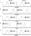
UCB tTreg were assessed for differentiation antigens directly after in vitro expansion (pre-infusion) and 8 days after adoptive transfer into patients (post-infusion). As in Figure 4, expanded UCB tTreg were identified in vivo by HLA mismatching. Cryopreserved samples of expanded UCB tTreg and day 8 PBMC were stained with antibodies to CD45RA (A), CD45RO (B), CCR7 (C), KLRG1 (D) and CD27 (E) and expression determined by flow cytometry.

Similar articles

Cited by

References

    1. Holtan SG, Pasquini M, Weisdorf DJ. Acute graft-versus-host disease: a bench-to-bedside update. Blood. 2014;124:363–73. - PMC - PubMed
    1. McDonald-Hyman C, Turka LA, Blazar BR. Advances and challenges in immunotherapy for solid organ and hematopoietic stem cell transplantation. Sci Transl Med. 2015;7:280rv2. - PMC - PubMed
    1. Bluestone JA, Thomson AW, Shevach EM, Weiner HL. What does the future hold for cell-based tolerogenic therapy? Nat Rev Immunol. 2007;7:650–4. - PubMed
    1. Shevach EM. Mechanisms of foxp3+ T regulatory cell-mediated suppression. Immunity. 2009;30:636–45. - PubMed
    1. Vignali DA, Collison LW, Workman CJ. How regulatory T cells work. Nature reviews Immunology. 2008;8:523–32. - PMC - PubMed

MeSH terms