. 2016 Nov 15;17(8):1950-1961.
doi: 10.1016/j.celrep.2016.10.062.

BORC Functions Upstream of Kinesins 1 and 3 to Coordinate Regional Movement of Lysosomes along Different Microtubule Tracks

Affiliations

BORC Functions Upstream of Kinesins 1 and 3 to Coordinate Regional Movement of Lysosomes along Different Microtubule Tracks

Carlos M Guardia et al. Cell Rep. .

Abstract

The multiple functions of lysosomes are critically dependent on their ability to undergo bidirectional movement along microtubules between the center and the periphery of the cell. Centrifugal and centripetal movement of lysosomes is mediated by kinesin and dynein motors, respectively. We recently described a multi-subunit complex named BORC that recruits the small GTPase Arl8 to lysosomes to promote their kinesin-dependent movement toward the cell periphery. Here, we show that BORC and Arl8 function upstream of two structurally distinct kinesin types: kinesin-1 (KIF5B) and kinesin-3 (KIF1Bβ and KIF1A). Remarkably, KIF5B preferentially moves lysosomes on perinuclear tracks enriched in acetylated α-tubulin, whereas KIF1Bβ and KIF1A drive lysosome movement on more rectilinear, peripheral tracks enriched in tyrosinated α-tubulin. These findings establish BORC as a master regulator of lysosome positioning through coupling to different kinesins and microtubule tracks. Common regulation by BORC enables coordinate control of lysosome movement in different regions of the cell.

Keywords: Arl8; BORC; SKIP; dynein; endosomes; intracellular trafficking; kinesin; lysosomes; microtubules; tubulin modifications.


Figures

Figure 1
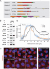
Figure 1. KIF5B and KIF1B are required for peripheral distribution of lysosomes
(A) Schematic representation of kinesins previously implicated in lysosome transport, indicating their structural domains and amino-acid numbers. CC, coiled-coil; FHA, forkhead-associated; PH, pleckstrin homology; UDR, undefined region. (B) Immunoblot analysis of siRNA KD. nt: non-targeting siRNA. The KIF1B and KIF2A siRNAs target all isoforms of these kinesins. GAPDH was used a loading control. The positions of molecular mass markers (in kDa) are indicated at left. (C) Confocal microscopy of HeLa cells transfected with siRNA pools for the indicated kinesins and immunostained for endogenous LAMTOR4 (lysosomal marker, red) and DAPI (nuclear marker, blue). Scale bar, 10 μm. (D) Lysosome distribution as a function of distance from the center of the nucleus, quantified from confocal images using the ImageJ plugin Radial Profile. Graphs show the average distribution ± SEM (standard error of the mean) of lysosomes in 30 different cells from three independent experiments. Student's t-tests (siRNA treatments vs. nt control) were performed at the last point of each distribution to determine the statistical significance of the differences in the cell periphery (arrow heads).
Figure 2
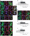
Figure 2. Overexpression of KIF5B-KLC2-SKIP, KIF1Bβ or KIF1A causes accumulation of lysosomes at the cell periphery
(A,B) HeLa cells expressing GFP-KIF5B, alone or in combinations with HA-KLC2 and SKIP-myc, were analyzed by immunofluorescence microscopy (A), and mean fractional distance ± SEM of lysosomes from the cell center was quantified from 30 cells in three independent experiments (B). (C-F) HeLa cells expressing FLAG-KIF1Bβ, GFP-KIF1A, or GFP-KIF1A deletion mutants were analyzed by immunofluorescence microscopy (C,E) and mean fractional distance ± SEM of lysosomes from the cell center was quantified as above (D,F). In A, C and E, lysosomes were stained with antibody to endogenous LAMTOR4, and nuclei were stained with DAPI (blue). Scale bars, 10 μm. Arrows point to cell protrusions where proteins co-localize.
Figure 3
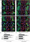
Figure 3. Peripheral accumulation of lysosomes driven by both KIF5B-KLC2-SKIP and KIF1A is dependent on Arl8a/b and BORC
(A-D) Immunofluorescence microscopy of lysosomes stained with antibody to endogenous LAMTOR4 in WT, Arl8b-KO, Arl8b-KO–Arl8a-KD, myrlysin-KO and diaskedin-KO HeLa cells expressing GFP-KIF5B+HA-KLC2+SKIP-myc or GFP-KIF1A. Nuclei were stained with DAPI (blue). Scale bars: 10 μm. Arrows point to cell protrusions where proteins co-localize. (E,F) Quantification of mean fractional distance ± SEM of lysosomes from the cell center from 30 cells in three independent experiments such as those in A-D.
Figure 4
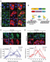
Figure 4. Dynein-mediated centripetal transport of lysosomes is independent of BORC
(A) A dominant-negative mutant of the dynactin p150 subunit (mCh-p150-CC1) was expressed in WT, myrlysin-KO and diaskedin-KO HeLa cells to inactivate dynein-mediated retrograde transport of lysosomes. Cells were stained with antibody to endogenous LAMTOR4 and analyzed by immunofluorescence microscopy. (B) Schematic representation of the constructs designed for accumulation of lysosomes at the cell periphery and their release after treatment with biotin. (C,D) Fluorescence microscopy and quantification of lysosome distribution (performed as explained in the legend to Figure 1D) showing changes in lysosome positioning after biotin-induced release from their peripheral sites in (C) WT and (D) myrlysin-KO HeLa cells. Student's t-tests (at 15 and 30 min of treatment vs. initial time point) were performed near the nucleus and in the periphery to determine the statistical significance of the differences in these regions of interest (arrow heads). In A, C and D, scale bars represent 10 μm. DAPI was used as a nuclear stain (blue). Arrows point to cell protrusions where the proteins co-localize.
Figure 5
Figure 5. Rigor KIF5B and KIF1A mutants associate with different microtubule tracks in HeLa cells
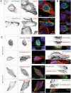
(A) HeLa cells were transfected with plasmids encoding the kinesin rigor mutants GFP-KIF5B-R and GFP-KIF1A-R (green) and immunostained for α-tubulin (red). (B) Co-expression of GRP-KIF5B-R (green) and mCh-KIF1A-R (red). Notice the association of each kinesin with different subsets of microtubules when expressed separately (A) or in the same cell (B). (C) HeLa cells expressing GFP-KIF5B-R and GFP-KIF1A-R (green) were immunostained with antibodies to acetylated or tyrosinated α-tubulin (red). Magnifications of the boxed regions are shown on the right panels. Scale bars, 10 μm. Nuclei were stained with DAPI (blue). Numbers in the merge images are Pearson's correlation coefficients.
Figure 6
Figure 6. Rigor KIF5B and KIF1A trap lysosomes in different regions of the cells
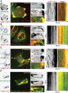
HeLa cells co-expressing LAMP-1-RFP (red) with GFP-tagged (A) KIF5B+HA-KLC2+SKIP-myc, (B) KIF5B-R+HA-KLC2+SKIP-myc, (C) KIF1A, or (D) KIF1A-R (green) were analyzed by live-cell imaging on a spinning-disk confocal microscope. Panels on the left and center correspond to a single frame from Movies S2 and S3. Panels on the right are kymographs from the boxed regions in the corresponding movies. Scale bars represent 10 μm. Arrows in the center panels show moving vesicles where LAMP-1-RFP and GFP-tagged kinesins co-localize.
Figure 7
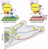
Figure 7. Model for BORC/Arl8-dependent coupling of lysosomes to different kinesins and microtubule tracks
The scheme depicts the interaction chains by which BORC and Arl8 promote the recruitment of both kinesin-1 and kinesin-3 motors to lysosomes. Recruitment of kinesin-1 involves interactions of Arl8 (both a or b isoforms) with the RUN domain of SKIP, of a WD motif in an unstructured region of SKIP with KLC2, and of KLC2 with KIF5B. Recruitment of kinesin-3, on the other hand, is mediated by a direct interaction between Arl8 (a or b) and the CC3 domain of the kinesin-3 KIF1Bβ or KIF1A proteins. Kinesin-1 and kinesin-3 are shown to mediate centrifugal lysosome transport in perinuclear and peripheral regions of the cytoplasm, respectively, though association with different microtubule tracks. The function of both kinesins is countered by BORC-independent centripetal transport mediated by dynein. The schemes are based on findings in this study and previous studies (Pu et al., 2015, Rosa-Ferreira and Munro, 2011, Dumont et al., 2010, Wu et al., 2013, Pernigo et al., 2013).

Similar articles

Cited by

References

    1. Bagshaw RD, Callahan JW, Mahuran DJ. The Arf-family protein, Arl8b, is involved in the spatial distribution of lysosomes. Biochem Biophys Res Commun. 2006;344:1186–1191. - PubMed
    1. Ballabio A. The awesome lysosome. EMBO Mol Med. 2016;8:73–76. - PMC - PubMed
    1. Bar-Peled L, Schweitzer LD, Zoncu R, Sabatini DM. Ragulator is a GEF for the rag GTPases that signal amino acid levels to mTORC1. Cell. 2012;150:1196–1208. - PMC - PubMed
    1. Bentley M, Decker H, Luisi J, Banker G. A novel assay reveals preferential binding between Rabs, kinesins, and specific endosomal subpopulations. J Cell Biol. 2015;208:273–281. - PMC - PubMed
    1. Bielska E, Schuster M, Roger Y, Berepiki A, Soanes DM, Talbot NJ, Steinberg G. Hook is an adapter that coordinates kinesin-3 and dynein cargo attachment on early endosomes. J Cell Biol. 2014;204:989–1007. - PMC - PubMed

LinkOut - more resources