

Review
doi: 10.1126/science.aaf8933.
Neural circuits for pain: Recent advances and current views
Affiliations
- PMID: 27811268
- PMCID: PMC11327866
- DOI: 10.1126/science.aaf8933
Item in Clipboard
Review
Neural circuits for pain: Recent advances and current views
Science.
.
Abstract
The mammalian nervous system encodes many different forms of pain, from those that arise as a result of short-term low-grade interactions with noxious thermal, chemical, or mechanical sources to more serious forms of pain induced by trauma and disease. In this Review, we highlight recent advances in our understanding of the neural circuits that encode these types of pain. Promising therapeutic strategies based on recent advances are also highlighted.
Copyright © 2016, American Association for the Advancement of Science.
Figures
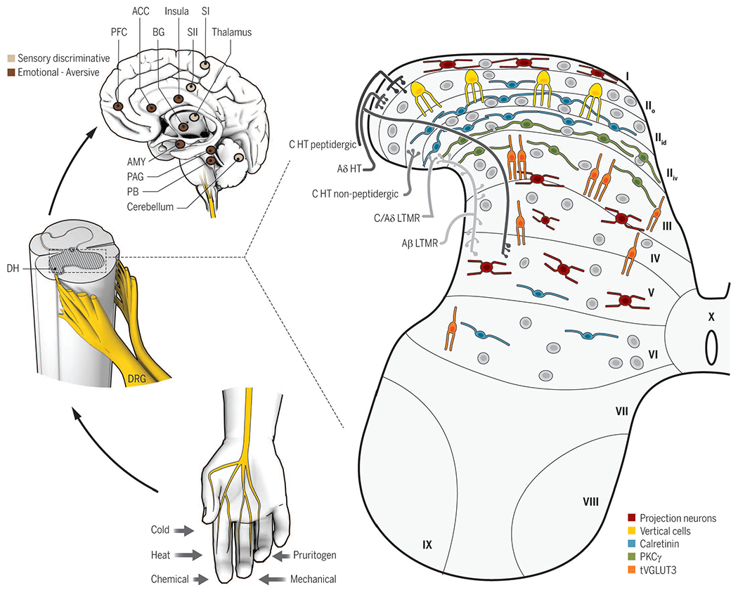
Cutaneous sensory neurons (DRG) are activated by a variety of stimuli (bottom left) and project to the spinal cord dorsal horn (DH, middle left). In the DH (right), the central terminals of high-threshold nociceptors (HT) are located in the most superficial laminae [lamina I to the dorsal part of inner lamina II (IIid)] and lamina V. Low-threshold mechanoreceptors (LTMR) preferentially end in the deep dorsal horn [ventral part of inner lamina II (IIiv) to lamina V]. The spinal cord is divided into 10 laminae (the DH is I to VI) and is composed of numerous neuronal populations. Some identified populations are organized in longitudinal layers (only excitatory neurons are represented): neurons transiently expressing VGLUT3 (tVGLUT3, orange) in laminae III and IV, PKCγ (green) in lamina IIiv, calretinin (blue) in outer lamina II (IIo) and lamina IIid, vertical cells (yellow) in lamina IIo, and projection neurons (red) in laminae I, IV, and V. Projection neurons send information to the brainstem and thalamus and then on to several brain regions implicated in sensory-discriminative (upper left, light brown) and emotional (upper left, dark brown) sensory perception. ACC, anterior cingular cortex; SI (II), primary (secondary) somatosensory cortex; PAG, periaqueductal gray area; PB, parabrachial nucleus; AMY, amygdala; PFC, prefrontal cortex; BG, basal ganglia.
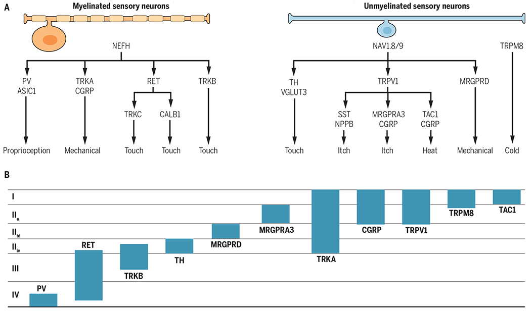
(A) The categories of myelinated and unmyelinated neurons and their respective functional roles are derived from large-scale transcriptional analyses, behavioral analyses, and the literature. (B) The locations of the central terminations of the primary sensory neuron categories are shown. The schematic is based on analyses of gene reporter mouse lines (PV, RET, TRKB, TH, MRGPRD, MRGPRA3, CGRP, and TRPM8) and immunohistological analyses (TRKA, TRPV1, TAC1). Myelinated neurons preferentially express neurofilament heavy chain (NEFH), and unmyelinated neurons preferentially express the sodium channels Nav1.8 and −1.9. Laminae are indicated on the left. RET, ret proto-oncogene; CALB1, calbindin 1; TAC1, tachykinin 1; SST, somatostatin; NPPB; natriuretic peptide type B.
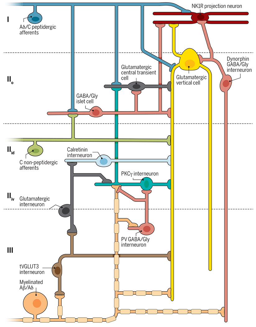
Peripheral nociceptors (blue) project onto excitatory interneurons in lamina IIo (central cell, dark gray; vertical cells, yellow) and onto neurokinin 1 receptor (NK1R) projection neurons (red) in lamina I. Nonpeptidergic afferents expressing MRGPRD (green) project to lamina IIid, including to excitatory vertical cells with ventrally directed elongated dendrites. Both primary afferents also contact inhibitory islet cells (horizontally elongated, pink). Stimulation of nociceptive afferents activates excitatory central cells, vertical cells, and NK1R projection neurons to mediate noxious pain. Inhibitory islet cells modulate this activity. Innocuous afferents (orange) project onto excitatory interneurons expressing tVGLUT3 in lamina III (brown), PKCγ in lamina IIiv (teal), and vertical cells in lamina IIo. Myelinated afferents also contact PV inhibitory interneurons in lamina III (radial, pink) and dynorphin inhibitory vertical cells in lamina IIo (vertical, pink). tVGLUT3 interneurons project onto excitatory vertical cell dendrites and intermediate excitatory interneurons in lamina III. Intermediate interneurons project onto PKCγ and calretinin excitatory interneurons in lamina IIiv. Inhibitory interneurons prevent A-fiber–mediated activation of the nociceptive network through feed-forward circuits that act on PKCγ interneurons, vertical cells, and NK1R projection neurons. After nerve injury, inhibition by PV and dynorphin interneurons is reduced, allowing A-fiber–mediated activation of a dorsally directed circuit that includes tVGLUT3 and PKCγ neurons. After inflammatory injury, reduction in a still-unknown mechanism of disinhibition allows A-fiber–mediated activation of a dorsally directed circuit that includes tVGLUT3 and lamina IIiv calretinin neurons.
Similar articles
-
The trigeminal system in birds and nociception.Cent Nerv Syst Agents Med Chem. 2009 Jun;9(2):150-8. doi: 10.2174/187152409788452072. Cent Nerv Syst Agents Med Chem. 2009. PMID: 20021348
-
Presynaptic inhibition of nociceptive neurotransmission by somatosensory neuron-secreted suppressors.Sci China Life Sci. 2017 Sep;60(9):1013-1018. doi: 10.1007/s11427-017-9061-y. Epub 2017 Jun 15. Sci China Life Sci. 2017. PMID: 28624955 Review.
-
[Neuropathic pain and neuroplasticity in functional imaging studies].Schmerz. 2010 Apr;24(2):137-45. doi: 10.1007/s00482-010-0902-6. Schmerz. 2010. PMID: 20376602 Review. German.
-
[Mechanisms of endogenous pain modulation illustrated by placebo analgesia : functional imaging findings].Schmerz. 2010 Apr;24(2):122-9. doi: 10.1007/s00482-010-0901-7. Schmerz. 2010. PMID: 20376600 Review. German.
-
Pain imaging in health and disease--how far have we come?J Clin Invest. 2010 Nov;120(11):3788-97. doi: 10.1172/JCI43498. Epub 2010 Nov 1. J Clin Invest. 2010. PMID: 21041961 Free PMC article. Review.
Cited by
-
Unveiling the Pain Relief Potential: Harnessing Analgesic Peptides from Animal Venoms.Pharmaceutics. 2023 Dec 13;15(12):2766. doi: 10.3390/pharmaceutics15122766. Pharmaceutics. 2023. PMID: 38140106 Free PMC article. Review.
-
Neuropeptide Y release in the rat spinal cord measured with Y1 receptor internalization is increased after nerve injury.Neuropharmacology. 2019 Nov 1;158:107732. doi: 10.1016/j.neuropharm.2019.107732. Epub 2019 Aug 2. Neuropharmacology. 2019. PMID: 31377198 Free PMC article.
-
Pentraxin-3 in the Spinal Dorsal Horn Upregulates Nectin-1 Expression in Neuropathic Pain after Spinal Nerve Damage in Male Mice.Brain Sci. 2022 May 15;12(5):648. doi: 10.3390/brainsci12050648. Brain Sci. 2022. PMID: 35625034 Free PMC article.
-
Microglia in Pain: Detrimental and Protective Roles in Pathogenesis and Resolution of Pain.Neuron. 2018 Dec 19;100(6):1292-1311. doi: 10.1016/j.neuron.2018.11.009. Neuron. 2018. PMID: 30571942 Free PMC article. Review.
-
Neck Pain: Do We Know Enough About the Sensorimotor Control System?Front Comput Neurosci. 2022 Jul 15;16:946514. doi: 10.3389/fncom.2022.946514. eCollection 2022. Front Comput Neurosci. 2022. PMID: 35910451 Free PMC article. Review.
References
-
- Bennett DL, Woods CG, Lancet Neurol. 13, 587–599 (2014). - PubMed
-
- Sabatowski R, Schäfer D, Kasper SM, Brunsch H, Radbruch L, Curr. Pharm. Des 10, 701–716 (2004). - PubMed
-
- Julius D, Carlson JR, Curr. Opin. Neurobiol 34, v–vi (2015). - PubMed
-
- Caterina MJ et al., Science 288, 306–313 (2000). - PubMed
-
- Vriens J et al., Neuron 70, 482–494 (2011). - PubMed
Publication types
MeSH terms
Grants and funding
LinkOut - more resources
Full Text Sources
Other Literature Sources
Medical
