. 2016 Nov 15;197(10):4053-4065.
doi: 10.4049/jimmunol.1600323. Epub 2016 Oct 19.

Secreted NS1 Protects Dengue Virus from Mannose-Binding Lectin-Mediated Neutralization

Affiliations

Secreted NS1 Protects Dengue Virus from Mannose-Binding Lectin-Mediated Neutralization

Somchai Thiemmeca et al. J Immunol. .

Abstract

Flavivirus nonstructural protein 1 (NS1) is a unique secreted nonstructural glycoprotein. Although it is absent from the flavivirus virion, intracellular and extracellular forms of NS1 have essential roles in viral replication and the pathogenesis of infection. The fate of NS1 in insect cells has been more controversial, with some reports suggesting it is exclusively cell associated. In this study, we confirm NS1 secretion from cells of insect origin and characterize its physical, biochemical, and functional properties in the context of dengue virus (DENV) infection. Unlike mammalian cell-derived NS1, which displays both high mannose and complex type N-linked glycans, soluble NS1 secreted from DENV-infected insect cells contains only high mannose glycans. Insect cell-derived secreted NS1 also has different physical properties, including smaller and more heterogeneous sizes and the formation of less stable NS1 hexamers. Both mammalian and insect cell-derived NS1 bind to complement proteins C1s, C4, and C4-binding protein, as well as to a novel partner, mannose-binding lectin. Binding of NS1 to MBL protects DENV against mannose-binding lectin-mediated neutralization by the lectin pathway of complement activation. As we detected secreted NS1 and DENV together in the saliva of infected Aedes aegypti mosquitoes, these findings suggest a mechanism of viral immune evasion at the very earliest phase of infection.


Figures

FIGURE 1
FIGURE 1. Detection of extracellular NS1 derived from DENV-infected insect cells
C6/36 (black bars), PscloneD (white bars) and BHK (grey bars) cells were infected with DENV at an MOI of 0.01, 0.1 or 1.0 for 2 days. The levels of secreted NS1 (A), cell viability (B), percentage of NS1- (C) and E- (D) expressing cells and DENV production (E) were determined by NS1-ELISA, flow cytometry analysis using propidium iodide exclusion, anti-NS1 mAb (clone 1F11) and anti-E mAb (clone 4G2) and FFA, respectively. The data are shown as mean ± standard deviation (SD) of three to four independent experiments. Asterisks denote levels of NS1 detected in supernatants that are statistically different (* P < 0.05). The correlation coefficients between secreted NS1 and DENV production in DENV-infected C6/36 (F), DENV-infected PscloneD (G) and DENV-infected BHK (H) were analyzed by linear regression. NS1 in 50-fold concentrated supernatants of DENV-infected C6/36, AP-61 and TRA-284 cells was analyzed by Western blotting using anti-DENV NS1 mAb clone 1F11 (I).
FIGURE 2
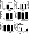
FIGURE 2. Tunicamycin treatment inhibits NS1 and virion release from DENV-infected insect cells
At 24 h after DENV infection, C6/36 cells were extensively washed to remove NS1-containing culture medium and treated with fresh medium containing either 0.1% DMSO or 10 μg/ml of tunicamycin in 0.1% DMSO for 6 h. Levels of NS1 in supernatants (A), percentage and mean fluorescent intensity (MFI) of NS1 positive cells (B) and percentages of E-positive cells (C), percentage of living cells (D), total cell number (E) and intracellular and extracellular DENV genome copies (F) were analyzed as described in Materials and Methods. The data are shown as mean ± SD from three independent experiments. Asterisks denote the means that are statistically different among indicated groups (* P < 0.05).
FIGURE 3
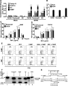
FIGURE 3. Oligomeric assembly, glycosylation pattern and stability of NS1 secreted from DENV-infected insect cells
Fifty-fold concentrated supernatants (lanes 1 and 3) and cell lysates (lanes 2 and 4) from DENV-infected C6/36 cells (lanes 1 and 2) and Vero cells (lanes 3 and 4) were separated by Native-PAGE (A, upper panel) and reducing 10% SDS-PAGE (A, lower panel) and Western blotted with 1F11 DENV NS1 specific mAb. B. Fifty-fold concentrated DENV-2 infected C6/36 (dashed line) and PscloneD (solid line) supernatants were passed over a Superdex 200 column and fractions were collected and quantitated for NS1 concentration by NS1-ELISA. Molecular weights of reference protein standards fractionated in parallel are depicted. C. Fifty-fold concentrated DENV-infected C6/36 or PscloneD supernatant was loaded on a 5% to 55% sucrose gradient. NS1 in each fraction was detected by Western blotting with 1F11 anti-NS1 mAb. Arrowheads indicate molecular weights of protein standards. D. Endoglycosidase (Endo H or PNGase F) treatment of extracellular NS1 derived from C6/36 (lanes 1, 2, 5 and 6) and PscloneD (lanes 3, 4, 7 and 8) cells prior to Western blot analysis with 1F11 anti-NS1 mAb. E-F. NS1 stability test.Supernatants from DENV-infected C6/36 and Vero cells were incubated in the presence of a protease inhibitor (10 mM PMSF) at 4°C or 37°C for 3 days. Amounts of NS1 at each time point of C6/36 cell-derived or Vero cell-derived at 4°C and 37°C were determined by ELISA and calculated by normalizing the zero-time point of each condition to 100 percent (E) and by Western blotting with 1F11 anti-NS1 mAb (F). Error bars indicate SD corresponding to three independent experiments. Values that are significantly different from the value for zero-time point are indicated by asterisks as follows: *, P < 0.05; **, P < 0.01; ***, P < 0.001.
FIGURE 4
FIGURE 4. Functional properties of extracellular NS1 derived from DENV-infected insect cells
A. Insect-cell derived NS1 binds human complement. Microtiter plates were coated with BSA, factor H, C1s proenzyme (ProC1s), C4 and C4BP (5 μg/ml) and incubated with culture supernatants of DENV-infected C6/36, PscloneD and S2-NS1 cells. Bound NS1 was detected with specific mAbs. Values [optical density (O.D.) at 492 nm.] of NS1 binding to each complement component were subtracted from those of BSA control. Error bars indicate SD corresponding to three independent experiments. Asterisks denote NS1 binding that is statistically different from corresponding mock controls (** P < 0.01 and *** P < 0.001). Levels of NS1 in the supernatants were determined by NS1-ELISA (B). C-D. Insect-cell derived NS1 binds to cell surfaces via GAGs. Serum-free supernatants of DENV-infected PscloneD (C and D), C6/36 (C and D) and S2-NS1 (D) cells containing indicated concentrations of NS1 were incubated with BHK (C) and HaCat (D) cells and NS1 binding was detected by 2G6 anti-DENV NS1 mAb. Binding also was performed in the presence of 10 mM heparin (C). Error bars indicate SD of the mean corresponding to three independent experiments. Asterisks denote the means that are statistically different from 0 μg/ml of NS1 (* P < 0.05, ** P < 0.01 and *** P < 0.001). E. NS1 secreted from insect cells recruits C4BP to the cell surface. C4BP or BSA (2.5 μg/ml) was incubated with serum-free supernatants of S2-NS1 cells and 106 BHK cells. Binding of NS1 and C4BP to the cell surface was assessed by mAbs to DENV NS1 or C4BP and analyzed by flow cytometry. Examples of contour plots from three independent experiments are depicted. The y-axis indicates the levels of cell surface-associated C4BP or NS1, and the x-axis shows the forward scatter (FSC) of the cell population. Percentages of cells positive for surface-NS1 or C4BP staining are shown in the right upper quadrant. F. Extracellular NS1 derived from insect cells recruits C4BP to degrade C4b on the cell surface. Serum-free supernatants (containing 12 μg/ml NS1) from DENV-infected PscloneD (lane 2), DENV-infected C6/36 (lanes 3 and 6) or S2-NS1 (lane 4) cells were preincubated with 2.5 μg/ml C4BP and the NS1-C4BP solution was added to BHK cells. After three washes, the cells were incubated with C4b and factor I for 15 min. Cofactor activity was determined in the supernatants by the appearance of C4b cleavage products, a 70-kDa α3-C4d and a 45-kDa C4d after 12% SDS-PAGE under reducing condition followed by Western blot with anti-C4d mAb. G. Diagram of factor I (FI)-mediated cleavage of C4b in the presence of a cofactor C4BP. FI cleaves the α′chain of C4b at two sites (designated as A and B). Cleavage at either one of the two sites generates two partial cleavage fragments, C4d-α4 (59 kDa) and α3-C4d (70 kDa). Further cleavage by FI yields the final product C4d (45 kDa).
FIGURE 5
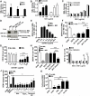
FIGURE 5. NS1 protects DENV from MBL-mediated neutralization
A-C. NS1 binds directly to MBL. Purified human MBL was coated on a microtiter plate and incubated with NS1 containing supernatants from DENV-infected C6/36, DENV-infected PscloneD and S2-NS1 cells (A) or with increasing concentrations of NS1 in culture supernatants of DENV-infected PscloneD and S2-NS1 cells (B) or with increasing concentrations of purified soluble NS1 from culture supernatants of DENV-infected C6/36 or Vero cells (C). Bound NS1 was detected with an anti-NS1 specific mAb. Error bars indicate SD for three independent experiments. D. Co-immunoprecipitation experiments. Serum-free supernatants from BHK-DENV2-Rep or control BHK cells were incubated with purified human MBL (0.75 μg) and Western blots were performed after immunoprecipitation with anti-DENV NS1 2G6 mAb-Sepharose. Immunoprecipitates were probed with a mouse anti-human MBL mAb after non-reduced (upper panel) or reduced (lower panel) SDS-PAGE. E-F. NS1 reduces MBL binding to DENV. DENV virions were captured on a microtiter plate using an anti-DENV prM protein-specific mAb and incubated with purified human MBL (3 μg/ml) with or without increasing concentrations of NS1 containing supernatants from S2-NS1 cells (E). MBL binding to captured DENV was detected with an anti-MBL specific mAb and inhibited by the presence of 20 mM EDTA. F. Culture supernatants from S2-NS1 cells (containing 12.5 μg/ml NS1) were incubated with anti-NS1 mAb (2E11)-Sepharose, anti-E mAb (4G2)-Sepharose or without Sepharose (no beads) prior to addition of purified human MBL and subjected to ELISA as described in E. MBL binding to DENV captured on a microtiter plate in GVB++ buffer (without NS1) or in buffer containing 20 mM EDTA were used as positive and negative controls, respectively. G-J. NS1 inhibits MBL-mediated neutralization of DENV. DENV virions were incubated with purified human MBL without or with indicated concentrations of NS1 in supernatants from S2-NS1 cells or with NS1 supernatants alone. The mixtures were then added to a BHK monolayer, and after carboxymethylcellulose overlay, cells were cultured for 3 days. After fixation and staining with anti-DENV E mAb, infected foci were counted (G). H. DENV virions were incubated with purified MBL in GVB++ buffer without or with NS1 containing supernatants (12 μg/ml) from S2-NS1 cells that were preincubated with anti-NS1 mAb (2E11)- or anti-E mAb (4G2)-Sepharose. The samples were added to a BHK monolayer and DENV-infected foci were determined as described in G. MBL-mediated neutralization of DENV was abolished by 20 mM EDTA. I. DENV virions were incubated with purified human MBL with or without indicated concentrations of purified soluble NS1 from DENV-infected C6/36 cells or with BSA control protein prior to the addition to a BHK monolayer to assess viral infectivity as described in G. J. DENV virions were incubated with MBL premixed with purified hexameric NS1 derived from BHK-DENV2-Rep, thyroglobulin or bovine serum albumin (BSA) at molar ratios of 1:1, 1:5, and 1:10 (MBL:protein). The samples were added to BHK monolayers, and after agarose overlay, cultured for 5 days. After fixation and staining of wells, plaques were counted. Soluble mannan (100 μg/ml) was used as an MBL inhibitor. K. Soluble NS1 facilitates DENV attachment to cells in the presence of human MBL. DENV was incubated with 80 μg/ml MBL (diluted with GVB++ buffer) alone or with MBL plus BSA or purified NS1 (50 μg/ml) derived from S2-NS1 cells or DENV-infected Vero cells. The mixture was then added to BHK cell suspensions for 2 h on ice. Cell-bound DENV was determined by real time RT-PCR. Error bars indicate SD corresponding to three independent experiments. Asterisks denote the means that are statistically different among indicated groups (* P < 0.05, ** P < 0.01 and *** P < 0.001).
FIGURE 6
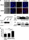
FIGURE 6. Both DENV and soluble NS1 are released during blood feeding from infected mosquitoes
A. Representative bright field and immunofluorescence images of the salivary glands of mock- and DENV-infected mosquitoes. Aedes aegypti female mosquitoes were challenged orally with DENV by feeding them with an infectious blood meal and incubated for 14 days. Salivary glands of mosquitoes were harvested, smeared, and fixed on glass slides. NS1 was detected using a mouse anti-DENV NS1 mAb 1F11 (1 μg/ml) followed by Cy3-conjugated goat anti-mouse IgG. Nuclei were stained with Hoechst, a DNA-specific dye. Salivary glands harvested from uninfected mosquitoes served as a negative control. Analysis was performed by confocal microscopy. B. Expression of NS1 in the salivary glands of DENV-infected mosquitoes was analyzed by Western blotting. Fifty-fold concentrated supernatants from DENV-infected C6/36 cells (DENV sup) and the salivary glands harvested from uninfected mosquitoes (Mock) served as positive and negative controls, respectively. C-D. At 14 days after initial challenge with DENV, a total of 600 to 1200 mosquitoes were fed with a second non-infectious blood meal, the presence of DENV and NS1 in the remaining blood were determined by RT-PCR (C), Western blotting with DENV NS1 specific mAb 1F11 (D) and NS1-ELISA (E), respectively. Error bars indicate SD corresponding to three independent experiments. Asterisks denote the means that are statistically different among indicated groups (*** P < 0.001).
FIGURE 7
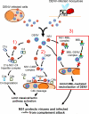
FIGURE 7. Model of complement antagonism by DENV NS1
Together with DENV virions, soluble NS1 is released from DENV-infected insect or mammalian cells. Attenuation of the classical and lectin pathways of complement activation in tissue is promoted by three mechanisms: 1) NS1 binds proC1s and C4 forming a tripartite complex promoting the cleavage of C4 to C4b which is rapidly inactivated by hydrolysis in the fluid phase (C4 degradation), thereby limiting the supply of native C4 (24). 2) NS1 binds and recruits C4BP to the cell surface to restrict the classical and lectin pathway C3 and C5 convertases via the cofactor activity of C4BP on cells (25). 3) NS1 competitively binds MBL and prevents the recognition of DENV by MBL, thereby protecting the virus from MBL-mediated neutralization. Overall, NS1 possesses complement evasion functions and protects DENV and DENV-infected cells from complement activation and inhibition.

Similar articles

Cited by

References

    1. WHO . Comprehensive Guidelines for Prevention and Control of Dengue and Dengue Haemorrhagic Fever, Revised and expanded edition ed. WHO Library Cataloguing-in-Publication data, World Health Organization, Regional Office for South-East Asia; 2011.
    1. Bhatt S, Gething PW, Brady OJ, Messina JP, Farlow AW, Moyes CL, Drake JM, Brownstein JS, Hoen AG, Sankoh O, Myers MF, George DB, Jaenisch T, Wint GR, Simmons CP, Scott TW, Farrar JJ, Hay SI. The global distribution and burden of dengue. Nature. 2013;496:504–507. - PMC - PubMed
    1. Brandt WE, Chiewslip D, Harris DL, Russell PK. Partial purification and characterization of a dengue virus soluble complement-fixing antigen. J Immunol. 1970;105:1565–1568. - PubMed
    1. Muller DA, Young PR. The flavivirus NS1 protein: molecular and structural biology, immunology, role in pathogenesis and application as a diagnostic biomarker. Antiviral Res. 2013;98:192–208. - PubMed
    1. Scaturro P, Cortese M, Chatel-Chaix L, Fischl W, Bartenschlager R. Dengue Virus Non-structural Protein 1 Modulates Infectious Particle Production via Interaction with the Structural Proteins. PLoS Pathog. 2015;11:e1005277. - PMC - PubMed

Publication types

Substances