. 2016 Oct 13:8:234.
doi: 10.3389/fnagi.2016.00234. eCollection 2016.

Nox-2-Mediated Phenotype Loss of Hippocampal Parvalbumin Interneurons Might Contribute to Postoperative Cognitive Decline in Aging Mice

Affiliations

Nox-2-Mediated Phenotype Loss of Hippocampal Parvalbumin Interneurons Might Contribute to Postoperative Cognitive Decline in Aging Mice

Li-Li Qiu et al. Front Aging Neurosci. .

Abstract

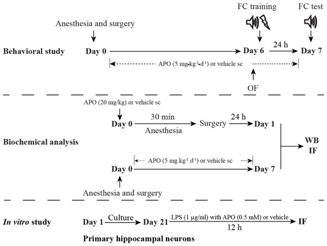
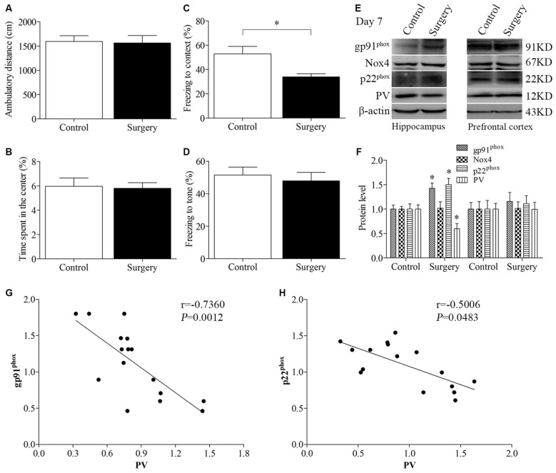
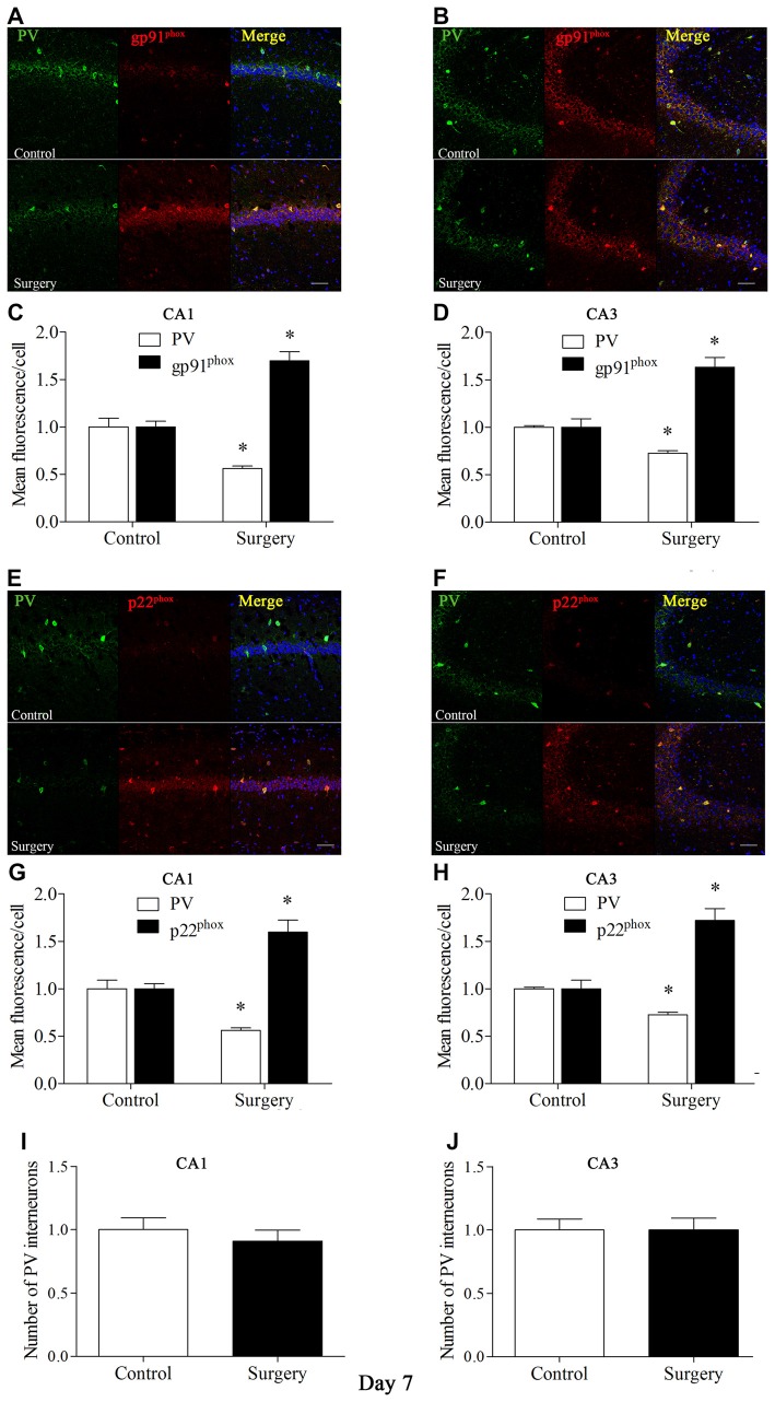
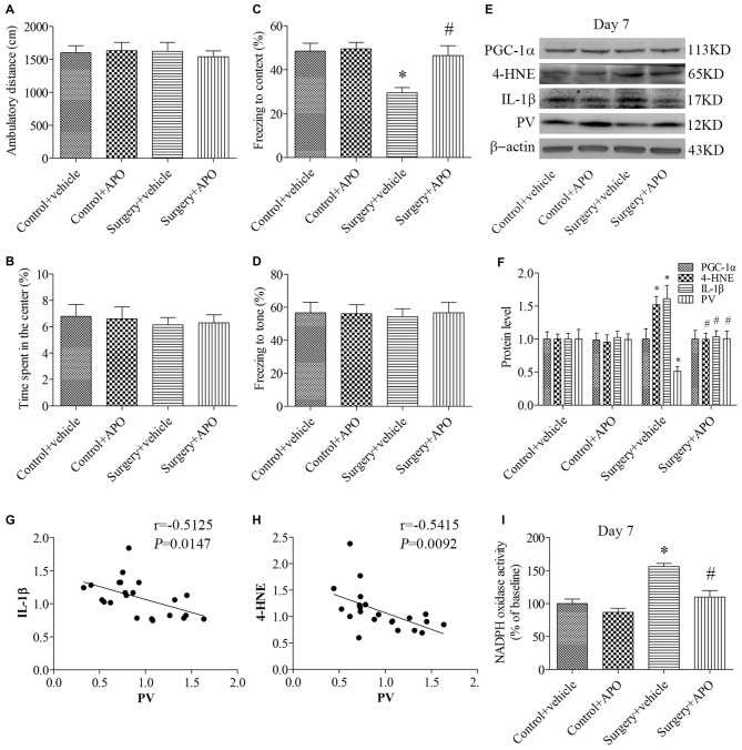
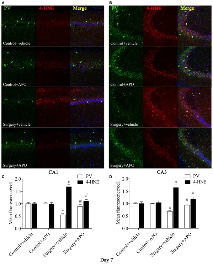
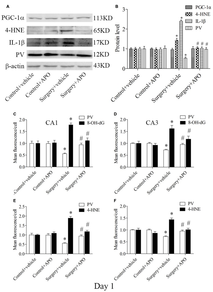
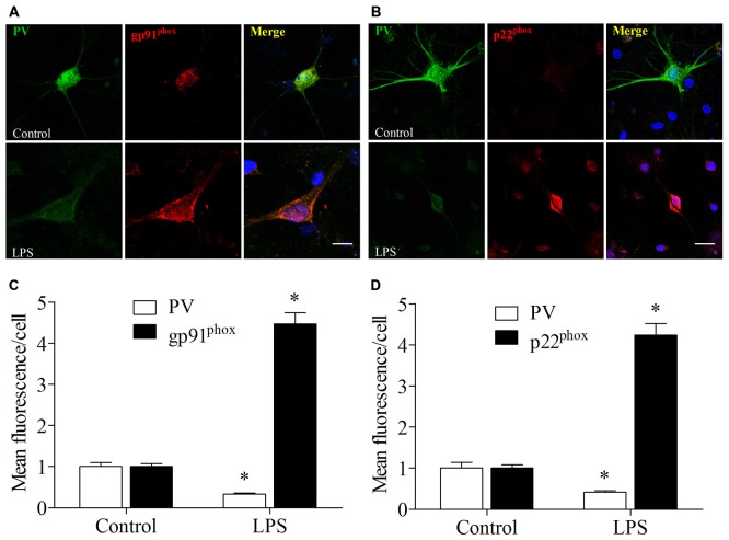
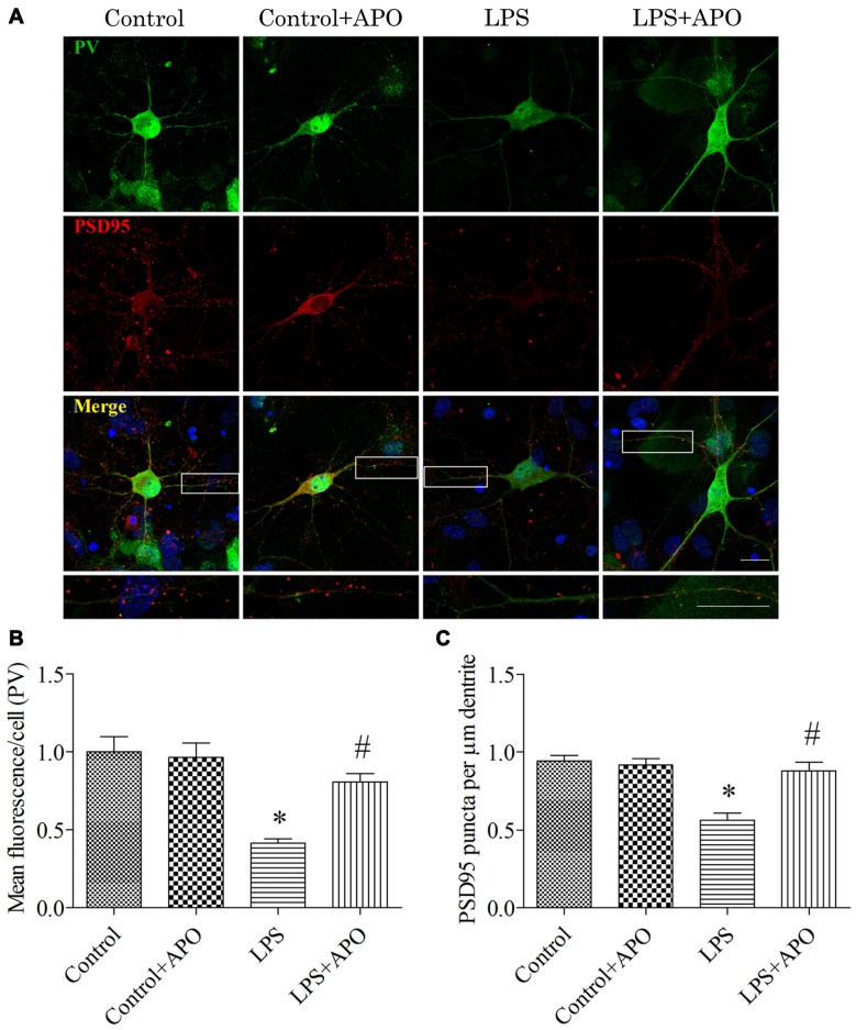
Postoperative cognitive decline (POCD) is a common complication following anesthesia and surgery, especially in elderly patients; however, the precise mechanisms of POCD remain unclear. Here, we investigated whether nicotinamide adenine dinucleotide phosphate (NADPH) oxidase mediated-abnormalities in parvalbumin (PV) interneurons play an important role in the pathophysiology of POCD. The animal model was established using isoflurane anesthesia and exploratory laparotomy in 16-month-old male C57BL/6 mice. For interventional experiments, mice were chronically treated with the NADPH oxidase inhibitor apocynin (APO). Open field and fear conditioning behavioral tests were performed on day 6 and 7 post-surgery, respectively. In a separate experiment, brain tissue was harvested and subjected to biochemical analysis. Primary hippocampal neurons challenged with lipopolysaccharide (LPS) in vitro were used to investigate the mechanisms underlying the oxidative stress-induced abnormalities in PV interneurons. Our results showed that anesthesia and surgery induced significant hippocampus-dependent memory impairment, which was accompanied by PV interneuron phenotype loss and increased expression of interleukin-1β (IL-1β), markers of oxidative stress and NADPH oxidase 2 (Nox2) in the hippocampus. In addition, LPS exposure increased Nox2 level and decreased the expression of PV and the number of excitatory synapses onto PV interneurons in the primary hippocampal neurons. Notably, treatment with APO reversed these abnormalities. Our study suggests that Nox2-derived reactive oxygen species (ROS) production triggers, at least in part, anesthesia- and surgery-induced hippocampal PV interneuron phenotype loss and consequent cognitive impairment in aging mice.

Keywords: Nox2; aging; apocynin; hippocampus; parvalbumin; postoperative cognitive decline.


Figures

Figure 1
Figure 1
Schematic timeline of the experimental procedure. For the behavioral study, the mice underwent anesthesia and surgery on day 0. After that, the mice were treated with 5 mg/kg apocynin (APO) or with an equal volume of vehicle s.c. at 1 h after the surgery, and then with the same treatment once daily for the next seven consecutive days. The open field test and fear conditioning training were performed on day 6. The fear conditioning test was performed on day 7. The mice used for the biochemical analysis were not subjected to the behavioral tests and were sacrificed on days 1 and 7, respectively. The primary hippocampal neuronal culture was used to investigate the effects of oxidative stress on parvalbumin (PV) interneurons in vitro study.
Figure 2
Figure 2
Impaired contextual fear conditioning behavioral phenotype, up-regulated NADPH oxidase 2 (Nox2), and down-regulated PV in the hippocampus on day 7 post-surgery. There was no significant difference in (A) the ambulatory distance and (B) time spent in the center in the open field test between the two groups on day 6 post-surgery. (C) The freezing time to context was decreased in the surgery group compared with the control group. (D) No significant difference was detected in the cued fear conditioning between the two groups. (E,F) Western blot results demonstrated that the expression of PV was decreased in the hippocampus but not in the prefrontal cortex in the surgery group compared with the control group. The expression of gp91phox and p22phox was increased in the hippocampus but not in the prefrontal cortex in the surgery group. There was no significant difference in the expression of Nox4 in the hippocampus or prefrontal cortex between the two groups. Correlation analysis showed that the expression of PV was negatively correlated with the (G) gp91phox and (H) p22phox expression in the hippocampus. The data are presented as the mean ± S.E.M. (10 mice in each group in A–D, 5 mice in each group in E–H). *p < 0.05 compared with the control group.
Figure 3
Figure 3
Immunofluorescent co-localization of PV and gp91phox or p22phox in areas Carbonic anhydrase 1 (CA1) and CA3 of the hippocampus on day 7 post-surgery. (A,B) Representative images of PV (green) and gp91phox (red) expression in areas CA1 and CA3. (C,D) Quantification of PV and gp91phox fluorescence in areas CA1 and CA3. (E,F) Representative images of PV (green) and p22phox (red) expression in areas CA1 and CA3. (G,H) Quantification of PV and p22phox fluorescence in areas CA1 and CA3. (I,J) Analysis of number of PV interneurons in areas CA1 and CA3. Scale bar = 50 μm. 6-diamidino-2-phenyl-indole (DAPI) staining is shown in blue. The data are presented as the mean ± S.E.M. (8 mice in each group). *p < 0.05 compared with the control group.
Figure 4
Figure 4
Rescuing effects of APO on surgery-induced impaired cognition, decreased PV expression, increased interleukin-1β (IL-1β) and 4-HNE expression and nicotinamide adenine dinucleotide phosphate (NADPH) oxidase activity in the hippocampus on day 7 post-surgery. There was no significant difference in (A) the ambulatory distance and (B) time spent in the center in the open field test among the four groups on day 6 post-surgery. (C) The freezing time to context was decreased in the surgery + vehicle group compared with the control + vehicle group, which was rescued by APO treatment. (D) No significant difference was detected in the cued fear conditioning among the four groups. (E,F) Western blot results demonstrated the decreased PV expression and increased IL-1β and 4-HNE expression in the hippocampus in the surgery + vehicle group compared with the control + vehicle group, which was rescued by APO treatment. There was no significant difference in the expression of proliferator-activated receptor coactivator 1α (PGC-1α) among the four groups. Correlation analysis showed that the PV expression was negatively correlated with the (G) IL-1β and (H) 4-HNE expression. (I) NADPH oxidase activity was increased in the surgery + vehicle group compared with the control + vehicle group, which was rescued by APO treatment. The data are presented as the mean ± S.E.M. (10 mice in each group in A–D, 5 mice in each group in E–I). *p < 0.05 compared with the control + vehicle group, #p < 0.05 compared with the surgery + vehicle group.
Figure 5
Figure 5
Immunofluorescent co-localization of PV and 8-OH-dG in areas Carbonic anhydrase 1 (CA1) and CA3 of the hippocampus on day 7 post-surgery. (A,B) Representative images of PV (green) and 8-OH-dG (red) expression in areas CA1 and CA3. (C,D) Quantification of PV and 8-OH-dG fluorescence in areas CA1 and CA3. Scale bar = 50 μm. DAPI staining is shown in blue. The data are presented as the mean ± S.E.M. (8 mice in each group). *p < 0.05 compared to the control + vehicle group, #p < 0.05 compared to the surgery + vehicle group.
Figure 6
Figure 6
Immunofluorescent co-localization of PV and 4-HNE in areas CA1 and CA3 of the hippocampus on day 7 post-surgery. (A,B) Representative images of PV (green) and 4-HNE (red) expression in areas CA1 and CA3. (C,D) Quantification of PV and 4-HNE fluorescence in areas CA1 and CA3. Scale bar = 50 μm. DAPI staining is shown in blue. The data are presented as the mean ± S.E.M. (8 mice in each group). *p < 0.05 compared to the control + vehicle group, #p < 0.05 compared to the surgery + vehicle group.
Figure 7
Figure 7
Hippocampal oxidative stress detected by Western blot and double-immunofluorescence staining in PV interneurons on day 1 post-surgery. (A) Representative Western blot images of PV, IL-1β, 4-HNE and PGC-1α in the hippocampus in the four groups; (B) Quantitative analysis. (C,D) Quantification of PV and 8-OH-dG fluorescence in areas CA1 and CA3. (E,F) Quantification of PV and 4-HNE fluorescence in areas CA1 and CA3. The data are presented as the mean ± S.E.M. (5 mice in each group in A,B, 8 mice in each group in C–F). *p < 0.05 compared to the control + vehicle group, #p < 0.05 compared to the surgery + vehicle group.
Figure 8
Figure 8
Lipopolysaccharide (LPS) exposure increased gp91phox and p22phox expression and decreased PV immunoreactivity in primary neuronal cultures. (A,B) Representative images of PV (green) and gp91phox (red) or p22phox (red) expression in primary hippocampal neuronal cultures. (C,D) Quantification of PV, gp91phox, and p22phox fluorescence in the primary hippocampal neuronal cultures. Scale bar = 20 μm. DAPI staining is shown in blue. The data are presented as the mean ± S.E.M. (12 cells in each group). *p < 0.05 compared to the control group.
Figure 9
Figure 9
Rescuing effects of APO on LPS-stimulated decreases in the PV expression and number of excitatory synapses onto PV interneurons in primary neuronal cultures. (A) Cultured hippocampal neurons stained for PV (green) and PSD95 (red); the boxed area is enlarged in the bottom image. (B) Quantification of PV fluorescence in the primary hippocampal neuronal cultures. (C) The number of PSD95-positive puncta per μm dendrite in the four groups. Scale bar = 20 μm. DAPI staining is shown in blue. The data are presented as the mean ± S.E.M. (12 cells in each group). *p < 0.05 compared to the control group, #p < 0.05 compared to the LPS group.

Similar articles

Cited by

References

    1. An L.-N., Yue Y., Guo W.-Z., Miao Y.-L., Mi W.-D., Zhang H., et al. . (2013). Surgical trauma induces iron accumulation and oxidative stress in a rodent model of postoperative cognitive dysfunction. Biol. Trace Elem. Res. 151, 277–283. 10.1007/s12011-012-9564-9 - DOI - PubMed
    1. Barrientos R. M., Hein A. M., Frank M. G., Watkins L. R., Maier S. F. (2012). Intracisternal interleukin-1 receptor antagonist prevents postoperative cognitive decline and neuroinflammatory response in aged rats. J. Neurosci. 32, 14641–14648. 10.1523/jneurosci.2173-12.2012 - DOI - PMC - PubMed
    1. Bartos M., Vida I., Jonas P. (2007). Synaptic mechanisms of synchronized gamma oscillations in inhibitory interneuron networks. Nat. Rev. Neurosci. 8, 45–56. 10.1038/nrn2044 - DOI - PubMed
    1. Beaudoin G. M., III, Lee S.-H., Singh D., Yuan Y., Ng Y.-G., Reichardt L. F., et al. . (2012). Culturing pyramidal neurons from the early postnatal mouse hippocampus and cortex. Nat. Protoc. 7, 1741–1754. 10.1038/nprot.2012.099 - DOI - PubMed
    1. Bedard K., Krause K.-H. (2007). The NOX family of ROS-generating NADPH oxidases: physiology and pathophysiology. Physiol. Rev. 87, 245–313. 10.1152/physrev.00044.2005 - DOI - PubMed

LinkOut - more resources